 Movie
Movie Controller

[English] 日本語
 Yorodumi
- EMDB-1317: Dodecameric structure and ATPase activity of the human TIP48/TIP4... -
+ Open data

- Basic information
| Entry | Database: EMDB / ID: EMD-1317 | |||||||||
|---|---|---|---|---|---|---|---|---|---|---|
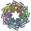
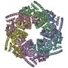
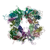
| Title | Dodecameric structure and ATPase activity of the human TIP48/TIP49 complex. | |||||||||
 Map data Map data | This is the masked 3D map of the TIP48/TIP49 complex | |||||||||
 Sample Sample |
| |||||||||
| Biological species |   Homo sapiens (human) Homo sapiens (human) | |||||||||
| Method | single particle reconstruction / negative staining / Resolution: 20.0 Å | |||||||||
 Authors Authors | PURI T / WENDLER P / SIGALA B / SAIBIL H / TSANEVA IR | |||||||||
 Citation Citation | Journal: J Mol Biol / Year: 2007 Title: Dodecameric structure and ATPase activity of the human TIP48/TIP49 complex. Authors: Teena Puri / Petra Wendler / Barbara Sigala / Helen Saibil / Irina R Tsaneva /  Abstract: TIP48 and TIP49 are two related and highly conserved eukaryotic AAA(+) proteins with an essential biological function and a critical role in major pathways that are closely linked to cancer. They are ...TIP48 and TIP49 are two related and highly conserved eukaryotic AAA(+) proteins with an essential biological function and a critical role in major pathways that are closely linked to cancer. They are found together as components of several highly conserved chromatin-modifying complexes. Both proteins show sequence homology to bacterial RuvB but the nature and mechanism of their biochemical role remain unknown. Recombinant human TIP48 and TIP49 were assembled into a stable high molecular mass equimolar complex and tested for activity in vitro. TIP48/TIP49 complex formation resulted in synergistic increase in ATPase activity but ATP hydrolysis was not stimulated in the presence of single-stranded, double-stranded or four-way junction DNA and no DNA helicase or branch migration activity could be detected. Complexes with catalytic defects in either TIP48 or TIP49 had no ATPase activity showing that both proteins within the TIP48/TIP49 complex are required for ATP hydrolysis. The structure of the TIP48/TIP49 complex was examined by negative stain electron microscopy. Three-dimensional reconstruction at 20 A resolution revealed that the TIP48/TIP49 complex consisted of two stacked hexameric rings with C6 symmetry. The top and bottom rings showed substantial structural differences. Interestingly, TIP48 formed oligomers in the presence of adenine nucleotides, whilst TIP49 did not. The results point to biochemical differences between TIP48 and TIP49, which may explain the structural differences between the two hexameric rings and could be significant for specialised functions that the proteins perform individually. | |||||||||
| History |
|
- Structure visualization
| Movie |
 Movie viewer Movie viewer |
|---|---|
| Structure viewer | EM map: SurfViewMolmilJmol/JSmol |
| Supplemental images |

- Downloads & links
-EMDB archive
| Map data | emd_1317.map.gz | 168.9 KB | EMDB map data format | |
|---|---|---|---|---|
| Header (meta data) | emd-1317-v30.xmlemd-1317.xml | 9.9 KB 9.9 KB | Display Display | EMDB header |
| Images | 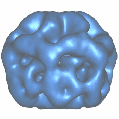 1317.gif 1317.gif | 61.8 KB | ||
| Archive directory |  http://ftp.pdbj.org/pub/emdb/structures/EMD-1317ftp://ftp.pdbj.org/pub/emdb/structures/EMD-1317 http://ftp.pdbj.org/pub/emdb/structures/EMD-1317ftp://ftp.pdbj.org/pub/emdb/structures/EMD-1317 | HTTPS FTP |
-Related structure data
| Similar structure data |
|---|
-Links
| EMDB pages | EMDB (EBI/PDBe) / EMDataResource |
|---|
-Map
| File | Download / File: emd_1317.map.gz / Format: CCP4 / Size: 10.2 MB / Type: IMAGE STORED AS FLOATING POINT NUMBER (4 BYTES) | ||||||||||||||||||||||||||||||||||||||||||||||||||||||||||||||||||||
|---|---|---|---|---|---|---|---|---|---|---|---|---|---|---|---|---|---|---|---|---|---|---|---|---|---|---|---|---|---|---|---|---|---|---|---|---|---|---|---|---|---|---|---|---|---|---|---|---|---|---|---|---|---|---|---|---|---|---|---|---|---|---|---|---|---|---|---|---|---|
| Annotation | This is the masked 3D map of the TIP48/TIP49 complex | ||||||||||||||||||||||||||||||||||||||||||||||||||||||||||||||||||||
| Projections & slices | Image control
Images are generated by Spider. | ||||||||||||||||||||||||||||||||||||||||||||||||||||||||||||||||||||
| Voxel size | X=Y=Z: 3.3 Å | ||||||||||||||||||||||||||||||||||||||||||||||||||||||||||||||||||||
| Density |
| ||||||||||||||||||||||||||||||||||||||||||||||||||||||||||||||||||||
| Symmetry | Space group: 1 | ||||||||||||||||||||||||||||||||||||||||||||||||||||||||||||||||||||
| Details | EMDB XML:
CCP4 map header:
| ||||||||||||||||||||||||||||||||||||||||||||||||||||||||||||||||||||
 Z (Sec.)
 Y (Row.)
 X (Col.)


-Supplemental data
- Sample components
-Entire : human TIP48-His6_ TIP49
| Entire | Name: human TIP48-His6_ TIP49 |
|---|---|
| Components |
|
-Supramolecule #1000: human TIP48-His6_ TIP49
| Supramolecule | Name: human TIP48-His6_ TIP49 / type: sample / ID: 1000 / Oligomeric state: double hexamer / Number unique components: 2 |
|---|---|
| Molecular weight | Experimental: 670 KDa / Theoretical: 609 KDa / Method: size exclusion chromatography |
-Macromolecule #1: TIP48-His6
| Macromolecule | Name: TIP48-His6 / type: protein_or_peptide / ID: 1 / Name.synonym: RUVBL2, 48 kDa TATA box-binding / Recombinant expression: Yes |
|---|---|
| Source (natural) | Organism: Homo sapiens (human) / Strain: IMAGE2819778 / synonym: human |
| Molecular weight | Experimental: 52 KDa |
| Recombinant expression | Organism: Escherichia coli BL21 Gold DE3 / Recombinant plasmid: pET21 |
-Macromolecule #2: TIP49
| Macromolecule | Name: TIP49 / type: protein_or_peptide / ID: 2 / Name.synonym: RUVBL1, 49 kDa TATA box-binding / Recombinant expression: Yes |
|---|---|
| Source (natural) | Organism: Homo sapiens (human) / Strain: IMAGE2823568 / synonym: human |
| Molecular weight | Experimental: 51.2 KDa / Theoretical: 55 KDa |
| Recombinant expression | Organism: Escherichia coli BL21 Gold DE3 / Recombinant plasmid: pET21 |
-Experimental details
-Structure determination
| Method | negative staining |
|---|---|
 Processing Processing | single particle reconstruction |
| Aggregation state | particle |
-Sample preparation
| Concentration | 0.063 mg/mL |
|---|---|
| Buffer | pH: 8 Details: 20mM Tris HCL pH 8.0, 100 mM NaCl, 5%(w/v)glycerol, 1mM DTT |
| Staining | Type: NEGATIVE Details: grids with adsorbed protein were stained twice with 2% (w/v) uranyl acetate |
| Grid | Details: carbon coated copper grids, 400 mesh, negatively glow discharged |
| Vitrification | Cryogen name: NONE |
- Electron microscopy
| Microscope | FEI TECNAI 12 |
|---|---|
| Electron beam | Acceleration voltage: 120 kV / Electron source: TUNGSTEN HAIRPIN |
| Electron optics | Illumination mode: FLOOD BEAM / Imaging mode: BRIGHT FIELDBright-field microscopy / Cs: 2.0 mm / Nominal defocus max: 1.014 µm / Nominal defocus min: 0.383 µm / Nominal magnification: 42000 |
| Sample stage | Specimen holder: side entry / Specimen holder model: OTHER |
| Temperature | Average: 273 K |
| Alignment procedure | Legacy - Astigmatism: corrected at 150,000 magnification |
| Date | Sep 3, 2004 |
| Image recording | Category: FILM / Film or detector model: KODAK SO-163 FILM / Digitization - Scanner: ZEISS SCAI / Digitization - Sampling interval: 14 µm / Number real images: 9 / Average electron dose: 20 e/Å2 / Od range: 1 / Bits/pixel: 8 |
-Image processing
| CTF correction | Details: phase flipping |
|---|---|
| Final reconstruction | Applied symmetry - Point group: C6 (6 fold cyclic) / Algorithm: OTHER / Resolution.type: BY AUTHOR / Resolution: 20.0 Å / Resolution method: FSC 0.5 CUT-OFF / Software - Name: Imagic, Spider / Number images used: 1765 |
