 Movie
Movie Controller

[English] 日本語
 Yorodumi
- EMDB-1300: Electron cryomicroscopy comparison of the architectures of the en... -
+ Open data

- Basic information
| Entry | Database: EMDB / ID: EMD-1300 | |||||||||
|---|---|---|---|---|---|---|---|---|---|---|
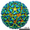
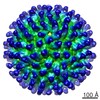
| Title | Electron cryomicroscopy comparison of the architectures of the enveloped bacteriophages phi6 and phi8. | |||||||||
 Map data Map data | 3D reconstruction of the bacteriophage Phi8 polymerase complex | |||||||||
 Sample Sample |
| |||||||||
| Function / homology | : / Major inner capsid protein P1 / identical protein binding / p1 Function and homology information Function and homology information | |||||||||
| Biological species |  Pseudomonas phage phi8 (bacteriophage) Pseudomonas phage phi8 (bacteriophage) | |||||||||
| Method |  single particle reconstruction / cryo EM / Resolution: 8.7 Å single particle reconstruction / cryo EM / Resolution: 8.7 Å | |||||||||
 Authors Authors | Jaalinoja HT / Huiskonen JT / Butcher SJ | |||||||||
 Citation Citation | Journal: Structure / Year: 2007 Title: Electron cryomicroscopy comparison of the architectures of the enveloped bacteriophages phi6 and phi8. Authors: Harri T Jäälinoja / Juha T Huiskonen / Sarah J Butcher /  Abstract: The enveloped dsRNA bacteriophages phi6 and phi8 are the two most distantly related members of the Cystoviridae family. Their structure and function are similar to that of the Reoviridae but their ...The enveloped dsRNA bacteriophages phi6 and phi8 are the two most distantly related members of the Cystoviridae family. Their structure and function are similar to that of the Reoviridae but their assembly can be conveniently studied in vitro. Electron cryomicroscopy and three-dimensional icosahedral reconstruction were used to determine the structures of the phi6 virion (14 A resolution), phi8 virion (18 A resolution), and phi8 core (8.5 A resolution). Spikes protrude 2 nm from the membrane bilayer in phi6 and 7 nm in phi8. In the phi6 nucleocapsid, 600 copies of P8 and 72 copies of P4 interact with the membrane, whereas in phi8 it is only P4 and 60 copies of a minor protein. The major polymerase complex protein P1 forms a dodecahedral shell from 60 asymmetric dimers in both viruses, but the alpha-helical fold has apparently diverged. These structural differences reflect the different host ranges and entry and assembly mechanisms of the two viruses. | |||||||||
| History |
|
- Structure visualization
| Movie |
Movie viewer |
|---|---|
| Structure viewer | EM map: SurfViewMolmilJmol/JSmol |
| Supplemental images |

- Downloads & links
-EMDB archive
| Map data | emd_1300.map.gz | 149.6 MB | EMDB map data format | |
|---|---|---|---|---|
| Header (meta data) | emd-1300-v30.xmlemd-1300.xml | 13.1 KB 13.1 KB | Display Display | EMDB header |
| Images | 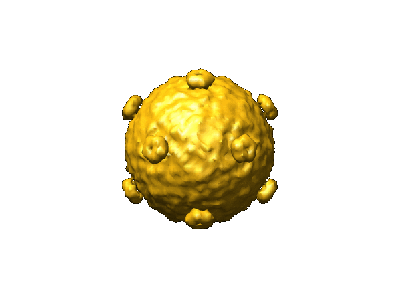 1300.gif 1300.gif | 18.7 KB | ||
| Masks | emd_1300_msk_1.mapemd_1300_msk_2.map | 152.7 MB 152.7 MB | Mask map | |
| Others | MASK_details.txt | 102 B | ||
| Archive directory |  http://ftp.pdbj.org/pub/emdb/structures/EMD-1300ftp://ftp.pdbj.org/pub/emdb/structures/EMD-1300 http://ftp.pdbj.org/pub/emdb/structures/EMD-1300ftp://ftp.pdbj.org/pub/emdb/structures/EMD-1300 | HTTPS FTP |
-Related structure data
| Related structure data |  4bx4M  1299C  1301C M: atomic model generated by this map C: citing same article ( |
|---|---|
| Similar structure data |
-Links
| EMDB pages | EMDB (EBI/PDBe) / EMDataResource |
|---|
-Map
| File | Download / File: emd_1300.map.gz / Format: CCP4 / Size: 298.2 MB / Type: IMAGE STORED AS SIGNED INTEGER (2 BYTES) | ||||||||||||||||||||||||||||||||||||||||||||||||||||||||||||||||||||
|---|---|---|---|---|---|---|---|---|---|---|---|---|---|---|---|---|---|---|---|---|---|---|---|---|---|---|---|---|---|---|---|---|---|---|---|---|---|---|---|---|---|---|---|---|---|---|---|---|---|---|---|---|---|---|---|---|---|---|---|---|---|---|---|---|---|---|---|---|---|
| Annotation | 3D reconstruction of the bacteriophage Phi8 polymerase complex | ||||||||||||||||||||||||||||||||||||||||||||||||||||||||||||||||||||
| Projections & slices | Image control
Images are generated by Spider. | ||||||||||||||||||||||||||||||||||||||||||||||||||||||||||||||||||||
| Voxel size | X=Y=Z: 1.4 Å | ||||||||||||||||||||||||||||||||||||||||||||||||||||||||||||||||||||
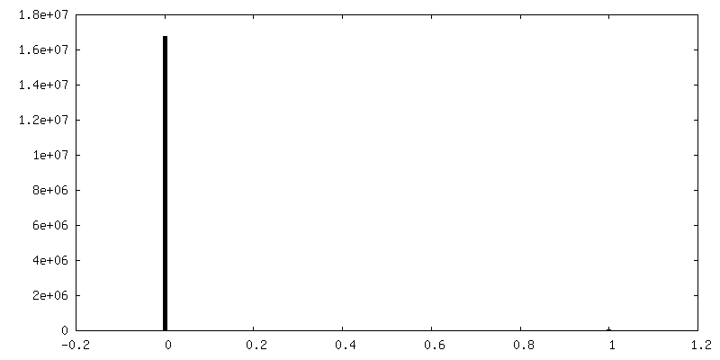
| Density |
| ||||||||||||||||||||||||||||||||||||||||||||||||||||||||||||||||||||
| Symmetry | Space group: 1 | ||||||||||||||||||||||||||||||||||||||||||||||||||||||||||||||||||||
| Details | EMDB XML:
CCP4 map header:
| ||||||||||||||||||||||||||||||||||||||||||||||||||||||||||||||||||||
 Z (Sec.)
 Y (Row.)
 X (Col.)


-Supplemental data
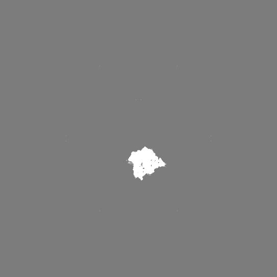
-Segmentation: Subunit A
| Annotation | Subunit A | ||||||||||||
|---|---|---|---|---|---|---|---|---|---|---|---|---|---|
| File | emd_1300_msk_1.map | ||||||||||||
| Projections & Slices |
| ||||||||||||
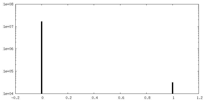
| Density Histograms |


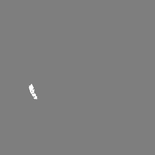
-Segmentation: Subunit A
| Annotation | Subunit A | ||||||||||||
|---|---|---|---|---|---|---|---|---|---|---|---|---|---|
| File | emd_1300_msk_2.map | ||||||||||||
| Projections & Slices |
| ||||||||||||
| Density Histograms |


-Others
| Details | [MASK_details.txt] mask_phi8_subunitA_emd1300.map> emd_1300_msk_1.map mask_phi8_subunitB_emd1300.map> emd_1300_msk_2.map |
|---|
- Sample components
-Entire : Bacteriophage Phi8 core
| Entire | Name: Bacteriophage Phi8 core |
|---|---|
| Components |
|
-Supramolecule #1000: Bacteriophage Phi8 core
| Supramolecule | Name: Bacteriophage Phi8 core / type: sample / ID: 1000 / Number unique components: 1 |
|---|
-Supramolecule #1: Pseudomonas phage phi8
| Supramolecule | Name: Pseudomonas phage phi8 / type: virus / ID: 1 / Name.synonym: Cystovirus Phi8 polymerase complex / NCBI-ID: 120086 / Sci species name: Pseudomonas phage phi8 / Virus type: VIRION / Virus isolate: SPECIES / Virus enveloped: No / Virus empty: No / Syn species name: Cystovirus Phi8 polymerase complex |
|---|---|
| Host (natural) | Organism: Pseudomonads syringae / synonym: BACTERIA(EUBACTERIA) |
| Virus shell | Shell ID: 1 / Name: Polymerase complex / Diameter: 500 Å / T number (triangulation number): 1 |
-Experimental details
-Structure determination
| Method | cryo EM |
|---|---|
 Processing Processing | single particle reconstruction |
| Aggregation state | particle |
-Sample preparation
| Buffer | Details: 10 mM potassium phosphate pH7.5, 1 mM MgCl2, 50 mM NaCl |
|---|---|
| Grid | Details: 400 mesh copper grid, Quantifoil R2/2 holey |
| Vitrification | Cryogen name: ETHANE / Chamber temperature: 90 K / Instrument: HOMEMADE PLUNGER / Details: Vitrification instrument: EMBL design Method: A small vial of ethane is placed inside a larger liquid nitrogen reservoir. The grid holding 3 microliters of the sample is held in place at the bottom of a plunger by the means of fine ...Method: A small vial of ethane is placed inside a larger liquid nitrogen reservoir. The grid holding 3 microliters of the sample is held in place at the bottom of a plunger by the means of fine tweezers. When the liquid ethane is ready, a piece of filter paper is then pressed against the sample to blot off excess buffer, sufficient to leave a thin layer on the grid. The filter paper is removed, and the plunger is allowed to drop into the liquid ethane. Once the grid enters the liquid ethane, the sample is rapidly frozen, and the grid is transferred under liquid nitrogen to a storage box immersed in liquid nitrogen for later use in the microscope. |
- Electron microscopy
| Microscope | FEI TECNAI F20 |
|---|---|
| Electron beam | Acceleration voltage: 200 kV / Electron source: FIELD EMISSION GUN |
| Electron optics | Calibrated magnification: 49300 / Illumination mode: FLOOD BEAM / Imaging mode: BRIGHT FIELDBright-field microscopy / Cs: 2.0 mm / Nominal defocus max: 3.3 µm / Nominal defocus min: 0.7 µm / Nominal magnification: 50000 |
| Sample stage | Specimen holder: Side entry liquid nitrogen-cooled cryo specimen holder Specimen holder model: GATAN LIQUID NITROGEN |
| Temperature | Min: 90 K / Max: 94 K / Average: 93 K |
| Image recording | Category: FILM / Film or detector model: KODAK SO-163 FILM / Digitization - Scanner: ZEISS SCAI / Digitization - Sampling interval: 7 µm / Number real images: 66 / Bits/pixel: 12 |
| Tilt angle min | 0 |
| Tilt angle max | 0 |
| Experimental equipment |  Model: Tecnai F20 / Image courtesy: FEI Company |
-Image processing
| CTF correction | Details: Each particle, wiener factor 0.1 |
|---|---|
| Final reconstruction | Applied symmetry - Point group: I (icosahedral) / Algorithm: OTHER / Resolution.type: BY AUTHOR / Resolution: 8.7 Å / Resolution method: FSC 0.5 CUT-OFF / Software - Name: pft2, em3dr2, POR, P3DR / Number images used: 12867 |
