 Movie
Movie Controller


+ Open data

- Basic information
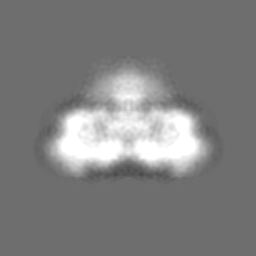
| Entry | Database: EMDB / ID: EMD-1275 | |||||||||
|---|---|---|---|---|---|---|---|---|---|---|
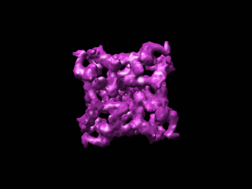
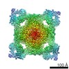
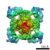
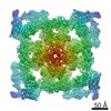
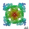
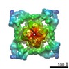
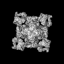
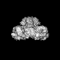
| Title | The pore structure of the closed RyR1 channel. | |||||||||
 Map data Map data | Z-axis of a 3D reconstruction coincides with the 4-fold axis of the channel complex | |||||||||
 Sample Sample |
| |||||||||
| Function / homology | ryanodine-sensitive calcium-release channel activity / RIH domain Function and homology information Function and homology information | |||||||||
| Biological species |   Oryctolagus cuniculus (rabbit) Oryctolagus cuniculus (rabbit) | |||||||||
| Method | single particle reconstruction / cryo EM / Resolution: 9.6 Å | |||||||||
 Authors Authors | Ludtke SJ / Serysheva II / Hamilton SL / Chiu W | |||||||||
 Citation Citation | Journal: Structure / Year: 2005 Title: The pore structure of the closed RyR1 channel. Authors: Steven J Ludtke / Irina I Serysheva / Susan L Hamilton / Wah Chiu /  Abstract: Using single particle electron cryomicroscopy, several helices in the membrane-spanning region of RyR1, including an inner transmembrane helix, a short pore helix, and a helix parallel to the ...Using single particle electron cryomicroscopy, several helices in the membrane-spanning region of RyR1, including an inner transmembrane helix, a short pore helix, and a helix parallel to the membrane on the cytoplasmic side, have been clearly resolved. Our model places a highly conserved glycine (G4934) at the hinge position of the bent inner helix and two rings of negative charges at the luminal and cytoplasmic mouths of the pore. The kinked inner helix closely resembles the inner helix of the open MthK channel, suggesting that kinking alone does not open RyR1, as proposed for K+ channels. | |||||||||
| History |
|
- Structure visualization
| Movie |
Movie viewer |
|---|---|
| Structure viewer | EM map: SurfViewMolmilJmol/JSmol |
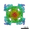
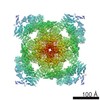
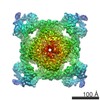
| Supplemental images |
- Downloads & links
-EMDB archive
| Map data | emd_1275.map.gz | 9.1 MB | EMDB map data format | |
|---|---|---|---|---|
| Header (meta data) | emd-1275-v30.xmlemd-1275.xml | 9.6 KB 9.6 KB | Display Display | EMDB header |
| Images |  1275.gif 1275.gif | 37.1 KB | ||
| Archive directory |  http://ftp.pdbj.org/pub/emdb/structures/EMD-1275ftp://ftp.pdbj.org/pub/emdb/structures/EMD-1275 http://ftp.pdbj.org/pub/emdb/structures/EMD-1275ftp://ftp.pdbj.org/pub/emdb/structures/EMD-1275 | HTTPS FTP |
-Related structure data
| Similar structure data |
|---|

-Links
| EMDB pages | EMDB (EBI/PDBe) / EMDataResource |
|---|
-Map
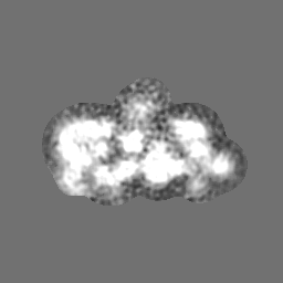
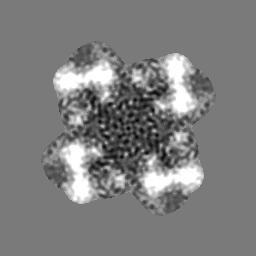
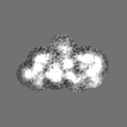
| File | Download / File: emd_1275.map.gz / Format: CCP4 / Size: 62.5 MB / Type: IMAGE STORED AS FLOATING POINT NUMBER (4 BYTES) | ||||||||||||||||||||||||||||||||||||||||||||||||||||||||||||||||||||
|---|---|---|---|---|---|---|---|---|---|---|---|---|---|---|---|---|---|---|---|---|---|---|---|---|---|---|---|---|---|---|---|---|---|---|---|---|---|---|---|---|---|---|---|---|---|---|---|---|---|---|---|---|---|---|---|---|---|---|---|---|---|---|---|---|---|---|---|---|---|
| Annotation | Z-axis of a 3D reconstruction coincides with the 4-fold axis of the channel complex | ||||||||||||||||||||||||||||||||||||||||||||||||||||||||||||||||||||
| Projections & slices | Image control
Images are generated by Spider. | ||||||||||||||||||||||||||||||||||||||||||||||||||||||||||||||||||||
| Voxel size | X=Y=Z: 1.81 Å | ||||||||||||||||||||||||||||||||||||||||||||||||||||||||||||||||||||
| Density |
| ||||||||||||||||||||||||||||||||||||||||||||||||||||||||||||||||||||
| Symmetry | Space group: 1 | ||||||||||||||||||||||||||||||||||||||||||||||||||||||||||||||||||||
| Details | EMDB XML:
CCP4 map header:
| ||||||||||||||||||||||||||||||||||||||||||||||||||||||||||||||||||||
 Z (Sec.)
 Y (Row.)
 X (Col.)


-Supplemental data
- Sample components
-Entire : ryanodine receptor 1
| Entire | Name: ryanodine receptor 1 |
|---|---|
| Components |
|
-Supramolecule #1000: ryanodine receptor 1
| Supramolecule | Name: ryanodine receptor 1 / type: sample / ID: 1000 Details: The sample was purified after solubilization with detergent from skeletal muscle cells Oligomeric state: homotetramer / Number unique components: 1 |
|---|---|
| Molecular weight | Theoretical: 565 KDa |
-Macromolecule #1: Ryanodine receptor type 1
| Macromolecule | Name: Ryanodine receptor type 1 / type: protein_or_peptide / ID: 1 / Name.synonym: Skeletal muscle calcium release channel / Details: detergent solubilized membrane protein / Number of copies: 4 / Oligomeric state: homotetramer / Recombinant expression: No |
|---|---|
| Source (natural) | Organism: Oryctolagus cuniculus (rabbit) / Strain: White New Zealand / synonym: rabbit / Tissue: fast twitch skeletal muscle / Cell: muscle cell / Organelle: sarcoplasmic reticulum / Location in cell: sarcoplasmic reticulum membrane |
| Molecular weight | Experimental: 565 KDa |
| Sequence | GO: ryanodine-sensitive calcium-release channel activity / InterPro: RIH domain |
-Experimental details
-Structure determination
| Method | cryo EM |
|---|---|
 Processing Processing | single particle reconstruction |
| Aggregation state | particle |
-Sample preparation
| Concentration | 2 mg/mL |
|---|---|
| Buffer | pH: 7.4 Details: 20 mM Mops, 300 mM KCl, 1 mM DTT,0.4% CHAPS,5%sucrose,1mM EGTA |
| Grid | Details: holey carbon 400 mesh Cu grid |
| Vitrification | Cryogen name: ETHANE / Chamber temperature: 101 K / Instrument: HOMEMADE PLUNGER Details: Vitrification instrument: Thin carbon film supported by holey carbon film Method: Blot for 3 second before plunging |
- Electron microscopy
| Microscope | JEOL 2010F |
|---|---|
| Electron beam | Acceleration voltage: 200 kV / Electron source: FIELD EMISSION GUN |
| Electron optics | Illumination mode: FLOOD BEAM / Imaging mode: BRIGHT FIELDBright-field microscopy / Cs: 2.0 mm / Nominal defocus max: 4.55 µm / Nominal defocus min: 1.37 µm / Nominal magnification: 60000 |
| Sample stage | Specimen holder: side entry / Specimen holder model: GATAN LIQUID NITROGEN |
| Temperature | Min: 96 K / Max: 97 K / Average: 96 K |
| Alignment procedure | Legacy - Astigmatism: objective lens astigmatism was corrected at 400,000X |
| Image recording | Category: CCD / Film or detector model: GENERIC GATAN (4k x 4k) / Digitization - Sampling interval: 1.81 µm / Number real images: 869 / Average electron dose: 15 e/Å2 / Bits/pixel: 8 |
| Tilt angle min | 0 |
| Tilt angle max | 0 |
-Image processing
| CTF correction | Details: CTF correction of each micrograph |
|---|---|
| Final two d classification | Number classes: 2294 |
| Final reconstruction | Applied symmetry - Point group: C4 (4 fold cyclic) / Algorithm: OTHER / Resolution.type: BY AUTHOR / Resolution: 9.6 Å / Resolution method: FSC 0.5 CUT-OFF / Software - Name: EMAN / Number images used: 28036 |
