 Movie
Movie Controller

[English] 日本語
 Yorodumi
- EMDB-1235: In situ structure of the complete Treponema primitia flagellar motor. -
+ Open data

- Basic information
| Entry | Database: EMDB / ID: EMD-1235 | |||||||||
|---|---|---|---|---|---|---|---|---|---|---|
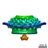
| Title | In situ structure of the complete Treponema primitia flagellar motor. | |||||||||
 Map data Map data | See header. As submitted, low values correspond to protein density, and it should be contoured at 1.3 standard deviations. | |||||||||
 Sample Sample |
| |||||||||
| Biological species |   Treponema primitia (bacteria) Treponema primitia (bacteria) | |||||||||
| Method | subtomogram averaging / cryo EM / Resolution: 70.0 Å | |||||||||
 Authors Authors | Murphy GE / Leadbetter JR / Jensen GJ | |||||||||
 Citation Citation | Journal: Nature / Year: 2006 Title: In situ structure of the complete Treponema primitia flagellar motor. Authors: Gavin E Murphy / Jared R Leadbetter / Grant J Jensen /  Abstract: The bacterial flagellar motor is an amazing nanomachine: built from approximately 25 different proteins, it uses an electrochemical ion gradient to drive rotation at speeds of up to 300 Hz (refs 1, 2) ...The bacterial flagellar motor is an amazing nanomachine: built from approximately 25 different proteins, it uses an electrochemical ion gradient to drive rotation at speeds of up to 300 Hz (refs 1, 2). The flagellar motor consists of a fixed, membrane-embedded, torque-generating stator and a typically bidirectional, spinning rotor that changes direction in response to chemotactic signals. Most structural analyses so far have targeted the purified rotor, and hence little is known about the stator and its interactions. Here we show, using electron cryotomography of whole cells, the in situ structure of the complete flagellar motor from the spirochaete Treponema primitia at 7 nm resolution. Twenty individual motor particles were computationally extracted from the reconstructions, aligned and then averaged. The stator assembly, revealed for the first time, possessed 16-fold symmetry and was connected directly to the rotor, C ring and a novel P-ring-like structure. The unusually large size of the motor suggested mechanisms for increasing torque and supported models wherein critical interactions occur atop the C ring, where our data suggest that both the carboxy-terminal and middle domains of FliG are found. | |||||||||
| History |
|
- Structure visualization
| Movie |
 Movie viewer Movie viewer |
|---|---|
| Structure viewer | EM map: SurfViewMolmilJmol/JSmol |
| Supplemental images |

- Downloads & links
-EMDB archive
| Map data | emd_1235.map.gz | 59.4 KB | EMDB map data format | |
|---|---|---|---|---|
| Header (meta data) | emd-1235-v30.xmlemd-1235.xml | 9.7 KB 9.7 KB | Display Display | EMDB header |
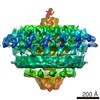
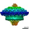
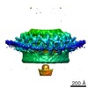
| Images | 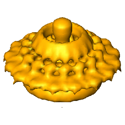 1235.gif 1235.gif | 47.8 KB | ||
| Archive directory |  http://ftp.pdbj.org/pub/emdb/structures/EMD-1235ftp://ftp.pdbj.org/pub/emdb/structures/EMD-1235 http://ftp.pdbj.org/pub/emdb/structures/EMD-1235ftp://ftp.pdbj.org/pub/emdb/structures/EMD-1235 | HTTPS FTP |
-Related structure data
| Similar structure data |
|---|

-Links
| EMDB pages | EMDB (EBI/PDBe) / EMDataResource |
|---|
-Map
| File | Download / File: emd_1235.map.gz / Format: CCP4 / Size: 335.9 KB / Type: IMAGE STORED AS SIGNED INTEGER (2 BYTES) | ||||||||||||||||||||||||||||||||||||||||||||||||||||||||||||||||||||
|---|---|---|---|---|---|---|---|---|---|---|---|---|---|---|---|---|---|---|---|---|---|---|---|---|---|---|---|---|---|---|---|---|---|---|---|---|---|---|---|---|---|---|---|---|---|---|---|---|---|---|---|---|---|---|---|---|---|---|---|---|---|---|---|---|---|---|---|---|---|
| Annotation | See header. As submitted, low values correspond to protein density, and it should be contoured at 1.3 standard deviations. | ||||||||||||||||||||||||||||||||||||||||||||||||||||||||||||||||||||
| Projections & slices | Image control
Images are generated by Spider. | ||||||||||||||||||||||||||||||||||||||||||||||||||||||||||||||||||||
| Voxel size | X=Y=Z: 19.6 Å | ||||||||||||||||||||||||||||||||||||||||||||||||||||||||||||||||||||
| Density |
| ||||||||||||||||||||||||||||||||||||||||||||||||||||||||||||||||||||
| Symmetry | Space group: 1 | ||||||||||||||||||||||||||||||||||||||||||||||||||||||||||||||||||||
| Details | EMDB XML:
CCP4 map header:
| ||||||||||||||||||||||||||||||||||||||||||||||||||||||||||||||||||||
 Z (Sec.)
 Y (Row.)
 X (Col.)


-Supplemental data
- Sample components
-Entire : Flagellar Motor
| Entire | Name: Flagellar MotorFlagellum |
|---|---|
| Components |
|
-Supramolecule #1000: Flagellar Motor
| Supramolecule | Name: Flagellar Motor / type: sample / ID: 1000 Details: The flagellar motor is built from more than 20 proteins, some of which are unknown. The stoichiometry is not certain. Number unique components: 1 |
|---|
-Supramolecule #1: Flagellar Motor
| Supramolecule | Name: Flagellar Motor / type: organelle_or_cellular_component / ID: 1 / Name.synonym: Basal Body Details: tens of megadaltons; T. primitia is a spirochete with a periplasmic flagella. It never exits the outer membrane. Recombinant expression: No / Database: NCBI |
|---|---|
| Source (natural) | Organism: Treponema primitia (bacteria) / Cell: Treponema primitia strain ZAS-2 / Location in cell: Plasma membrane |
-Experimental details
-Structure determination
| Method | cryo EM |
|---|---|
 Processing Processing | subtomogram averaging |
-Sample preparation
| Buffer | Details: T. primitia cells were frozen in their 4YACo growth media (Leadbetter et al., Science 1999) |
|---|---|
| Grid | Details: 200 |
| Vitrification | Cryogen name: ETHANE / Chamber humidity: 100 % / Chamber temperature: 22 K / Instrument: OTHER / Details: Vitrification instrument: Vitrobot Method: Typically a 2 second blot, -2 offset and a 1 s drain. |
- Electron microscopy
| Microscope | FEI TECNAI F30 |
|---|---|
| Electron beam | Acceleration voltage: 300 kV / Electron source: FIELD EMISSION GUN |
| Electron optics | Calibrated magnification: 30600 / Illumination mode: FLOOD BEAM / Imaging mode: BRIGHT FIELDBright-field microscopy / Cs: 2 mm / Nominal magnification: 22500 |
| Specialist optics | Energy filter - Name: GIF 3000 / Energy filter - Lower energy threshold: 0.0 eV / Energy filter - Upper energy threshold: 20.0 eV |
| Sample stage | Specimen holder: FEI Polara / Specimen holder model: GATAN HELIUM / Tilt series - Axis1 - Min angle: 63 ° / Tilt series - Axis1 - Max angle: 63 ° |
| Temperature | Min: 82 K / Max: 82 K / Average: 82 K |
| Alignment procedure | Legacy - Astigmatism: obj lens astigmatism corrected at working magnification Legacy - Electron beam tilt params: 0 |
| Image recording | Category: CCD / Film or detector model: GATAN ULTRASCAN 1000 (2k x 2k) / Average electron dose: 110 e/Å2 |
| Experimental equipment |  Model: Tecnai F30 / Image courtesy: FEI Company |
-Image processing
| CTF correction | Details: none |
|---|---|
| Final reconstruction | Algorithm: OTHER / Resolution.type: BY AUTHOR / Resolution: 70.0 Å / Resolution method: FSC 0.5 CUT-OFF / Software - Name: IMOD Details: Final map is the C16 symmetrized average calculated from 20 individual 3D motor particles. |
| Details | Motor particles were picked from 15 different tomograms. The above parameters are the typical values. Average number of tilts used in the 3D reconstructions: 125. Average tomographic tilt angle increment: 1. |
