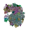
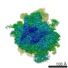
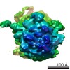
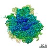
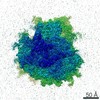
Journal: Science / Year: 2003 Title: Visualizing tmRNA entry into a stalled ribosome. Authors: Mikel Valle / Reynald Gillet / Sukhjit Kaur / Anke Henne / V Ramakrishnan / Joachim Frank / Abstract: Bacterial ribosomes stalled on defective messenger RNAs (mRNAs) are rescued by tmRNA, an approximately 300-nucleotide-long molecule that functions as both transfer RNA (tRNA) and mRNA. Translation ...Bacterial ribosomes stalled on defective messenger RNAs (mRNAs) are rescued by tmRNA, an approximately 300-nucleotide-long molecule that functions as both transfer RNA (tRNA) and mRNA. Translation then switches from the defective message to a short open reading frame on tmRNA that tags the defective nascent peptide chain for degradation. However, the mechanism by which tmRNA can enter and move through the ribosome is unknown. We present a cryo-electron microscopy study at approximately 13 to 15 angstroms of the entry of tmRNA into the ribosome. The structure reveals how tmRNA could move through the ribosome despite its complicated topology and also suggests roles for proteins S1 and SmpB in the function of tmRNA.
History
Deposition
Mar 24, 2005
-
Header (metadata) release
Apr 11, 2005
-
Map release
Apr 11, 2005
-
Update
Oct 24, 2012
-
Current status
Oct 24, 2012
Processing site: PDBe / Status: Released
-
Structure visualization
Movie
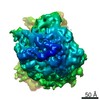
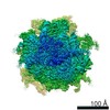
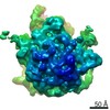
Surface view with section colored by density value
Category: FILM / Film or detector model: KODAK SO-163 FILM / Digitization - Scanner: ZEISS SCAI / Digitization - Sampling interval: 14 µm / Number real images: 44 / Average electron dose: 15 e/Å2 / Od range: 1.2 / Bits/pixel: 12
Experimental equipment
Model: Tecnai F20 / Image courtesy: FEI Company
-
Image processing
CTF correction
Details: defocus groups
Final angle assignment
Details: SPIDER definition
Final reconstruction
Applied symmetry - Point group: C1 (asymmetric) / Algorithm: OTHER / Resolution.type: BY AUTHOR / Resolution: 13.0 Å / Resolution method: OTHER / Software - Name: SPIDER / Number images used: 27644
-
Atomic model buiding 1
Software
Name: manual
Details
Protocol: Rigid Body. The domains were separately fitted by manual docking using program O
Refinement
Protocol: RIGID BODY FIT / Target criteria: cross correlation coefficient
Output model
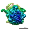
PDB-1zc8: Coordinates of tmRNA, SmpB, EF-Tu and h44 fitted into Cryo-EM map of the 70S ribosome and tmRNA complex
+
About Yorodumi
-
News
-
Feb 9, 2022. New format data for meta-information of EMDB entries
New format data for meta-information of EMDB entries
Version 3 of the EMDB header file is now the official format.
The previous official version 1.9 will be removed from the archive.
In the structure databanks used in Yorodumi, some data are registered as the other names, "COVID-19 virus" and "2019-nCoV". Here are the details of the virus and the list of structure data.
Jan 31, 2019. EMDB accession codes are about to change! (news from PDBe EMDB page)
EMDB accession codes are about to change! (news from PDBe EMDB page)
The allocation of 4 digits for EMDB accession codes will soon come to an end. Whilst these codes will remain in use, new EMDB accession codes will include an additional digit and will expand incrementally as the available range of codes is exhausted. The current 4-digit format prefixed with “EMD-” (i.e. EMD-XXXX) will advance to a 5-digit format (i.e. EMD-XXXXX), and so on. It is currently estimated that the 4-digit codes will be depleted around Spring 2019, at which point the 5-digit format will come into force.
The EM Navigator/Yorodumi systems omit the EMD- prefix.
Related info.:Q: What is EMD? / ID/Accession-code notation in Yorodumi/EM Navigator
Yorodumi is a browser for structure data from EMDB, PDB, SASBDB, etc.
This page is also the successor to EM Navigator detail page, and also detail information page/front-end page for Omokage search.
The word "yorodu" (or yorozu) is an old Japanese word meaning "ten thousand". "mi" (miru) is to see.
Related info.:EMDB / PDB / SASBDB / Comparison of 3 databanks / Yorodumi Search / Aug 31, 2016. New EM Navigator & Yorodumi / Yorodumi Papers / Jmol/JSmol / Function and homology information / Changes in new EM Navigator and Yorodumi
 Movie
Movie Controller


 Open data
 Basic information
 Map data
 Sample
 Function and homology information
 trans-translation /

 Authors
 Citation
 Structure visualization
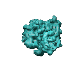
 Downloads & links
 1122.gif
 http://ftp.pdbj.org/pub/emdb/structures/EMD-1122


 Sample components
 Processing
 Electron microscopy
