 Movie
Movie Controller

[English] 日本語
 Yorodumi
- EMDB-1095: A mutant chaperonin with rearranged inter-ring electrostatic cont... -
+ Open data

- Basic information
| Entry | Database: EMDB / ID: EMD-1095 | |||||||||
|---|---|---|---|---|---|---|---|---|---|---|
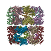
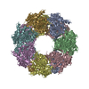
| Title | A mutant chaperonin with rearranged inter-ring electrostatic contacts and temperature-sensitive dissociation. | |||||||||
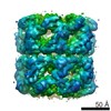
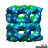
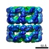
 Map data Map data | Map of the E461K mutant of GroEL made from 1477 images picked from 7 cryo micrographs. Big-endian bypte order. | |||||||||
 Sample Sample |
| |||||||||
| Biological species |  | |||||||||
| Method | single particle reconstruction / cryo EM / negative staining / Resolution: 25.0 Å | |||||||||
 Authors Authors | Sewell BT / Best RB / Chen S / Roseman AM / Farr GW / Horwich AR / Saibil HR | |||||||||
 Citation Citation | Journal: Nat Struct Mol Biol / Year: 2004 Title: A mutant chaperonin with rearranged inter-ring electrostatic contacts and temperature-sensitive dissociation. Authors: B Trevor Sewell / Robert B Best / Shaoxia Chen / Alan M Roseman / George W Farr / Arthur L Horwich / Helen R Saibil /  Abstract: The chaperonin GroEL assists protein folding through ATP-dependent, cooperative movements that alternately create folding chambers in its two rings. The substitution E461K at the interface between ...The chaperonin GroEL assists protein folding through ATP-dependent, cooperative movements that alternately create folding chambers in its two rings. The substitution E461K at the interface between these two rings causes temperature-sensitive, defective protein folding in Escherichia coli. To understand the molecular defect, we have examined the mutant chaperonin by cryo-EM. The normal out-of-register alignment of contacts between subunits of opposing wild-type rings is changed in E461K to an in-register one. This is associated with loss of cooperativity in ATP binding and hydrolysis. Consistent with the loss of negative cooperativity between rings, the cochaperonin GroES binds simultaneously to both E461K rings. These GroES-bound structures were unstable at higher temperature, dissociating into complexes of single E461K rings associated with GroES. Lacking the allosteric signal from the opposite ring, these complexes cannot release their GroES and become trapped, dead-end states. | |||||||||
| History |
|
- Structure visualization
| Movie |
 Movie viewer Movie viewer |
|---|---|
| Structure viewer | EM map: SurfViewMolmilJmol/JSmol |
| Supplemental images |
 UCSF Chimera
- Downloads & links
-EMDB archive
| Map data | emd_1095.map.gz | 93.8 KB | EMDB map data format | |
|---|---|---|---|---|
| Header (meta data) | emd-1095-v30.xmlemd-1095.xml | 11.5 KB 11.5 KB | Display Display | EMDB header |
| Images |  1095.gif 1095.gif | 41.5 KB | ||
| Archive directory |  http://ftp.pdbj.org/pub/emdb/structures/EMD-1095ftp://ftp.pdbj.org/pub/emdb/structures/EMD-1095 http://ftp.pdbj.org/pub/emdb/structures/EMD-1095ftp://ftp.pdbj.org/pub/emdb/structures/EMD-1095 | HTTPS FTP |
-Validation report
| Summary document | emd_1095_validation.pdf.gz | 204.2 KB | Display | EMDB validaton report |
|---|---|---|---|---|
| Full document | emd_1095_full_validation.pdf.gz | 203.3 KB | Display | |
| Data in XML | emd_1095_validation.xml.gz | 4.9 KB | Display | |
| Arichive directory | https://ftp.pdbj.org/pub/emdb/validation_reports/EMD-1095ftp://ftp.pdbj.org/pub/emdb/validation_reports/EMD-1095 | HTTPS FTP |
-Related structure data
| Similar structure data |
|---|


-Links
| EMDB pages | EMDB (EBI/PDBe) / EMDataResource |
|---|
-Map
| File | Download / File: emd_1095.map.gz / Format: CCP4 / Size: 1001 KB / Type: IMAGE STORED AS FLOATING POINT NUMBER (4 BYTES) | ||||||||||||||||||||||||||||||||||||||||||||||||||||||||||||||||||||
|---|---|---|---|---|---|---|---|---|---|---|---|---|---|---|---|---|---|---|---|---|---|---|---|---|---|---|---|---|---|---|---|---|---|---|---|---|---|---|---|---|---|---|---|---|---|---|---|---|---|---|---|---|---|---|---|---|---|---|---|---|---|---|---|---|---|---|---|---|---|
| Annotation | Map of the E461K mutant of GroEL made from 1477 images picked from 7 cryo micrographs. Big-endian bypte order. | ||||||||||||||||||||||||||||||||||||||||||||||||||||||||||||||||||||
| Voxel size | X=Y=Z: 6.67 Å | ||||||||||||||||||||||||||||||||||||||||||||||||||||||||||||||||||||
| Density |
| ||||||||||||||||||||||||||||||||||||||||||||||||||||||||||||||||||||
| Symmetry | Space group: 1 | ||||||||||||||||||||||||||||||||||||||||||||||||||||||||||||||||||||
| Details | EMDB XML:
CCP4 map header:
| ||||||||||||||||||||||||||||||||||||||||||||||||||||||||||||||||||||
-Supplemental data
- Sample components
-Entire : E461K mutant of GroEL
| Entire | Name: E461K mutant of GroEL |
|---|---|
| Components |
|
-Supramolecule #1000: E461K mutant of GroEL
| Supramolecule | Name: E461K mutant of GroEL / type: sample / ID: 1000 / Oligomeric state: homo tetradecamer / Number unique components: 1 |
|---|---|
| Molecular weight | Experimental: 840 KDa / Theoretical: 840 KDa / Method: sequencing, gel-filtration |
-Macromolecule #1: E461K mutant of GroEL
| Macromolecule | Name: E461K mutant of GroEL / type: protein_or_peptide / ID: 1 / Name.synonym: E461K / Details: homo tetradecamer / Number of copies: 14 / Oligomeric state: homo tetradecamer / Recombinant expression: Yes |
|---|---|
| Source (natural) | Organism: |
| Molecular weight | Experimental: 840 KDa / Theoretical: 840 KDa |
| Recombinant expression | Organism: |
-Experimental details
-Structure determination
| Method | negative staining, cryo EM |
|---|---|
 Processing Processing | single particle reconstruction |
| Aggregation state | particle |
-Sample preparation
| Concentration | 0.84 mg/mL |
|---|---|
| Buffer | pH: 7.4 / Details: 20 mM Tris, 5 mM KCl, 10 mM MgCl2, pH 7.4 |
| Staining | Type: NEGATIVE Details: grids were flash frozen by plunging into liquid ethane at -170 degrees C. No stain was used. |
| Grid | Details: copper grid, holey carbon films |
| Vitrification | Cryogen name: ETHANE / Instrument: HOMEMADE PLUNGER / Details: Vitrification instrument: Home made plunger Method: grids were blotted prior to plunging for approximately two seconds |
- Electron microscopy
| Microscope | JEOL 2010HT |
|---|---|
| Temperature | Min: 100 K / Max: 100 K / Average: 100 K |
| Image recording | Category: FILM / Film or detector model: KODAK SO-163 FILM / Digitization - Scanner: OTHER / Digitization - Sampling interval: 10 µm / Number real images: 7 / Average electron dose: 10 e/Å2 Details: Scanned on a Leafscan 45 at 2540 pixels per inch. Subsequently pixels were averaged in 2x2 blocks. Bits/pixel: 16 |
| Electron beam | Acceleration voltage: 200 kV / Electron source: TUNGSTEN HAIRPIN |
| Electron optics | Calibrated magnification: 30000 / Illumination mode: FLOOD BEAM / Imaging mode: BRIGHT FIELD / Nominal defocus max: 2.0 µm / Nominal defocus min: 1.0 µm / Nominal magnification: 30000 |
| Sample stage | Specimen holder: Oxford CT3500 / Specimen holder model: OTHER |
-Image processing
| Details | Side views only were used in the reconstruction |
|---|---|
| Final reconstruction | Applied symmetry - Point group: C7 (7 fold cyclic) / Algorithm: OTHER / Resolution.type: BY AUTHOR / Resolution: 25.0 Å / Resolution method: FSC 0.5 CUT-OFF / Software - Name: Spider Details: Seven fold symmetry was imposed on the reconstruction. Number images used: 1477 |
| Final angle assignment | Details: Spider euler angles file as follows: 1 3 90.0 90.000 0.00000E+00 2 3 90.0 90.000 3.2143 3 3 90.0 90.000 6.4286 4 3 90.0 90.000 9.6429 5 3 90.0 90.000 12.857 6 3 90.0 90.000 16.071 7 3 90.0 ...Details: Spider euler angles file as follows: 1 3 90.0 90.000 0.00000E+00 2 3 90.0 90.000 3.2143 3 3 90.0 90.000 6.4286 4 3 90.0 90.000 9.6429 5 3 90.0 90.000 12.857 6 3 90.0 90.000 16.071 7 3 90.0 90.000 19.286 8 3 90.0 90.000 22.500 9 3 90.0 90.000 25.714 10 3 90.0 90.000 28.929 11 3 90.0 90.000 32.143 12 3 90.0 90.000 35.357 13 3 90.0 90.000 38.572 14 3 90.0 90.000 41.786 15 3 90.0 90.000 45.000 16 3 90.0 90.000 48.214 17 3 90.0 96.429 0.00000E+00 18 3 90.0 96.429 3.2143 19 3 90.0 96.429 6.4286 20 3 90.0 96.429 9.6429 21 3 90.0 96.429 12.857 22 3 90.0 96.429 16.071 23 3 90.0 96.429 19.286 24 3 90.0 96.429 22.500 25 3 90.0 96.429 25.714 26 3 90.0 96.429 28.929 27 3 90.0 96.429 32.143 28 3 90.0 96.429 35.357 29 3 90.0 96.429 38.572 30 3 90.0 96.429 41.786 31 3 90.0 96.429 45.000 32 3 90.0 96.429 48.214 33 3 90.0 102.857 0.0 34 3 90.0 102.857 6.4825 35 3 90.0 102.857 12.857 36 3 90.0 102.857 19.2885 37 3 90.0 102.857 25.714 38 3 90.0 102.857 32.1425 39 3 90.0 102.857 38.571 40 3 90.0 102.857 45.0 |
| Final two d classification | Number classes: 40 |
-Atomic model buiding 1
| Initial model | PDB ID: |
|---|---|
| Details | The seven membered rings of GroEL (1EMS) were docked separately into the reconstructed density. This was done manually using the programme O. |
| Refinement | Protocol: RIGID BODY FIT |

