 Movie
Movie Controller

[English] 日本語
 Yorodumi
- EMDB-1076: Structure of the 80S ribosome from Saccharomyces cerevisiae--tRNA... -
+ Open data

- Basic information
| Entry | Database: EMDB / ID: EMD-1076 | |||||||||
|---|---|---|---|---|---|---|---|---|---|---|
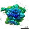
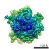
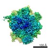
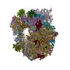
| Title | Structure of the 80S ribosome from Saccharomyces cerevisiae--tRNA-ribosome and subunit-subunit interactions. | |||||||||
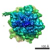
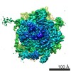
 Map data Map data | This is a volume of 80S ribosome | |||||||||
 Sample Sample |
| |||||||||
| Biological species |  | |||||||||
| Method | single particle reconstruction / cryo EM / Resolution: 15.4 Å | |||||||||
 Authors Authors | Spahn CM | |||||||||
 Citation Citation | Journal: Cell / Year: 2001 Title: Structure of the 80S ribosome from Saccharomyces cerevisiae--tRNA-ribosome and subunit-subunit interactions. Authors: C M Spahn / R Beckmann / N Eswar / P A Penczek / A Sali / G Blobel / J Frank /  Abstract: A cryo-EM reconstruction of the translating yeast 80S ribosome was analyzed. Computationally separated rRNA and protein densities were used for docking of appropriately modified rRNA models and ...A cryo-EM reconstruction of the translating yeast 80S ribosome was analyzed. Computationally separated rRNA and protein densities were used for docking of appropriately modified rRNA models and homology models of yeast ribosomal proteins. The core of the ribosome shows a remarkable degree of conservation. However, some significant differences in functionally important regions and dramatic changes in the periphery due to expansion segments and additional ribosomal proteins are evident. As in the bacterial ribosome, bridges between the subunits are mainly formed by RNA contacts. Four new bridges are present at the periphery. The position of the P site tRNA coincides precisely with its prokaryotic counterpart, with mainly rRNA contributing to its molecular environment. This analysis presents an exhaustive inventory of an eukaryotic ribosome at the molecular level. | |||||||||
| History |
|
- Structure visualization
| Movie |
 Movie viewer Movie viewer |
|---|---|
| Structure viewer | EM map: SurfViewMolmilJmol/JSmol |
| Supplemental images |

- Downloads & links
-EMDB archive
| Map data | emd_1076.map.gz | 7 MB | EMDB map data format | |
|---|---|---|---|---|
| Header (meta data) | emd-1076-v30.xmlemd-1076.xml | 8.4 KB 8.4 KB | Display Display | EMDB header |
| Images |  1076.gif 1076.gif | 44.1 KB | ||
| Archive directory |  http://ftp.pdbj.org/pub/emdb/structures/EMD-1076ftp://ftp.pdbj.org/pub/emdb/structures/EMD-1076 http://ftp.pdbj.org/pub/emdb/structures/EMD-1076ftp://ftp.pdbj.org/pub/emdb/structures/EMD-1076 | HTTPS FTP |
-Validation report
| Summary document | emd_1076_validation.pdf.gz | 241.9 KB | Display | EMDB validaton report |
|---|---|---|---|---|
| Full document | emd_1076_full_validation.pdf.gz | 241.1 KB | Display | |
| Data in XML | emd_1076_validation.xml.gz | 5.8 KB | Display | |
| Arichive directory | https://ftp.pdbj.org/pub/emdb/validation_reports/EMD-1076ftp://ftp.pdbj.org/pub/emdb/validation_reports/EMD-1076 | HTTPS FTP |
-Related structure data
| Similar structure data |
|---|


-Links
| EMDB pages | EMDB (EBI/PDBe) / EMDataResource |
|---|---|
| Related items in Molecule of the Month |

-Map
| File | Download / File: emd_1076.map.gz / Format: CCP4 / Size: 7.3 MB / Type: IMAGE STORED AS FLOATING POINT NUMBER (4 BYTES) | ||||||||||||||||||||||||||||||||||||||||||||||||||||||||||||||||||||
|---|---|---|---|---|---|---|---|---|---|---|---|---|---|---|---|---|---|---|---|---|---|---|---|---|---|---|---|---|---|---|---|---|---|---|---|---|---|---|---|---|---|---|---|---|---|---|---|---|---|---|---|---|---|---|---|---|---|---|---|---|---|---|---|---|---|---|---|---|---|
| Annotation | This is a volume of 80S ribosome | ||||||||||||||||||||||||||||||||||||||||||||||||||||||||||||||||||||
| Voxel size | X=Y=Z: 2.93 Å | ||||||||||||||||||||||||||||||||||||||||||||||||||||||||||||||||||||
| Density |
| ||||||||||||||||||||||||||||||||||||||||||||||||||||||||||||||||||||
| Symmetry | Space group: 1 | ||||||||||||||||||||||||||||||||||||||||||||||||||||||||||||||||||||
| Details | EMDB XML:
CCP4 map header:
| ||||||||||||||||||||||||||||||||||||||||||||||||||||||||||||||||||||
-Supplemental data
- Sample components
-Entire : the 80S Ribosome from Saccharomyces cerevisiae
| Entire | Name: the 80S Ribosome from Saccharomyces cerevisiae |
|---|---|
| Components |
|
-Supramolecule #1000: the 80S Ribosome from Saccharomyces cerevisiae
| Supramolecule | Name: the 80S Ribosome from Saccharomyces cerevisiae / type: sample / ID: 1000 / Number unique components: 3 |
|---|---|
| Molecular weight | Theoretical: 3.2 MDa |
-Supramolecule #1: 80S ribosome
| Supramolecule | Name: 80S ribosome / type: complex / ID: 1 / Recombinant expression: No / Ribosome-details: ribosome-eukaryote: ALL |
|---|---|
| Source (natural) | Organism: |
-Macromolecule #1: tRNA
| Macromolecule | Name: tRNA / type: rna / ID: 1 / Classification: TRANSFER / Structure: DOUBLE HELIX / Synthetic?: No |
|---|---|
| Source (natural) | Organism: |
-Macromolecule #2: Sec61
| Macromolecule | Name: Sec61 / type: protein_or_peptide / ID: 2 / Recombinant expression: Yes |
|---|---|
| Source (natural) | Organism: |
| Recombinant expression | Organism: |
-Experimental details
-Structure determination
| Method | cryo EM |
|---|---|
 Processing Processing | single particle reconstruction |
| Aggregation state | particle |
-Sample preparation
| Vitrification | Cryogen name: ETHANE |
|---|
- Electron microscopy
| Microscope | FEI TECNAI F20 |
|---|---|
| Temperature | Average: 93 K |
| Image recording | Category: FILM / Film or detector model: KODAK SO-163 FILM / Digitization - Scanner: ZEISS SCAI / Bits/pixel: 12 |
| Electron beam | Acceleration voltage: 200 kV / Electron source:  FIELD EMISSION GUN FIELD EMISSION GUN |
| Electron optics | Calibrated magnification: 52000 / Illumination mode: FLOOD BEAM / Imaging mode: BRIGHT FIELD / Cs: 2.0 mm / Nominal defocus max: 6.0 µm / Nominal defocus min: 1.7 µm / Nominal magnification: 50000 |
| Sample stage | Specimen holder: cryo-transfer / Specimen holder model: GATAN LIQUID NITROGEN |
| Experimental equipment |  Model: Tecnai F20 / Image courtesy: FEI Company |
-Image processing
| CTF correction | Details: defocus group volumes |
|---|---|
| Final reconstruction | Applied symmetry - Point group: C1 (asymmetric) / Algorithm: OTHER / Resolution.type: BY AUTHOR / Resolution: 15.4 Å / Resolution method: FSC 0.5 CUT-OFF / Software - Name: SPIDER |
