Movie
Movie Controller

[English] 日本語
 Yorodumi
- EMDB-1074: Three-dimensional structure of I(to); Kv4.2-KChIP2 ion channels b... -
+ Open data

- Basic information
| Entry | Database: EMDB / ID: EMD-1074 | |||||||||
|---|---|---|---|---|---|---|---|---|---|---|
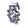
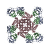
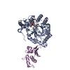
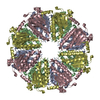
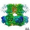
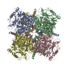
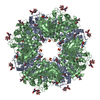
| Title | Three-dimensional structure of I(to); Kv4.2-KChIP2 ion channels by electron microscopy at 21 Angstrom resolution. | |||||||||
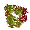
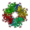
 Map data Map data | This is the 3D map of the Kv4.2-KChIP complex | |||||||||
 Sample Sample |
| |||||||||
| Biological species |   Homo sapiens (human) Homo sapiens (human) | |||||||||
| Method | single particle reconstruction / negative staining / Resolution: 21.0 Å | |||||||||
 Authors Authors | Kim LA / Furst J / Gutierrez D / Butler MH / Xu S / Goldstein SAN / Grigorieff N | |||||||||
 Citation Citation | Journal: Neuron / Year: 2004 Title: Three-dimensional structure of I(to); Kv4.2-KChIP2 ion channels by electron microscopy at 21 Angstrom resolution. Authors: Leo A Kim / Johannes Furst / David Gutierrez / Margaret H Butler / Shuhua Xu / Steve A N Goldstein / Nikolaus Grigorieff /  Abstract: Regulatory KChIP2 subunits assemble with pore-forming Kv4.2 subunits in 4:4 complexes to produce native voltage-gated potassium (Kv) channels like cardiac I(to) and neuronal I(A) subtypes. Here, ...Regulatory KChIP2 subunits assemble with pore-forming Kv4.2 subunits in 4:4 complexes to produce native voltage-gated potassium (Kv) channels like cardiac I(to) and neuronal I(A) subtypes. Here, negative stain electron microscopy (EM) and single particle averaging reveal KChIP2 to create a novel approximately 35 x 115 x 115 Angstrom, intracellular fenestrated rotunda: four peripheral columns that extend down from the membrane-embedded portion of the channel to enclose the Kv4.2 "hanging gondola" (a platform held beneath the transmembrane conduction pore by four internal columns). To reach the pore from the cytosol, ions traverse one of four external fenestrae to enter the rotundal vestibule and then cross one of four internal windows in the gondola. | |||||||||
| History |
|
- Structure visualization
| Movie |
 Movie viewer Movie viewer |
|---|---|
| Structure viewer | EM map: SurfViewMolmilJmol/JSmol |
| Supplemental images |

- Downloads & links
-EMDB archive
| Map data | emd_1074.map.gz | 2.6 MB | EMDB map data format | |
|---|---|---|---|---|
| Header (meta data) | emd-1074-v30.xmlemd-1074.xml | 11.7 KB 11.7 KB | Display Display | EMDB header |
| Images |  1074.gif 1074.gif | 26.5 KB | ||
| Archive directory |  http://ftp.pdbj.org/pub/emdb/structures/EMD-1074ftp://ftp.pdbj.org/pub/emdb/structures/EMD-1074 http://ftp.pdbj.org/pub/emdb/structures/EMD-1074ftp://ftp.pdbj.org/pub/emdb/structures/EMD-1074 | HTTPS FTP |
-Related structure data
| Similar structure data |
|---|
-Links
| EMDB pages | EMDB (EBI/PDBe) / EMDataResource |
|---|
-Map
| File | Download / File: emd_1074.map.gz / Format: CCP4 / Size: 2.7 MB / Type: IMAGE STORED AS FLOATING POINT NUMBER (4 BYTES) | ||||||||||||||||||||||||||||||||||||||||||||||||||||||||||||||||||||
|---|---|---|---|---|---|---|---|---|---|---|---|---|---|---|---|---|---|---|---|---|---|---|---|---|---|---|---|---|---|---|---|---|---|---|---|---|---|---|---|---|---|---|---|---|---|---|---|---|---|---|---|---|---|---|---|---|---|---|---|---|---|---|---|---|---|---|---|---|---|
| Annotation | This is the 3D map of the Kv4.2-KChIP complex | ||||||||||||||||||||||||||||||||||||||||||||||||||||||||||||||||||||
| Voxel size | X=Y=Z: 4.67 Å | ||||||||||||||||||||||||||||||||||||||||||||||||||||||||||||||||||||
| Density |
| ||||||||||||||||||||||||||||||||||||||||||||||||||||||||||||||||||||
| Symmetry | Space group: 1 | ||||||||||||||||||||||||||||||||||||||||||||||||||||||||||||||||||||
| Details | EMDB XML:
CCP4 map header:
| ||||||||||||||||||||||||||||||||||||||||||||||||||||||||||||||||||||
-Supplemental data
- Sample components
-Entire : Human Kv4.2-KChIP2 Ion Channel Complex
| Entire | Name: Human Kv4.2-KChIP2 Ion Channel Complex |
|---|---|
| Components |
|
-Supramolecule #1000: Human Kv4.2-KChIP2 Ion Channel Complex
| Supramolecule | Name: Human Kv4.2-KChIP2 Ion Channel Complex / type: sample / ID: 1000 Details: The sample was solubilised in 0.7% CHAPS and was monodisperse Oligomeric state: 4 Kv4.2 subunits bind to 4 KChIP2 subunits Number unique components: 2 |
|---|---|
| Molecular weight | Experimental: 440 KDa / Theoretical: 400 KDa / Method: SDS-PAGE |
-Macromolecule #1: Kv4.2
| Macromolecule | Name: Kv4.2 / type: protein_or_peptide / ID: 1 / Details: 4 monomers form a membrane-embedded channel / Number of copies: 4 / Oligomeric state: tetramer / Recombinant expression: Yes |
|---|---|
| Source (natural) | Organism: Homo sapiens (human) / synonym: Human / Location in cell: Cell membrane |
| Molecular weight | Experimental: 72 MDa / Theoretical: 75 MDa |
| Recombinant expression | Organism: COS7 / Recombinant plasmid: pRAT |
-Macromolecule #2: KChIP2
| Macromolecule | Name: KChIP2 / type: ligand / ID: 2 Details: 4 monomers bind on the cytoplasmic side of the channel Number of copies: 4 / Oligomeric state: Monomer / Recombinant expression: Yes |
|---|---|
| Molecular weight | Experimental: 28 MDa / Theoretical: 35 MDa |
| Recombinant expression | Organism: COS7 / Recombinant plasmid: pRAT |
-Experimental details
-Structure determination
| Method | negative staining |
|---|---|
 Processing Processing | single particle reconstruction |
| Aggregation state | particle |
-Sample preparation
| Concentration | 0.1 mg/mL |
|---|---|
| Buffer | pH: 7.4 Details: 0.7% CHAPS, 100 mM NaCl, 40 mM KCl, 0.01 mM leupeptin/pepstatin, 1 mM EDTA, 20 mM HEPES-KOH (pH 7.4) |
| Staining | Type: NEGATIVE Details: Grids with adsorbed protein were washed 3 times in detergent-free buffer and then floated on 1% uranyl acetate for 10 seconds |
| Grid | Details: 400 mesh copper grid |
| Vitrification | Cryogen name: NONE |
- Electron microscopy
| Microscope | FEI/PHILIPS CM120T |
|---|---|
| Electron beam | Acceleration voltage: 120 kV / Electron source: LAB6 |
| Electron optics | Illumination mode: FLOOD BEAM / Imaging mode: BRIGHT FIELDBright-field microscopy / Cs: 2.0 mm / Nominal defocus max: 1.5 µm / Nominal defocus min: 1.5 µm / Nominal magnification: 60000 |
| Sample stage | Specimen holder: Eucentric, room temperature / Specimen holder model: OTHER |
| Temperature | Average: 300 K |
| Alignment procedure | Legacy - Astigmatism: carbon film grain |
| Date | Jun 1, 2003 |
| Image recording | Category: FILM / Film or detector model: KODAK SO-163 FILM / Digitization - Scanner: ZEISS SCAI / Digitization - Sampling interval: 4.67 µm / Number real images: 63 / Average electron dose: 10 e/Å2 Details: Images were scanned at 7 microns resolution and 4 x 4 pixels were averaged Od range: 1.4 / Bits/pixel: 8 |
| Tilt angle min | 0 |
| Tilt angle max | 0 |
-Image processing
| CTF correction | Details: Each particle |
|---|---|
| Final angle assignment | Details: theta 90 degrees, phi 90 degrees |
| Final reconstruction | Applied symmetry - Point group: C4 (4 fold cyclic) / Algorithm: OTHER / Resolution.type: BY AUTHOR / Resolution: 21.0 Å / Resolution method: FSC 0.5 CUT-OFF / Software - Name: IMAGIC, FREALIGN / Details: FREALIGN reconstruction in Fourier space / Number images used: 8164 |
| Details | particles were selected manually |