 Movie
Movie Controller

[English] 日本語
 Yorodumi
- EMDB-8901: Structure of TRPM2 ion channel receptor by single particle electr... -
+ Open data

- Basic information
| Entry | Database: EMDB / ID: EMD-8901 | |||||||||
|---|---|---|---|---|---|---|---|---|---|---|
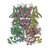
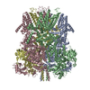
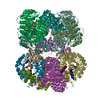
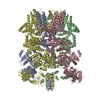
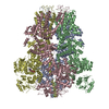
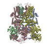
| Title | Structure of TRPM2 ion channel receptor by single particle electron cryo-microscopy, Apo state | |||||||||
 Map data Map data | Ion channel receptor | |||||||||
 Sample Sample |
| |||||||||
| Function / homology |  Function and homology information Function and homology information TRP channels / ligand-gated monoatomic cation channel activity / Neutrophil degranulation / ADP-D-ribose binding / mono-ADP-D-ribose binding / ligand-gated calcium channel activity / calcium ion transmembrane transport / calcium channel activity / monoatomic ion channel activity / protein homotetramerization ...TRP channels / ligand-gated monoatomic cation channel activity / Neutrophil degranulation / ADP-D-ribose binding / mono-ADP-D-ribose binding / ligand-gated calcium channel activity / calcium ion transmembrane transport / calcium channel activity / monoatomic ion channel activity / protein homotetramerization / calcium ion binding / plasma membrane TRP channels / ligand-gated monoatomic cation channel activity / Neutrophil degranulation / ADP-D-ribose binding / mono-ADP-D-ribose binding / ligand-gated calcium channel activity / calcium ion transmembrane transport / calcium channel activity / monoatomic ion channel activity / protein homotetramerization ...TRP channels / ligand-gated monoatomic cation channel activity / Neutrophil degranulation / ADP-D-ribose binding / mono-ADP-D-ribose binding / ligand-gated calcium channel activity / calcium ion transmembrane transport / calcium channel activity / monoatomic ion channel activity / protein homotetramerization / calcium ion binding / plasma membraneSimilarity search - Function | |||||||||
| Biological species |  Danio rerio (zebrafish) Danio rerio (zebrafish) | |||||||||
| Method | single particle reconstruction / cryo EM / Resolution: 3.8 Å | |||||||||
 Authors Authors | Du J / Lu W / Huang Y / Winkler P / Sun W | |||||||||
 Citation Citation | Journal: Nature / Year: 2018 Title: Architecture of the TRPM2 channel and its activation mechanism by ADP-ribose and calcium. Authors: Yihe Huang / Paige A Winkler / Weinan Sun / Wei Lü / Juan Du /  Abstract: Transient receptor potential melastatin 2 (TRPM2) is a calcium-permeable, non-selective cation channel that has an essential role in diverse physiological processes such as core body temperature ...Transient receptor potential melastatin 2 (TRPM2) is a calcium-permeable, non-selective cation channel that has an essential role in diverse physiological processes such as core body temperature regulation, immune response and apoptosis. TRPM2 is polymodal and can be activated by a wide range of stimuli, including temperature, oxidative stress and NAD-related metabolites such as ADP-ribose (ADPR). Its activation results in both Ca entry across the plasma membrane and Ca release from lysosomes, and has been linked to diseases such as ischaemia-reperfusion injury, bipolar disorder and Alzheimer's disease. Here we report the cryo-electron microscopy structures of the zebrafish TRPM2 in the apo resting (closed) state and in the ADPR/Ca-bound active (open) state, in which the characteristic NUDT9-H domains hang underneath the MHR1/2 domain. We identify an ADPR-binding site located in the bi-lobed structure of the MHR1/2 domain. Our results provide an insight into the mechanism of activation of the TRPM channel family and define a framework for the development of therapeutic agents to treat neurodegenerative diseases and temperature-related pathological conditions. | |||||||||
| History |
|
- Structure visualization
| Movie |
Movie viewer |
|---|---|
| Structure viewer | EM map: SurfViewMolmilJmol/JSmol |
| Supplemental images |

- Downloads & links
-EMDB archive
| Map data | emd_8901.map.gz | 95.6 MB | EMDB map data format | |
|---|---|---|---|---|
| Header (meta data) | emd-8901-v30.xmlemd-8901.xml | 10.4 KB 10.4 KB | Display Display | EMDB header |
| Images | 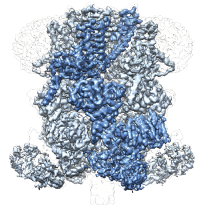 emd_8901.png emd_8901.png | 311.3 KB | ||
| Archive directory |  http://ftp.pdbj.org/pub/emdb/structures/EMD-8901ftp://ftp.pdbj.org/pub/emdb/structures/EMD-8901 http://ftp.pdbj.org/pub/emdb/structures/EMD-8901ftp://ftp.pdbj.org/pub/emdb/structures/EMD-8901 | HTTPS FTP |
-Related structure data
| Related structure data |  6drkMC  7999C  6drjC C: citing same article ( M: atomic model generated by this map |
|---|---|
| Similar structure data |


-Links
| EMDB pages | EMDB (EBI/PDBe) / EMDataResource |
|---|---|
| Related items in Molecule of the Month |


-Map
| File | Download / File: emd_8901.map.gz / Format: CCP4 / Size: 103 MB / Type: IMAGE STORED AS FLOATING POINT NUMBER (4 BYTES) | ||||||||||||||||||||||||||||||||||||||||||||||||||||||||||||
|---|---|---|---|---|---|---|---|---|---|---|---|---|---|---|---|---|---|---|---|---|---|---|---|---|---|---|---|---|---|---|---|---|---|---|---|---|---|---|---|---|---|---|---|---|---|---|---|---|---|---|---|---|---|---|---|---|---|---|---|---|---|
| Annotation | Ion channel receptor | ||||||||||||||||||||||||||||||||||||||||||||||||||||||||||||
| Projections & slices | Image control
Images are generated by Spider. | ||||||||||||||||||||||||||||||||||||||||||||||||||||||||||||
| Voxel size | X=Y=Z: 1.074 Å | ||||||||||||||||||||||||||||||||||||||||||||||||||||||||||||
| Density |
| ||||||||||||||||||||||||||||||||||||||||||||||||||||||||||||
| Symmetry | Space group: 1 | ||||||||||||||||||||||||||||||||||||||||||||||||||||||||||||
| Details | EMDB XML:
CCP4 map header:
| ||||||||||||||||||||||||||||||||||||||||||||||||||||||||||||
 Z (Sec.)
 Y (Row.)
 X (Col.)


-Supplemental data
- Sample components
-Entire : Ion channel 1
| Entire | Name: Ion channel 1 |
|---|---|
| Components |
|
-Supramolecule #1: Ion channel 1
| Supramolecule | Name: Ion channel 1 / type: complex / ID: 1 / Parent: 0 / Macromolecule list: all |
|---|---|
| Source (natural) | Organism: Danio rerio (zebrafish) |
| Recombinant expression | Organism: Mammalia (mammals) |
| Molecular weight | Experimental: 500 KDa |
-Macromolecule #1: Transient receptor potential cation channel, subfamily M, member 2
| Macromolecule | Name: Transient receptor potential cation channel, subfamily M, member 2 type: protein_or_peptide / ID: 1 / Number of copies: 4 / Enantiomer: LEVO |
|---|---|
| Source (natural) | Organism: Danio rerio (zebrafish) |
| Molecular weight | Theoretical: 168.512578 KDa |
| Recombinant expression | Organism:  Homo sapiens (human) Homo sapiens (human) |
| Sequence | String: MLGTSGVKIH PNGNSNQLGV QLENVKLTSL FKKLDKRCSL ASWIKENIKK KECCFYVEDG REGICKCGYP KVQHCDEAIK PEDYMGEQW DKHRHVRETP TDAFGDISFG GLGQKTGKYV RVSSDTSCEN LYQLMTEQWK LRSPNLLISV TGGAKNFYIK T HLKDKFRR ...String: MLGTSGVKIH PNGNSNQLGV QLENVKLTSL FKKLDKRCSL ASWIKENIKK KECCFYVEDG REGICKCGYP KVQHCDEAIK PEDYMGEQW DKHRHVRETP TDAFGDISFG GLGQKTGKYV RVSSDTSCEN LYQLMTEQWK LRSPNLLISV TGGAKNFYIK T HLKDKFRR GLIKVAQTTG AWILTGGTHA GVMKHVGMAV RDYTLSSGSM EGQIVVIGVA PWGVIHNRST LIHPEGRFPA YY SLDEQGQ GRLSCLDINH THFLLVDDGT QGHYGVEIEL RARLEKLISK LSLGNRESGV TIPVVCVVLD GGPGTLNTIY NSM LNHTPC VVLEGSGRLA DVIAHVASVP VSKVTMALIN RLLKRFFMQE YKNFTELQII EWTKKIQDIL RMPHLLTVFR IDED KNYDV DVAILQALLK ASRSDEHAGR HCWERQLELA VAWNRVDIAE SEIFTEESQW TSSDLHPAMF SALVGDKPEF VRLLL ENGV CVREFLEREE TLCELYSHLP SCFFLRKLAK RVQGGKMRRG QEPLPGSRKV CLSHVSEEVR HLLGSFTQPL YIASRY KPT KDDVRLKVPS KGALDLPCSG EEWSADTVWD PGRDLFLWAV VQNNRELAEI GWEQCRDCIA AALAASKILR KLAQESG ED DSEEATEMLE LANHYEKQAI GVFSECHSWD AQRAQKLLIR ISPSWGRSTC LWLALEAHDK SFIAHSGVQA LLTQIWCG E LSVDNPHWKV LLCMIFFPLI YTGFLTFRRD EDIQRQAERT EQQKLAMESV FAGQSDGKIK RHLRGFSQKS ELKPLNCSS RLMSFLKSPQ VKFYWNIASY FGFLWLFAVV LMIDFQTSPS WRELLLYVWL TSLVCEEIRQ LYHDFDGSGF RRKAKMYIKD LWNILDVLS IVLFIAGLIC RLQASDTVFY IGKVILCIDF IIFCLRLMAI FSISRTLGPK IIIVRRMMLD LFFFMFLLSI W VVAYGVAK QGILIENEER LNWIIRGAVY EPYITIFGNF PTNIDNTLFD ISSCSVNASD PLKPKCPMLN ADNTPVFPEW LT IMMLCVY LLFANILLLN LLIAIFNYTF QEVQDNTDTI WKFQRYELIK EYHSRPALPP PFILLSHLIL FIRGVFLRDL PQR HKNFRQ ELEQTEEEEL LSWEAYMKDN YLASTRQDES QSVEHRIHDT AEKVGAMSEL LEREQEMVSA TMAKRLARLE EQVS ESAKA LRWIIDALKS QGCKSKVQPP LMRSKSSDRD DGDSSGQETD DEEAPHMFAR QLQYPDSTVR RFPVPEEKVS WEVNF SPYQ PPVYNQQDSS ESDTSALDKH RNPGGRTGIR GKGALNTLGP NHILHPIFTR WRDAEHKVLE FLAVWEDAEK RWALLG GPA QPDEPLAQVL ERILGKKLNE KTKTLLKAGE EVYKGYVDDS RNTDNAWVET SIITLHCDKN TPLMADLNHM VESSLSS HQ PLQWREVSSD ACRCSYQREA LRQIAHHHNT YF |
-Experimental details
-Structure determination
| Method | cryo EM |
|---|---|
 Processing Processing | single particle reconstruction |
| Aggregation state | particle |
-Sample preparation
| Buffer | pH: 8 |
|---|---|
| Vitrification | Cryogen name: ETHANE |
- Electron microscopy
| Microscope | FEI TITAN KRIOS |
|---|---|
| Electron beam | Acceleration voltage: 300 kV / Electron source: FIELD EMISSION GUN |
| Electron optics | Illumination mode: OTHER / Imaging mode: BRIGHT FIELDBright-field microscopy |
| Image recording | Film or detector model: GATAN K2 SUMMIT (4k x 4k) / Average electron dose: 45.0 e/Å2 |
| Experimental equipment |  Model: Titan Krios / Image courtesy: FEI Company |
-Image processing
| Initial angle assignment | Type: ANGULAR RECONSTITUTION |
|---|---|
| Final angle assignment | Type: MAXIMUM LIKELIHOOD |
| Final reconstruction | Resolution.type: BY AUTHOR / Resolution: 3.8 Å / Resolution method: FSC 0.143 CUT-OFF / Number images used: 183041 |
