 Movie
Movie Controller

[English] 日本語
 Yorodumi
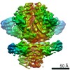
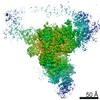
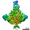
- EMDB-6500: Negative stain EM reconstruction of HIV-1 Env AMC008 SOSIP.v4 in ... -
+ Open data

- Basic information
| Entry | Database: EMDB / ID: EMD-6500 | |||||||||
|---|---|---|---|---|---|---|---|---|---|---|
| Title | Negative stain EM reconstruction of HIV-1 Env AMC008 SOSIP.v4 in complex with Fabs 35O22 and PGV04 | |||||||||
 Map data Map data | Reconstruction of HIV-1 Env AMC008 SOSIP.v4 in complex with broadly-neutralzing antibody Fabs 35O22 and PGV04 | |||||||||
 Sample Sample |
| |||||||||
 Keywords Keywords |  HIV-1 / Env / SOSIP / broadly-neutralizing antibody / trimer HIV-1 / Env / SOSIP / broadly-neutralizing antibody / trimer | |||||||||
| Biological species |   Human immunodeficiency virus 1 / Human immunodeficiency virus 1 /  Homo sapiens (human) Homo sapiens (human) | |||||||||
| Method | single particle reconstruction / negative staining / Resolution: 15.0 Å | |||||||||
 Authors Authors | de Taeye SW / Ozorowski G / de la Pena AT / Guttman M / Julien J-P / van den Kerkhof TLGM / Burger J / Pritchard LK / Pugach P / Yasmeen A ...de Taeye SW / Ozorowski G / de la Pena AT / Guttman M / Julien J-P / van den Kerkhof TLGM / Burger J / Pritchard LK / Pugach P / Yasmeen A / Bontjer I / Torres JL / Arendt H / DeStefano J / Koff WC / Schuitemaker H / Eggink D / Berkhout B / Dean H / LaBranche C / Crispin M / Montefiori DC / Klasse PJ / Lee KK / Moore JP / Wilson IA / Ward AB / Sanders RW | |||||||||
 Citation Citation | Journal: Cell / Year: 2015 Title: Immunogenicity of Stabilized HIV-1 Envelope Trimers with Reduced Exposure of Non-neutralizing Epitopes. Authors: Steven W de Taeye / Gabriel Ozorowski / Alba Torrents de la Peña / Miklos Guttman / Jean-Philippe Julien / Tom L G M van den Kerkhof / Judith A Burger / Laura K Pritchard / Pavel Pugach / ...Authors: Steven W de Taeye / Gabriel Ozorowski / Alba Torrents de la Peña / Miklos Guttman / Jean-Philippe Julien / Tom L G M van den Kerkhof / Judith A Burger / Laura K Pritchard / Pavel Pugach / Anila Yasmeen / Jordan Crampton / Joyce Hu / Ilja Bontjer / Jonathan L Torres / Heather Arendt / Joanne DeStefano / Wayne C Koff / Hanneke Schuitemaker / Dirk Eggink / Ben Berkhout / Hansi Dean / Celia LaBranche / Shane Crotty / Max Crispin / David C Montefiori / P J Klasse / Kelly K Lee / John P Moore / Ian A Wilson / Andrew B Ward / Rogier W Sanders /    Abstract: The envelope glycoprotein trimer mediates HIV-1 entry into cells. The trimer is flexible, fluctuating between closed and more open conformations and sometimes sampling the fully open, CD4-bound form. ...The envelope glycoprotein trimer mediates HIV-1 entry into cells. The trimer is flexible, fluctuating between closed and more open conformations and sometimes sampling the fully open, CD4-bound form. We hypothesized that conformational flexibility and transient exposure of non-neutralizing, immunodominant epitopes could hinder the induction of broadly neutralizing antibodies (bNAbs). We therefore modified soluble Env trimers to stabilize their closed, ground states. The trimer variants were indeed stabilized in the closed conformation, with a reduced ability to undergo receptor-induced conformational changes and a decreased exposure of non-neutralizing V3-directed antibody epitopes. In rabbits, the stabilized trimers induced similar autologous Tier-1B or Tier-2 NAb titers to those elicited by the corresponding wild-type trimers but lower levels of V3-directed Tier-1A NAbs. Stabilized, closed trimers might therefore be useful components of vaccines aimed at inducing bNAbs. | |||||||||
| History |
|
- Structure visualization
| Movie |
 Movie viewer Movie viewer |
|---|---|
| Structure viewer | EM map: SurfViewMolmilJmol/JSmol |
| Supplemental images |

- Downloads & links
-EMDB archive
| Map data | emd_6500.map.gz | 14.4 MB | EMDB map data format | |
|---|---|---|---|---|
| Header (meta data) | emd-6500-v30.xmlemd-6500.xml | 14.5 KB 14.5 KB | Display Display | EMDB header |
| Images | emd_6500.tif | 156.3 KB | ||
| Archive directory |  http://ftp.pdbj.org/pub/emdb/structures/EMD-6500ftp://ftp.pdbj.org/pub/emdb/structures/EMD-6500 http://ftp.pdbj.org/pub/emdb/structures/EMD-6500ftp://ftp.pdbj.org/pub/emdb/structures/EMD-6500 | HTTPS FTP |
-Related structure data
| Similar structure data |
|---|


-Links
| EMDB pages | EMDB (EBI/PDBe) / EMDataResource |
|---|
-Map
| File | Download / File: emd_6500.map.gz / Format: CCP4 / Size: 15.3 MB / Type: IMAGE STORED AS FLOATING POINT NUMBER (4 BYTES) | ||||||||||||||||||||||||||||||||||||||||||||||||||||||||||||||||||||
|---|---|---|---|---|---|---|---|---|---|---|---|---|---|---|---|---|---|---|---|---|---|---|---|---|---|---|---|---|---|---|---|---|---|---|---|---|---|---|---|---|---|---|---|---|---|---|---|---|---|---|---|---|---|---|---|---|---|---|---|---|---|---|---|---|---|---|---|---|---|
| Annotation | Reconstruction of HIV-1 Env AMC008 SOSIP.v4 in complex with broadly-neutralzing antibody Fabs 35O22 and PGV04 | ||||||||||||||||||||||||||||||||||||||||||||||||||||||||||||||||||||
| Voxel size | X=Y=Z: 1.57 Å | ||||||||||||||||||||||||||||||||||||||||||||||||||||||||||||||||||||
| Density |
| ||||||||||||||||||||||||||||||||||||||||||||||||||||||||||||||||||||
| Symmetry | Space group: 1 | ||||||||||||||||||||||||||||||||||||||||||||||||||||||||||||||||||||
| Details | EMDB XML:
CCP4 map header:
| ||||||||||||||||||||||||||||||||||||||||||||||||||||||||||||||||||||
-Supplemental data
- Sample components
-Entire : HIV-1 Env AMC008 SOSIP.v4 in complex with broadly neutralizing an...
| Entire | Name: HIV-1 Env AMC008 SOSIP.v4 in complex with broadly neutralizing antibody Fabs 35O22 and PGV04 |
|---|---|
| Components |
|
-Supramolecule #1000: HIV-1 Env AMC008 SOSIP.v4 in complex with broadly neutralizing an...
| Supramolecule | Name: HIV-1 Env AMC008 SOSIP.v4 in complex with broadly neutralizing antibody Fabs 35O22 and PGV04 type: sample / ID: 1000 Details: All components were purified by size-exclusion chromatography prior to complex formation. Oligomeric state: 3 of each Fab bound to a trimer of HIV-1 Env SOSIP.v4 Number unique components: 3 |
|---|---|
| Molecular weight | Theoretical: 720 KDa Method: Estimation based on average molecular weight of Fab and average mass of SOSIP trimer including glycan mass |
-Macromolecule #1: HIV-1 AMC008 Env SOSIP.v4
| Macromolecule | Name: HIV-1 AMC008 Env SOSIP.v4 / type: protein_or_peptide / ID: 1 / Name.synonym: HIV-1 gp140 / Number of copies: 1 / Oligomeric state: Trimer / Recombinant expression: Yes |
|---|---|
| Source (natural) | Organism: Human immunodeficiency virus 1 / Strain: AMC008 |
| Molecular weight | Theoretical: 420 KDa |
| Recombinant expression | Organism: Homo sapiens (human) / Recombinant cell: HEK293F |
-Macromolecule #2: Anti-HIV-1 antibody PGV04 Fab
| Macromolecule | Name: Anti-HIV-1 antibody PGV04 Fab / type: protein_or_peptide / ID: 2 / Number of copies: 3 / Oligomeric state: Heterodimer of light and heavy chain / Recombinant expression: Yes |
|---|---|
| Source (natural) | Organism: Homo sapiens (human) / synonym: Human |
| Molecular weight | Theoretical: 50 KDa |
| Recombinant expression | Organism: Homo sapiens (human) / Recombinant cell: HEK293F |
-Macromolecule #3: Anti-HIV-1 antibody 35O22 Fab
| Macromolecule | Name: Anti-HIV-1 antibody 35O22 Fab / type: protein_or_peptide / ID: 3 / Number of copies: 3 / Oligomeric state: Heterodimer of light and heavy chain / Recombinant expression: Yes |
|---|---|
| Source (natural) | Organism: Homo sapiens (human) / synonym: Human |
| Molecular weight | Theoretical: 50 KDa |
| Recombinant expression | Organism: Homo sapiens (human) / Recombinant cell: HEK293F |
-Experimental details
-Structure determination
| Method | negative staining |
|---|---|
 Processing Processing | single particle reconstruction |
| Aggregation state | particle |
-Sample preparation
| Concentration | 0.03 mg/mL |
|---|---|
| Buffer | pH: 7.4 / Details: 50 mM Tris-HCl, 150 mM NaCl |
| Staining | Type: NEGATIVE Details: Grids were stained with 2% w/v uranyl formate for 60 seconds. |
| Grid | Details: 400 mesh copper grid with thin carbon support, glow-discharged |
| Vitrification | Cryogen name: NONE / Instrument: OTHER |
- Electron microscopy
| Microscope | OTHER |
|---|---|
| Electron beam | Acceleration voltage: 200 kV / Electron source: FIELD EMISSION GUN |
| Electron optics | Illumination mode: FLOOD BEAM / Imaging mode: BRIGHT FIELDBright-field microscopy / Nominal defocus max: 1.0 µm / Nominal defocus min: 1.0 µm / Nominal magnification: 92000 |
| Sample stage | Specimen holder model: SIDE ENTRY, EUCENTRIC / Tilt angle min: -50 |
| Alignment procedure | Legacy - Astigmatism: Objective lens astigmatism was corrected at 92,000 times magnification. |
| Details | Tilt series of -50, -40, -30, -20, -10, 0 degrees |
| Date | Feb 19, 2015 |
| Image recording | Category: CCD / Film or detector model: FEI CETA (4k x 4k) / Number real images: 152 / Average electron dose: 25 e/Å2 |
| Tilt angle max | 0 |
-Image processing
| Final reconstruction | Algorithm: OTHER / Resolution.type: BY AUTHOR / Resolution: 15.0 Å / Resolution method: OTHER / Software - Name: EMAN2, SPARX / Number images used: 24522 |
|---|---|
| Details | Particles were selected using an automatic selection program and aligned into class averages using Iterative MSA/MRA. Classes containing fully decorated (3 molecules of each Fab per trimer) complexes were used to generate a common lines model that was later refined using particles from those classes. |
