 Movie
Movie Controller


+ Open data

- Basic information
| Entry | Database: EMDB / ID: EMD-6395 | |||||||||
|---|---|---|---|---|---|---|---|---|---|---|
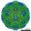
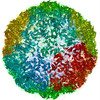
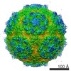
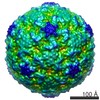
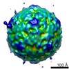
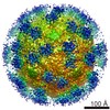
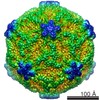
| Title | Structure of Ljungan virus: insight into picornavirus packaging | |||||||||
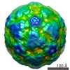
 Map data Map data | Reconstruction of Ljungan virus at lower resolution without post-refinement in Relion | |||||||||
 Sample Sample |
| |||||||||
 Keywords Keywords | Ljungan virus / Picornavirus / assembly / pathogen | |||||||||
| Biological species |  Ljungan virus Ljungan virus | |||||||||
| Method | single particle reconstruction / cryo EM / negative staining / Resolution: 4.5 Å | |||||||||
 Authors Authors | Zhu L / Wang XX / Ren JS / Porta C / Wenham H / Ekstrom J-O / Panjwani A / Knowles NJ / Kotecha A / Siebert A ...Zhu L / Wang XX / Ren JS / Porta C / Wenham H / Ekstrom J-O / Panjwani A / Knowles NJ / Kotecha A / Siebert A / Lindberg M / Fry EE / Rao ZH / Tuthill TJ / Stuart DI | |||||||||
 Citation Citation | Journal: Nat Commun / Year: 2015 Title: Structure of Ljungan virus provides insight into genome packaging of this picornavirus. Authors: Ling Zhu / Xiangxi Wang / Jingshan Ren / Claudine Porta / Hannah Wenham / Jens-Ola Ekström / Anusha Panjwani / Nick J Knowles / Abhay Kotecha / C Alistair Siebert / A Michael Lindberg / ...Authors: Ling Zhu / Xiangxi Wang / Jingshan Ren / Claudine Porta / Hannah Wenham / Jens-Ola Ekström / Anusha Panjwani / Nick J Knowles / Abhay Kotecha / C Alistair Siebert / A Michael Lindberg / Elizabeth E Fry / Zihe Rao / Tobias J Tuthill / David I Stuart /    Abstract: Picornaviruses are responsible for a range of human and animal diseases, but how their RNA genome is packaged remains poorly understood. A particularly poorly studied group within this family are ...Picornaviruses are responsible for a range of human and animal diseases, but how their RNA genome is packaged remains poorly understood. A particularly poorly studied group within this family are those that lack the internal coat protein, VP4. Here we report the atomic structure of one such virus, Ljungan virus, the type member of the genus Parechovirus B, which has been linked to diabetes and myocarditis in humans. The 3.78-Å resolution cryo-electron microscopy structure shows remarkable features, including an extended VP1 C terminus, forming a major protuberance on the outer surface of the virus, and a basic motif at the N terminus of VP3, binding to which orders some 12% of the viral genome. This apparently charge-driven RNA attachment suggests that this branch of the picornaviruses uses a different mechanism of genome encapsidation, perhaps explored early in the evolution of picornaviruses. | |||||||||
| History |
|
- Structure visualization
| Movie |
 Movie viewer Movie viewer |
|---|---|
| Structure viewer | EM map: SurfViewMolmilJmol/JSmol |
| Supplemental images |

- Downloads & links
-EMDB archive
| Map data | emd_6395.map.gz | 139.1 MB | EMDB map data format | |
|---|---|---|---|---|
| Header (meta data) | emd-6395-v30.xmlemd-6395.xml | 10.4 KB 10.4 KB | Display Display | EMDB header |
| Images | emd_6395.tif | 198.8 KB | ||
| Archive directory |  http://ftp.pdbj.org/pub/emdb/structures/EMD-6395ftp://ftp.pdbj.org/pub/emdb/structures/EMD-6395 http://ftp.pdbj.org/pub/emdb/structures/EMD-6395ftp://ftp.pdbj.org/pub/emdb/structures/EMD-6395 | HTTPS FTP |
-Validation report
| Summary document | emd_6395_validation.pdf.gz | 78.7 KB | Display | EMDB validaton report |
|---|---|---|---|---|
| Full document | emd_6395_full_validation.pdf.gz | 77.9 KB | Display | |
| Data in XML | emd_6395_validation.xml.gz | 493 B | Display | |
| Arichive directory | https://ftp.pdbj.org/pub/emdb/validation_reports/EMD-6395ftp://ftp.pdbj.org/pub/emdb/validation_reports/EMD-6395 | HTTPS FTP |
-Related structure data


-Links
| EMDB pages | EMDB (EBI/PDBe) / EMDataResource |
|---|
-Map
| File | Download / File: emd_6395.map.gz / Format: CCP4 / Size: 173.8 MB / Type: IMAGE STORED AS FLOATING POINT NUMBER (4 BYTES) | ||||||||||||||||||||||||||||||||||||||||||||||||||||||||||||
|---|---|---|---|---|---|---|---|---|---|---|---|---|---|---|---|---|---|---|---|---|---|---|---|---|---|---|---|---|---|---|---|---|---|---|---|---|---|---|---|---|---|---|---|---|---|---|---|---|---|---|---|---|---|---|---|---|---|---|---|---|---|
| Annotation | Reconstruction of Ljungan virus at lower resolution without post-refinement in Relion | ||||||||||||||||||||||||||||||||||||||||||||||||||||||||||||
| Voxel size | X=Y=Z: 1.35 Å | ||||||||||||||||||||||||||||||||||||||||||||||||||||||||||||
| Density |
| ||||||||||||||||||||||||||||||||||||||||||||||||||||||||||||
| Symmetry | Space group: 1 | ||||||||||||||||||||||||||||||||||||||||||||||||||||||||||||
| Details | EMDB XML:
CCP4 map header:
| ||||||||||||||||||||||||||||||||||||||||||||||||||||||||||||
-Supplemental data
- Sample components
-Entire : Ljungan virus (type: 87-012)
| Entire | Name: Ljungan virus (type: 87-012) |
|---|---|
| Components |
|
-Supramolecule #1000: Ljungan virus (type: 87-012)
| Supramolecule | Name: Ljungan virus (type: 87-012) / type: sample / ID: 1000 / Number unique components: 1 |
|---|---|
| Molecular weight | Experimental: 6 MDa / Theoretical: 6 MDa / Method: Sedimentation |
-Supramolecule #1: Ljungan virus
| Supramolecule | Name: Ljungan virus / type: virus / ID: 1 / NCBI-ID: 172314 / Sci species name: Ljungan virus / Virus type: VIRION / Virus isolate: SPECIES / Virus enveloped: No / Virus empty: No |
|---|---|
| Host (natural) | Organism:  |
| Host system | Organism: Chlorocebus aethiops (grivet) / Recombinant cell: kidney (Vero) cell |
| Molecular weight | Experimental: 6 MDa / Theoretical: 6 MDa |
| Virus shell | Shell ID: 1 / Diameter: 320 Å / T number (triangulation number): 1 |
-Experimental details
-Structure determination
| Method | negative staining, cryo EM |
|---|---|
 Processing Processing | single particle reconstruction |
| Aggregation state | particle |
-Sample preparation
| Concentration | 1 mg/mL |
|---|---|
| Buffer | pH: 7.4 / Details: PBS |
| Staining | Type: NEGATIVE Details: Grids with adsorbed viruses were floated on 1% w/v uranyl acetate for 20 seconds. |
| Grid | Details: 200 mesh gold grid with thin carbon support, glow-discharged in amylamine atmosphere |
| Vitrification | Cryogen name: ETHANE / Chamber humidity: 90 % / Instrument: FEI VITROBOT MARK III / Method: Blot for 3 seconds before plunging |
- Electron microscopy
| Microscope | FEI POLARA 300 |
|---|---|
| Alignment procedure | Legacy - Astigmatism: Objective lens astigmatism was corrected at 160,000 times magnification. |
| Specialist optics | Energy filter - Name: FEI |
| Date | Feb 2, 2015 |
| Image recording | Category: CCD / Film or detector model: GATAN K2 (4k x 4k) / Number real images: 288 |
| Electron beam | Acceleration voltage: 300 kV / Electron source:  FIELD EMISSION GUN FIELD EMISSION GUN |
| Electron optics | Illumination mode: FLOOD BEAM / Imaging mode: BRIGHT FIELD / Cs: 2.0 mm |
| Sample stage | Specimen holder model: GATAN HELIUM |
| Experimental equipment |  Model: Tecnai Polara / Image courtesy: FEI Company |
-Image processing
| Final reconstruction | Resolution.type: BY AUTHOR / Resolution: 4.5 Å / Resolution method: OTHER / Software - Name: RELION / Number images used: 5558 |
|---|---|
| Final two d classification | Number classes: 20 |
