 Movie
Movie Controller

[English] 日本語
 Yorodumi
- EMDB-6358: Negative stain (RCT) surface of full-length glucagon receptor FAB... -
+ Open data

- Basic information
| Entry | Database: EMDB / ID: EMD-6358 | |||||||||
|---|---|---|---|---|---|---|---|---|---|---|
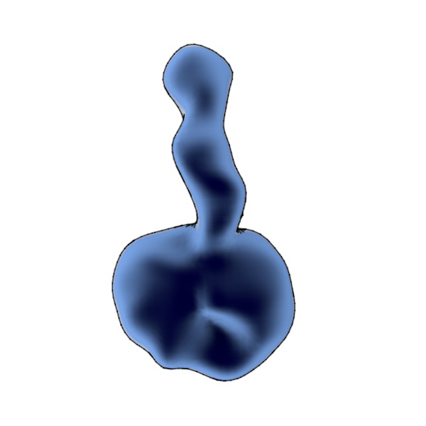
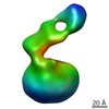
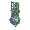
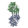
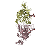
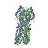
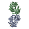
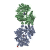
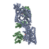
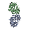
| Title | Negative stain (RCT) surface of full-length glucagon receptor FAB complex | |||||||||
 Map data Map data | RCT of full-length glucagon receptor FAB complex, alternative orientation | |||||||||
 Sample Sample |
| |||||||||
 Keywords Keywords |  Glucagon Receptor / GPCR Glucagon Receptor / GPCR | |||||||||
| Biological species |  Homo sapiens (human) / Homo sapiens (human) /  Mus musculus (house mouse) Mus musculus (house mouse) | |||||||||
| Method | single particle reconstruction / negative staining / Resolution: 40.0 Å | |||||||||
 Authors Authors | Yang L / Yang D / de Graaf C / Moeller A / West GM / Dharmarajan V / Wang C / Siu FY / Song G / Reedtz-Runge S ...Yang L / Yang D / de Graaf C / Moeller A / West GM / Dharmarajan V / Wang C / Siu FY / Song G / Reedtz-Runge S / Pascal BD / Wu B / Potter CS / Zhou H / Griffin PR / Carragher B / Yang H / Wang MW / Stevens RC / Jiang H | |||||||||
 Citation Citation | Journal: Nat Commun / Year: 2015 Title: Conformational states of the full-length glucagon receptor. Authors: Linlin Yang / Dehua Yang / Chris de Graaf / Arne Moeller / Graham M West / Venkatasubramanian Dharmarajan / Chong Wang / Fai Y Siu / Gaojie Song / Steffen Reedtz-Runge / Bruce D Pascal / ...Authors: Linlin Yang / Dehua Yang / Chris de Graaf / Arne Moeller / Graham M West / Venkatasubramanian Dharmarajan / Chong Wang / Fai Y Siu / Gaojie Song / Steffen Reedtz-Runge / Bruce D Pascal / Beili Wu / Clinton S Potter / Hu Zhou / Patrick R Griffin / Bridget Carragher / Huaiyu Yang / Ming-Wei Wang / Raymond C Stevens / Hualiang Jiang /     Abstract: Class B G protein-coupled receptors are composed of an extracellular domain (ECD) and a seven-transmembrane (7TM) domain, and their signalling is regulated by peptide hormones. Using a hybrid ...Class B G protein-coupled receptors are composed of an extracellular domain (ECD) and a seven-transmembrane (7TM) domain, and their signalling is regulated by peptide hormones. Using a hybrid structural biology approach together with the ECD and 7TM domain crystal structures of the glucagon receptor (GCGR), we examine the relationship between full-length receptor conformation and peptide ligand binding. Molecular dynamics (MD) and disulfide crosslinking studies suggest that apo-GCGR can adopt both an open and closed conformation associated with extensive contacts between the ECD and 7TM domain. The electron microscopy (EM) map of the full-length GCGR shows how a monoclonal antibody stabilizes the ECD and 7TM domain in an elongated conformation. Hydrogen/deuterium exchange (HDX) studies and MD simulations indicate that an open conformation is also stabilized by peptide ligand binding. The combined studies reveal the open/closed states of GCGR and suggest that glucagon binds to GCGR by a conformational selection mechanism. | |||||||||
| History |
|
- Structure visualization
| Movie |
 Movie viewer Movie viewer |
|---|---|
| Structure viewer | EM map: SurfViewMolmilJmol/JSmol |
| Supplemental images |
- Downloads & links
-EMDB archive
| Map data | emd_6358.map.gz | 424 KB | EMDB map data format | |
|---|---|---|---|---|
| Header (meta data) | emd-6358-v30.xmlemd-6358.xml | 11.1 KB 11.1 KB | Display Display | EMDB header |
| Images |  emd_6358.jpg emd_6358.jpg | 27.8 KB | ||
| Archive directory |  http://ftp.pdbj.org/pub/emdb/structures/EMD-6358ftp://ftp.pdbj.org/pub/emdb/structures/EMD-6358 http://ftp.pdbj.org/pub/emdb/structures/EMD-6358ftp://ftp.pdbj.org/pub/emdb/structures/EMD-6358 | HTTPS FTP |
-Related structure data
-Links
| EMDB pages | EMDB (EBI/PDBe) / EMDataResource |
|---|
-Map
| File | Download / File: emd_6358.map.gz / Format: CCP4 / Size: 1.9 MB / Type: IMAGE STORED AS FLOATING POINT NUMBER (4 BYTES) | ||||||||||||||||||||||||||||||||||||||||||||||||||||||||||||||||||||
|---|---|---|---|---|---|---|---|---|---|---|---|---|---|---|---|---|---|---|---|---|---|---|---|---|---|---|---|---|---|---|---|---|---|---|---|---|---|---|---|---|---|---|---|---|---|---|---|---|---|---|---|---|---|---|---|---|---|---|---|---|---|---|---|---|---|---|---|---|---|
| Annotation | RCT of full-length glucagon receptor FAB complex, alternative orientation | ||||||||||||||||||||||||||||||||||||||||||||||||||||||||||||||||||||
| Voxel size | X=Y=Z: 2.7275 Å | ||||||||||||||||||||||||||||||||||||||||||||||||||||||||||||||||||||
| Density |
| ||||||||||||||||||||||||||||||||||||||||||||||||||||||||||||||||||||
| Symmetry | Space group: 1 | ||||||||||||||||||||||||||||||||||||||||||||||||||||||||||||||||||||
| Details | EMDB XML:
CCP4 map header:
| ||||||||||||||||||||||||||||||||||||||||||||||||||||||||||||||||||||
-Supplemental data
- Sample components
-Entire : Full-length GCGR in complex with the antigen-binding fragment (Fa...
| Entire | Name: Full-length GCGR in complex with the antigen-binding fragment (Fab) of the monoclonal antibody mAb2330 |
|---|---|
| Components |
|
-Supramolecule #1000: Full-length GCGR in complex with the antigen-binding fragment (Fa...
| Supramolecule | Name: Full-length GCGR in complex with the antigen-binding fragment (Fab) of the monoclonal antibody mAb2330 type: sample / ID: 1000 / Oligomeric state: heterodimer / Number unique components: 2 |
|---|---|
| Molecular weight | Theoretical: 95 KDa |
-Macromolecule #1: glucagon receptor
| Macromolecule | Name: glucagon receptor / type: protein_or_peptide / ID: 1 / Name.synonym: GCGR / Number of copies: 1 / Oligomeric state: monomer / Recombinant expression: Yes |
|---|---|
| Source (natural) | Organism: Homo sapiens (human) / synonym: human / Location in cell: plasma membrane |
| Molecular weight | Experimental: 95 KDa / Theoretical: 95 KDa |
| Recombinant expression | Organism:  Cricetulus griseus (Chinese hamster) / Recombinant cell: CHO-K1 Cricetulus griseus (Chinese hamster) / Recombinant cell: CHO-K1 |
-Macromolecule #2: antibody mAb2330 Fab
| Macromolecule | Name: antibody mAb2330 Fab / type: protein_or_peptide / ID: 2 / Number of copies: 1 / Recombinant expression: No / Database: NCBI |
|---|---|
| Source (natural) | Organism: Mus musculus (house mouse) / synonym: mouse |
-Experimental details
-Structure determination
| Method | negative staining |
|---|---|
 Processing Processing | single particle reconstruction |
| Aggregation state | particle |
-Sample preparation
| Concentration | 0.00095 mg/mL |
|---|---|
| Buffer | pH: 7.5 / Details: HEPES LMNG |
| Staining | Type: NEGATIVE / Details: stained 3 times with 2% w/v uranyl formate |
| Grid | Details: thin carbon over holes |
| Vitrification | Cryogen name: NONE / Instrument: OTHER |
- Electron microscopy
| Microscope | FEI TECNAI F20 |
|---|---|
| Electron beam | Acceleration voltage: 200 kV / Electron source: FIELD EMISSION GUN |
| Electron optics | Calibrated magnification: 62000 / Illumination mode: FLOOD BEAM / Imaging mode: BRIGHT FIELDBright-field microscopy / Nominal magnification: 62000 |
| Sample stage | Specimen holder model: SIDE ENTRY, EUCENTRIC / Tilt angle min: -50 |
| Date | Feb 20, 2014 |
| Image recording | Category: CCD / Film or detector model: TVIPS TEMCAM-F416 (4k x 4k) / Number real images: 648 / Average electron dose: 45 e/Å2 |
| Tilt angle max | 0 |
| Experimental equipment |  Model: Tecnai F20 / Image courtesy: FEI Company |
-Image processing
| Final two d classification | Number classes: 1 |
|---|---|
| Final reconstruction | Algorithm: OTHER / Resolution.type: BY AUTHOR / Resolution: 40.0 Å / Resolution method: OTHER / Software - Name: Appion, Xmipp, Spider / Number images used: 343 |
| Details | RCT |
