negative regulation of protein modification process / detection of chemical stimulus / methyl accepting chemotaxis protein complex / protein histidine kinase binding / positive regulation of post-translational protein modification / bacterial-type flagellum-dependent swimming motility / cell tip / regulation of bacterial-type flagellum-dependent cell motility / aerotaxis / protein histidine kinase activity ...negative regulation of protein modification process / detection of chemical stimulus / methyl accepting chemotaxis protein complex / protein histidine kinase binding / positive regulation of post-translational protein modification / bacterial-type flagellum-dependent swimming motility / cell tip / regulation of bacterial-type flagellum-dependent cell motility / aerotaxis / protein histidine kinase activity / regulation of chemotaxis / thermotaxis / signal complex assembly / histidine kinase / phosphorelay sensor kinase activity / phosphorelay signal transduction system / establishment of localization in cell / cellular response to amino acid stimulus / protein homooligomerization / chemotaxis / transmembrane signaling receptor activity / protein domain specific binding / phosphorylation / signal transduction / protein homodimerization activity / ATP binding / identical protein binding / plasma membrane / cytosol / cytoplasm Similarity search - Function
Methyl-accepting chemotaxis protein II / Chemotaxis protein CheA / Chemotaxis protein CheW / Chemotaxis protein CheA / Chemotaxis protein CheW / Methyl-accepting chemotaxis protein 2 Similarity search - Component
Biological species
Escherichia coli (E. coli)
Method
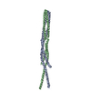
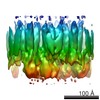
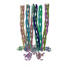
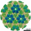
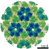
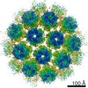
subtomogram averaging / cryo EM / Resolution: 17.5 Å
Journal: Elife / Year: 2015 Title: CryoEM and computer simulations reveal a novel kinase conformational switch in bacterial chemotaxis signaling. Authors: C Keith Cassidy / Benjamin A Himes / Frances J Alvarez / Jun Ma / Gongpu Zhao / Juan R Perilla / Klaus Schulten / Peijun Zhang / Abstract: Chemotactic responses in bacteria require large, highly ordered arrays of sensory proteins to mediate the signal transduction that ultimately controls cell motility. A mechanistic understanding of ...Chemotactic responses in bacteria require large, highly ordered arrays of sensory proteins to mediate the signal transduction that ultimately controls cell motility. A mechanistic understanding of the molecular events underlying signaling, however, has been hampered by the lack of a high-resolution structural description of the extended array. Here, we report a novel reconstitution of the array, involving the receptor signaling domain, histidine kinase CheA, and adaptor protein CheW, as well as a density map of the core-signaling unit at 11.3 Å resolution, obtained by cryo-electron tomography and sub-tomogram averaging. Extracting key structural constraints from our density map, we computationally construct and refine an atomic model of the core array structure, exposing novel interfaces between the component proteins. Using all-atom molecular dynamics simulations, we further reveal a distinctive conformational change in CheA. Mutagenesis and chemical cross-linking experiments confirm the importance of the conformational dynamics of CheA for chemotactic function.
History
Deposition
Apr 17, 2015
-
Header (metadata) release
May 20, 2015
-
Map release
Dec 9, 2015
-
Update
Dec 9, 2015
-
Current status
Dec 9, 2015
Processing site: RCSB / Status: Released
-
Structure visualization
Movie
Surface view with section colored by density value
Macromolecule #3: Methyl-accepting chemotaxis protein II
Macromolecule
Name: Methyl-accepting chemotaxis protein II / type: protein_or_peptide / ID: 3 / Name.synonym: tarCF Details: Cytoplasmic fragment of wild-type aspartate receptor Number of copies: 6 / Oligomeric state: trimer of dimers / Recombinant expression: Yes
Source (natural)
Organism: Escherichia coli (E. coli) / Location in cell: Inner Membrane
Specimen holder model: OTHER / Tilt series - Axis1 - Min angle: -70 ° / Tilt series - Axis1 - Max angle: 70 °
Date
Jan 7, 2009
Image recording
Category: CCD / Film or detector model: GATAN ULTRASCAN 4000 (4k x 4k) / Number real images: 60 / Average electron dose: 60 e/Å2 / Bits/pixel: 16
Experimental equipment
Model: Tecnai Polara / Image courtesy: FEI Company
-
Image processing
Crystal parameters
Plane group: P 1
CTF correction
Details: TomoCTF (strip-based periodogram)
Final reconstruction
Algorithm: OTHER / Resolution.type: BY AUTHOR / Resolution: 17.5 Å / Resolution method: OTHER / Software - Name: IMOD / Number subtomograms used: 400
Details
Subtomograms were initially selected using template matching. Positions were refined using alignment to class averages. Classification was also used to select symmetry centers and to remove high variance outliers. Cross-correlation with missing-wedge compensation was used for alignment, with SVD and HAC used for statistical analysis.
In the structure databanks used in Yorodumi, some data are registered as the other names, "COVID-19 virus" and "2019-nCoV". Here are the details of the virus and the list of structure data.
Jan 31, 2019. EMDB accession codes are about to change! (news from PDBe EMDB page)
EMDB accession codes are about to change! (news from PDBe EMDB page)
The allocation of 4 digits for EMDB accession codes will soon come to an end. Whilst these codes will remain in use, new EMDB accession codes will include an additional digit and will expand incrementally as the available range of codes is exhausted. The current 4-digit format prefixed with “EMD-” (i.e. EMD-XXXX) will advance to a 5-digit format (i.e. EMD-XXXXX), and so on. It is currently estimated that the 4-digit codes will be depleted around Spring 2019, at which point the 5-digit format will come into force.
The EM Navigator/Yorodumi systems omit the EMD- prefix.
Related info.:Q: What is EMD? / ID/Accession-code notation in Yorodumi/EM Navigator
Yorodumi is a browser for structure data from EMDB, PDB, SASBDB, etc.
This page is also the successor to EM Navigator detail page, and also detail information page/front-end page for Omokage search.
The word "yorodu" (or yorozu) is an old Japanese word meaning "ten thousand". "mi" (miru) is to see.
Related info.:EMDB / PDB / SASBDB / Comparison of 3 databanks / Yorodumi Search / Aug 31, 2016. New EM Navigator & Yorodumi / Yorodumi Papers / Jmol/JSmol / Function and homology information / Changes in new EM Navigator and Yorodumi
 Movie
Movie Controller

 Yorodumi
 Open data
 Basic information
 Map data
 Sample
 Keywords
 Signal transduction /
  Function and homology information
 Authors
 Citation
 Structure visualization
 Downloads & links
 emd_6320.png
 http://ftp.pdbj.org/pub/emdb/structures/EMD-6320
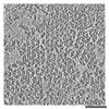


 Sample components
 Processing
 Electron microscopy