 Movie
Movie Controller

[English] 日本語
 Yorodumi
- EMDB-6318: Three-dimensional structure of AcrAB and MacA-TolC alpha-hybrid d... -
+ Open data

- Basic information
| Entry | Database: EMDB / ID: EMD-6318 | |||||||||
|---|---|---|---|---|---|---|---|---|---|---|
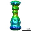
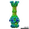
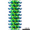
| Title | Three-dimensional structure of AcrAB and MacA-TolC alpha-hybrid dimer complex | |||||||||
 Map data Map data | Reconstruction of negatively stained AcrB-transmembrane linker-AcrA-AcrA and MacA-TolC alpha hybrid dimer complex, which supports direct interaction between AcrA and TolC | |||||||||
 Sample Sample |
| |||||||||
 Keywords Keywords | multidrug efflux pump /  multidrug resistance / resistance-nodulation-division family multidrug resistance / resistance-nodulation-division family | |||||||||
| Biological species |  Escherichia coli (E. coli) Escherichia coli (E. coli) | |||||||||
| Method | single particle reconstruction / negative staining / Resolution: 24.0 Å | |||||||||
 Authors Authors | Kim J-S / Jeong H / Song S / Kim H-Y / Lee K / Hyun J / Ha N-C | |||||||||
 Citation Citation | Journal: Mol Cells / Year: 2015 Title: Structure of the tripartite multidrug efflux pump AcrAB-TolC suggests an alternative assembly mode. Authors: Jin-Sik Kim / Hyeongseop Jeong / Saemee Song / Hye-Yeon Kim / Kangseok Lee / Jaekyung Hyun / Nam-Chul Ha /  Abstract: Escherichia coli AcrAB-TolC is a multidrug efflux pump that expels a wide range of toxic substrates. The dynamic nature of the binding or low affinity between the components has impeded elucidation ...Escherichia coli AcrAB-TolC is a multidrug efflux pump that expels a wide range of toxic substrates. The dynamic nature of the binding or low affinity between the components has impeded elucidation of how the three components assemble in the functional state. Here, we created fusion proteins composed of AcrB, a transmembrane linker, and two copies of AcrA. The fusion protein exhibited acridine pumping activity, suggesting that the protein reflects the functional structure in vivo. To discern the assembling mode with TolC, the AcrBA fusion protein was incubated with TolC or a chimeric protein containing the TolC aperture tip region. Three-dimensional structures of the complex proteins were determined through transmission electron microscopy. The overall structure exemplifies the adaptor bridging model, wherein the funnel-like AcrA hexamer forms an intermeshing cogwheel interaction with the α-barrel tip region of TolC, and a direct interaction between AcrB and TolC is not allowed. These observations provide a structural blueprint for understanding multidrug resistance in pathogenic Gram-negative bacteria. | |||||||||
| History |
|
- Structure visualization
| Movie |
 Movie viewer Movie viewer |
|---|---|
| Structure viewer | EM map: SurfViewMolmilJmol/JSmol |
| Supplemental images |

- Downloads & links
-EMDB archive
| Map data | emd_6318.map.gz | 7.9 MB | EMDB map data format | |
|---|---|---|---|---|
| Header (meta data) | emd-6318-v30.xmlemd-6318.xml | 10.3 KB 10.3 KB | Display Display | EMDB header |
| Images |  400_6318.gif 400_6318.gif 80_6318.gif 80_6318.gif | 28.8 KB 2.6 KB | ||
| Archive directory |  http://ftp.pdbj.org/pub/emdb/structures/EMD-6318ftp://ftp.pdbj.org/pub/emdb/structures/EMD-6318 http://ftp.pdbj.org/pub/emdb/structures/EMD-6318ftp://ftp.pdbj.org/pub/emdb/structures/EMD-6318 | HTTPS FTP |
-Related structure data


-Links
| EMDB pages | EMDB (EBI/PDBe) / EMDataResource |
|---|
-Map
| File | Download / File: emd_6318.map.gz / Format: CCP4 / Size: 62.5 MB / Type: IMAGE STORED AS FLOATING POINT NUMBER (4 BYTES) | ||||||||||||||||||||||||||||||||||||||||||||||||||||||||||||
|---|---|---|---|---|---|---|---|---|---|---|---|---|---|---|---|---|---|---|---|---|---|---|---|---|---|---|---|---|---|---|---|---|---|---|---|---|---|---|---|---|---|---|---|---|---|---|---|---|---|---|---|---|---|---|---|---|---|---|---|---|---|
| Annotation | Reconstruction of negatively stained AcrB-transmembrane linker-AcrA-AcrA and MacA-TolC alpha hybrid dimer complex, which supports direct interaction between AcrA and TolC | ||||||||||||||||||||||||||||||||||||||||||||||||||||||||||||
| Voxel size | X=Y=Z: 2.1 Å | ||||||||||||||||||||||||||||||||||||||||||||||||||||||||||||
| Density |
| ||||||||||||||||||||||||||||||||||||||||||||||||||||||||||||
| Symmetry | Space group: 1 | ||||||||||||||||||||||||||||||||||||||||||||||||||||||||||||
| Details | EMDB XML:
CCP4 map header:
| ||||||||||||||||||||||||||||||||||||||||||||||||||||||||||||
-Supplemental data
- Sample components
-Entire : AcrB-TM#5-AcrA-AcrA and MacA-TolC alpha hybrid dimer complex
| Entire | Name: AcrB-TM#5-AcrA-AcrA and MacA-TolC alpha hybrid dimer complex |
|---|---|
| Components |
|
-Supramolecule #1000: AcrB-TM#5-AcrA-AcrA and MacA-TolC alpha hybrid dimer complex
| Supramolecule | Name: AcrB-TM#5-AcrA-AcrA and MacA-TolC alpha hybrid dimer complex type: sample / ID: 1000 Oligomeric state: AcrB trimer, AcrA hexamer and MacA-TolC trimer Number unique components: 1 |
|---|---|
| Molecular weight | Theoretical: 700 KDa |
-Macromolecule #1: AcrAB and MacA-TolC alpha hybrid dimer complex
| Macromolecule | Name: AcrAB and MacA-TolC alpha hybrid dimer complex / type: protein_or_peptide / ID: 1 Details: AcrAB fusion protein was generated by fusing functional AcrA dimer to the C-terminus of AcrB with a short transmembrane linker, and MacA-TolC alpha hybrid dimer was bound to the fusion ...Details: AcrAB fusion protein was generated by fusing functional AcrA dimer to the C-terminus of AcrB with a short transmembrane linker, and MacA-TolC alpha hybrid dimer was bound to the fusion protein in order to generate the complete tripartite complex Number of copies: 12 Oligomeric state: Dodecamer (3 x AcrB, 6 x AcrA, 3 x MacA-TolC) Recombinant expression: Yes |
|---|---|
| Source (natural) | Organism: Escherichia coli (E. coli) |
| Molecular weight | Theoretical: 700 KDa |
| Recombinant expression | Organism: Escherichia coli (E. coli) / Recombinant plasmid: pET22b |
-Experimental details
-Structure determination
| Method | negative staining |
|---|---|
 Processing Processing | single particle reconstruction |
| Aggregation state | particle |
-Sample preparation
| Concentration | 0.01 mg/mL |
|---|---|
| Buffer | pH: 7.5 / Details: 20mM HEPES, 150mM NaCl, 0.02% DDM, 10% glycerol |
| Staining | Type: NEGATIVE Details: Grid was stained with 0.75% uranyl formate for 60 seconds, followed by blotting of excess solution using a piece of filter paper |
| Grid | Details: 300-mesh copper EM-grid with covered with thin carbon film, glow discharged in air |
| Vitrification | Cryogen name: NONE / Instrument: OTHER |
- Electron microscopy
| Microscope | FEI TECNAI SPIRIT |
|---|---|
| Electron beam | Acceleration voltage: 120 kV / Electron source: LAB6 |
| Electron optics | Calibrated magnification: 66667 / Illumination mode: FLOOD BEAM / Imaging mode: BRIGHT FIELDBright-field microscopy / Cs: 6.3 mm / Nominal defocus max: 1.2 µm / Nominal defocus min: 0.7 µm / Nominal magnification: 52000 |
| Sample stage | Specimen holder model: SIDE ENTRY, EUCENTRIC |
| Date | Mar 10, 2014 |
| Image recording | Category: CCD / Film or detector model: GATAN ULTRASCAN 4000 (4k x 4k) / Digitization - Sampling interval: 14 µm / Number real images: 100 |
| Experimental equipment |  Model: Tecnai Spirit / Image courtesy: FEI Company |
-Image processing
| CTF correction | Details: CTF correction to each particle |
|---|---|
| Final reconstruction | Algorithm: OTHER / Resolution.type: BY AUTHOR / Resolution: 24.0 Å / Resolution method: OTHER / Software - Name: EMAN2 / Number images used: 1563 |
| Details | Particles were manually selected and boxed in 256 x 256 pixel boxes using e2boxer.py. Boxed particles were used to estimate and correct for CTF using e2ctf.py. Initial model from selected class averages was generated using e2initialmodel.py, and the 3D refinement was performed using e2refine_easy.py. |
