 Movie
Movie Controller


+ Open data

- Basic information
| Entry | Database: EMDB / ID: EMD-6120 | |||||||||
|---|---|---|---|---|---|---|---|---|---|---|
| Title | Structure of a mycobacterial protein | |||||||||
 Map data Map data | Reconstruction of mycobacterial protein | |||||||||
 Sample Sample |
| |||||||||
| Function / homology | ESX-1 secretion-associated protein EspB, PE domain / ESX-1 secreted protein B PE domain / protein secretion by the type VII secretion system / PPE superfamily / biological process involved in interaction with host / extracellular region / identical protein binding / ESX-1 secretion-associated protein EspB Function and homology information Function and homology information | |||||||||
| Biological species |   Mycobacterium tuberculosis (bacteria) Mycobacterium tuberculosis (bacteria) | |||||||||
| Method | single particle reconstruction / negative staining / Resolution: 30.0 Å | |||||||||
 Authors Authors | Solomonson M / Setiaputra D / Makepeace KAT / Lameignere E / Petrotchenko EV / Conrady DG / Bergeron JR / Vuckovic M / DiMaio F / Borchers CH ...Solomonson M / Setiaputra D / Makepeace KAT / Lameignere E / Petrotchenko EV / Conrady DG / Bergeron JR / Vuckovic M / DiMaio F / Borchers CH / Yip CK / Strynadka NCJ | |||||||||
 Citation Citation | Journal: Structure / Year: 2015 Title: Structure of EspB from the ESX-1 type VII secretion system and insights into its export mechanism. Authors: Matthew Solomonson / Dheva Setiaputra / Karl A T Makepeace / Emilie Lameignere / Evgeniy V Petrotchenko / Deborah G Conrady / Julien R Bergeron / Marija Vuckovic / Frank DiMaio / Christoph H ...Authors: Matthew Solomonson / Dheva Setiaputra / Karl A T Makepeace / Emilie Lameignere / Evgeniy V Petrotchenko / Deborah G Conrady / Julien R Bergeron / Marija Vuckovic / Frank DiMaio / Christoph H Borchers / Calvin K Yip / Natalie C J Strynadka /   Abstract: Mycobacterium tuberculosis (Mtb) uses the ESX-1 type VII secretion system to export virulence proteins across its lipid-rich cell wall, which helps permeabilize the host's macrophage phagosomal ...Mycobacterium tuberculosis (Mtb) uses the ESX-1 type VII secretion system to export virulence proteins across its lipid-rich cell wall, which helps permeabilize the host's macrophage phagosomal membrane, facilitating the escape and cell-to-cell spread of Mtb. ESX-1 membranolytic activity depends on a set of specialized secreted Esp proteins, the structure and specific roles of which are not currently understood. Here, we report the X-ray and electron microscopic structures of the ESX-1-secreted EspB. We demonstrate that EspB adopts a PE/PPE-like fold that mediates oligomerization with apparent heptameric symmetry, generating a barrel-shaped structure with a central pore that we propose contributes to the macrophage killing functions of EspB. Our structural data also reveal unexpected direct interactions between the EspB bipartite secretion signal sequence elements that form a unified aromatic surface. These findings provide insight into how specialized proteins encoded within the ESX-1 locus are targeted for secretion, and for the first time indicate an oligomerization-dependent role for Esp virulence factors. | |||||||||
| History |
|
- Structure visualization
| Movie |
Movie viewer |
|---|---|
| Structure viewer | EM map: SurfViewMolmilJmol/JSmol |
| Supplemental images |
 UCSF Chimera
- Downloads & links
-EMDB archive
| Map data | emd_6120.map.gz | 954 KB | EMDB map data format | |
|---|---|---|---|---|
| Header (meta data) | emd-6120-v30.xmlemd-6120.xml | 11.2 KB 11.2 KB | Display Display | EMDB header |
| Images | 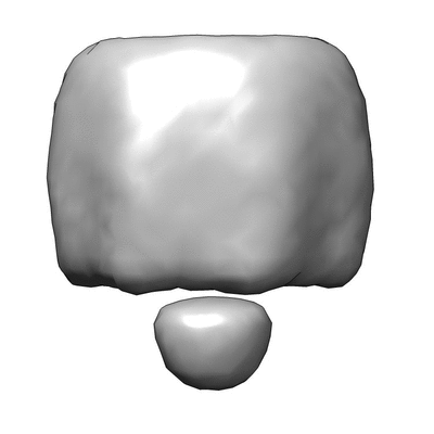 400_6120.gif 400_6120.gif 80_6120.gif 80_6120.gif | 29 KB 3.1 KB | ||
| Archive directory |  http://ftp.pdbj.org/pub/emdb/structures/EMD-6120ftp://ftp.pdbj.org/pub/emdb/structures/EMD-6120 http://ftp.pdbj.org/pub/emdb/structures/EMD-6120ftp://ftp.pdbj.org/pub/emdb/structures/EMD-6120 | HTTPS FTP |
-Validation report
| Summary document | emd_6120_validation.pdf.gz | 298.8 KB | Display | EMDB validaton report |
|---|---|---|---|---|
| Full document | emd_6120_full_validation.pdf.gz | 298.3 KB | Display | |
| Data in XML | emd_6120_validation.xml.gz | 4.8 KB | Display | |
| Arichive directory | https://ftp.pdbj.org/pub/emdb/validation_reports/EMD-6120ftp://ftp.pdbj.org/pub/emdb/validation_reports/EMD-6120 | HTTPS FTP |
-Related structure data
| Related structure data |  3j83MC  4wj1C  4wj2C M: atomic model generated by this map C: citing same article ( |
|---|---|
| Similar structure data |


-Links
| EMDB pages | EMDB (EBI/PDBe) / EMDataResource |
|---|
-Map
| File | Download / File: emd_6120.map.gz / Format: CCP4 / Size: 1001 KB / Type: IMAGE STORED AS FLOATING POINT NUMBER (4 BYTES) | ||||||||||||||||||||||||||||||||||||||||||||||||||||||||||||||||||||
|---|---|---|---|---|---|---|---|---|---|---|---|---|---|---|---|---|---|---|---|---|---|---|---|---|---|---|---|---|---|---|---|---|---|---|---|---|---|---|---|---|---|---|---|---|---|---|---|---|---|---|---|---|---|---|---|---|---|---|---|---|---|---|---|---|---|---|---|---|---|
| Annotation | Reconstruction of mycobacterial protein | ||||||||||||||||||||||||||||||||||||||||||||||||||||||||||||||||||||
| Voxel size | X=Y=Z: 4.7 Å | ||||||||||||||||||||||||||||||||||||||||||||||||||||||||||||||||||||
| Density |
| ||||||||||||||||||||||||||||||||||||||||||||||||||||||||||||||||||||
| Symmetry | Space group: 1 | ||||||||||||||||||||||||||||||||||||||||||||||||||||||||||||||||||||
| Details | EMDB XML:
CCP4 map header:
| ||||||||||||||||||||||||||||||||||||||||||||||||||||||||||||||||||||
-Supplemental data
- Sample components
-Entire : Mycobacterial protein
| Entire | Name: Mycobacterial protein |
|---|---|
| Components |
|
-Supramolecule #1000: Mycobacterial protein
| Supramolecule | Name: Mycobacterial protein / type: sample / ID: 1000 / Number unique components: 1 |
|---|---|
| Molecular weight | Experimental: 230 KDa / Theoretical: 230 KDa Method: size exclusion chromatography coupled to multi-angle light scattering |
-Macromolecule #1: Mycobacterial protein
| Macromolecule | Name: Mycobacterial protein / type: protein_or_peptide / ID: 1 / Recombinant expression: Yes |
|---|---|
| Source (natural) | Organism: Mycobacterium tuberculosis (bacteria) |
| Recombinant expression | Organism: |
-Experimental details
-Structure determination
| Method | negative staining |
|---|---|
 Processing Processing | single particle reconstruction |
| Aggregation state | particle |
-Sample preparation
| Concentration | 0.01 mg/mL |
|---|---|
| Buffer | pH: 7.5 / Details: 20 mM HEPES, 150 mM NaCl |
| Staining | Type: NEGATIVE Details: Protein was adsorbed onto grid, quickly blotted, and floated on 1% uranyl formate solution for 30 seconds |
| Grid | Details: 200 mesh gold grid with thin carbon support, glow discharged |
| Vitrification | Cryogen name: NONE / Instrument: OTHER |
- Electron microscopy
| Microscope | FEI TECNAI SPIRIT |
|---|---|
| Temperature | Average: 298 K |
| Details | Low dose |
| Date | Aug 28, 2013 |
| Image recording | Category: CCD / Film or detector model: FEI EAGLE (4k x 4k) / Digitization - Sampling interval: 1.3 µm / Number real images: 50 / Average electron dose: 25 e/Å2 / Bits/pixel: 16 |
| Electron beam | Acceleration voltage: 120 kV / Electron source: LAB6 |
| Electron optics | Calibrated magnification: 65900 / Illumination mode: FLOOD BEAM / Imaging mode: BRIGHT FIELD / Nominal defocus max: 1.2 µm / Nominal defocus min: 0.8 µm / Nominal magnification: 49000 |
| Sample stage | Specimen holder: side entry room temperature tomography holder Specimen holder model: SIDE ENTRY, EUCENTRIC |
| Experimental equipment |  Model: Tecnai Spirit / Image courtesy: FEI Company |
-Image processing
| Final reconstruction | Resolution.type: BY AUTHOR / Resolution: 30.0 Å / Resolution method: OTHER / Software - Name: Spider, EMAN2 Details: Initial reconstructions were determined from a side-view 2D average based on observed 7-fold symmetry of the top view using a rotational extrusion method. The initial model was then refined ...Details: Initial reconstructions were determined from a side-view 2D average based on observed 7-fold symmetry of the top view using a rotational extrusion method. The initial model was then refined in 6 iterations with EMAN2 using untilted particles. Number images used: 1455 |
|---|---|
| Final two d classification | Number classes: 2 |
-Atomic model buiding 1
| Initial model | PDB ID: Chain - Chain ID: A |
|---|---|
| Software | Name: Rosetta |
| Details | Homology model generated from PDB coordinates and used for symmetric docking in Rosetta. |
| Refinement | Space: REAL / Protocol: RIGID BODY FIT / Target criteria: RMSD / Rosetta score |
| Output model | PDB-3j83: |
