 Movie
Movie Controller


+ Open data

- Basic information
| Entry | Database: EMDB / ID: EMD-6102 | |||||||||
|---|---|---|---|---|---|---|---|---|---|---|
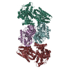
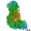
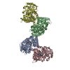
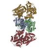
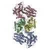
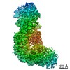
| Title | Electron cryo-microscopy of DNGR-1 in complex with F-actin | |||||||||
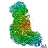
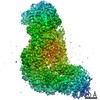
 Map data Map data | Reconstruction of DNGR1 in complex with F-actin | |||||||||
 Sample Sample |
| |||||||||
 Keywords Keywords | DNGR-1 /  Actin / Damage-associated molecular patterns Actin / Damage-associated molecular patterns | |||||||||
| Function / homology |  Function and homology information Function and homology informationpositive regulation of cytokine production => GO:0001819 / positive regulation of norepinephrine uptake / cellular response to cytochalasin B / bBAF complex / npBAF complex / postsynaptic actin cytoskeleton organization / regulation of transepithelial transport / brahma complex / nBAF complex / structural constituent of postsynaptic actin cytoskeleton ...positive regulation of cytokine production => GO:0001819 / positive regulation of norepinephrine uptake / cellular response to cytochalasin B / bBAF complex / npBAF complex / postsynaptic actin cytoskeleton organization / regulation of transepithelial transport / brahma complex / nBAF complex / structural constituent of postsynaptic actin cytoskeleton / morphogenesis of a polarized epithelium / Formation of annular gap junctions / GBAF complex / Gap junction degradation / postsynaptic actin cytoskeleton / protein localization to adherens junction / regulation of G0 to G1 transition / dense body / Cell-extracellular matrix interactions / Tat protein binding / Folding of actin by CCT/TriC / regulation of double-strand break repair / regulation of nucleotide-excision repair / RSC-type complex / apical protein localization / Prefoldin mediated transfer of substrate to CCT/TriC / adherens junction assembly / RHOF GTPase cycle / Adherens junctions interactions / tight junction / Sensory processing of sound by outer hair cells of the cochlea / Sensory processing of sound by inner hair cells of the cochlea / SWI/SNF complex / Interaction between L1 and Ankyrins / regulation of mitotic metaphase/anaphase transition / regulation of norepinephrine uptake / positive regulation of double-strand break repair / positive regulation of T cell differentiation / NuA4 histone acetyltransferase complex / regulation of synaptic vesicle endocytosis / apical junction complex / maintenance of blood-brain barrier / establishment or maintenance of cell polarity / cortical cytoskeleton / positive regulation of double-strand break repair via homologous recombination / positive regulation of stem cell population maintenance / nitric-oxide synthase binding / Recycling pathway of L1 / regulation of cyclin-dependent protein serine/threonine kinase activity / regulation of G1/S transition of mitotic cell cycle / negative regulation of cell differentiation / brush border / kinesin binding / calyx of Held / EPH-ephrin mediated repulsion of cells / RHO GTPases Activate WASPs and WAVEs / RHO GTPases activate IQGAPs / positive regulation of myoblast differentiation / regulation of protein localization to plasma membrane / EPHB-mediated forward signaling / substantia nigra development / receptor-mediated endocytosis / axonogenesis / negative regulation of protein binding / cell motility / actin filament / RHO GTPases Activate Formins / Translocation of SLC2A4 (GLUT4) to the plasma membrane / regulation of transmembrane transporter activity / positive regulation of cell differentiation / FCGR3A-mediated phagocytosis / adherens junction / Hydrolases; Acting on acid anhydrides; Acting on acid anhydrides to facilitate cellular and subcellular movement / DNA Damage Recognition in GG-NER / tau protein binding / Signaling by high-kinase activity BRAF mutants / Schaffer collateral - CA1 synapse / MAP2K and MAPK activation / B-WICH complex positively regulates rRNA expression / structural constituent of cytoskeleton / cytoplasmic ribonucleoprotein granule / kinetochore / Regulation of actin dynamics for phagocytic cup formation / platelet aggregation / nuclear matrix / VEGFA-VEGFR2 Pathway / UCH proteinases / Signaling by RAF1 mutants / Signaling by moderate kinase activity BRAF mutants / Paradoxical activation of RAF signaling by kinase inactive BRAF / Signaling downstream of RAS mutants / nucleosome / cell-cell junction / Signaling by BRAF and RAF1 fusions / actin cytoskeleton / presynapse / lamellipodium / Clathrin-mediated endocytosis / Factors involved in megakaryocyte development and platelet production / HATs acetylate histonesSimilarity search - Function | |||||||||
| Biological species |  Mus musculus (house mouse) Mus musculus (house mouse) | |||||||||
| Method | single particle reconstruction / cryo EM / Resolution: 7.7 Å | |||||||||
 Authors Authors | Hanc P / Fujii T / Yamada Y / Huotari J / Schulz O / Ahrens S / Kjaer S / Way M / Namba K / Reis e Sousa C | |||||||||
 Citation Citation | Journal: Immunity / Year: 2015 Title: Structure of the Complex of F-Actin and DNGR-1, a C-Type Lectin Receptor Involved in Dendritic Cell Cross-Presentation of Dead Cell-Associated Antigens. Authors: Pavel Hanč / Takashi Fujii / Salvador Iborra / Yurika Yamada / Jatta Huotari / Oliver Schulz / Susan Ahrens / Svend Kjær / Michael Way / David Sancho / Keiichi Namba / Caetano Reis e Sousa /    Abstract: DNGR-1 is a C-type lectin receptor that binds F-actin exposed by dying cells and facilitates cross-presentation of dead cell-associated antigens by dendritic cells. Here we present the structure of ...DNGR-1 is a C-type lectin receptor that binds F-actin exposed by dying cells and facilitates cross-presentation of dead cell-associated antigens by dendritic cells. Here we present the structure of DNGR-1 bound to F-actin at 7.7 Å resolution. Unusually for F-actin binding proteins, the DNGR-1 ligand binding domain contacts three actin subunits helically arranged in the actin filament, bridging over two protofilaments, as well as two neighboring actin subunits along one protofilament. Mutation of residues predicted to mediate ligand binding led to loss of DNGR-1-dependent cross-presentation of dead cell-associated antigens, formally demonstrating that the latter depends on F-actin recognition. Notably, DNGR-1 has relatively modest affinity for F-actin but multivalent interactions allow a marked increase in binding strength. Our findings shed light on modes of actin binding by cellular proteins and reveal how extracellular detection of cytoskeletal components by dedicated receptors allows immune monitoring of loss of cellular integrity. | |||||||||
| History |
|
- Structure visualization
| Movie |
Movie viewer |
|---|---|
| Structure viewer | EM map: SurfViewMolmilJmol/JSmol |
| Supplemental images |

- Downloads & links
-EMDB archive
| Map data | emd_6102.map.gz | 3.6 MB | EMDB map data format | |
|---|---|---|---|---|
| Header (meta data) | emd-6102-v30.xmlemd-6102.xml | 10.7 KB 10.7 KB | Display Display | EMDB header |
| Images |  400_6102.gif 400_6102.gif 80_6102.gif 80_6102.gif | 40.5 KB 4.3 KB | ||
| Archive directory |  http://ftp.pdbj.org/pub/emdb/structures/EMD-6102ftp://ftp.pdbj.org/pub/emdb/structures/EMD-6102 http://ftp.pdbj.org/pub/emdb/structures/EMD-6102ftp://ftp.pdbj.org/pub/emdb/structures/EMD-6102 | HTTPS FTP |
-Related structure data
| Related structure data | 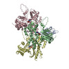 3j82MC M: atomic model generated by this map C: citing same article ( |
|---|---|
| Similar structure data |
-Links
| EMDB pages | EMDB (EBI/PDBe) / EMDataResource |
|---|---|
| Related items in Molecule of the Month |


-Map
| File | Download / File: emd_6102.map.gz / Format: CCP4 / Size: 3.7 MB / Type: IMAGE STORED AS FLOATING POINT NUMBER (4 BYTES) | ||||||||||||||||||||||||||||||||||||||||||||||||||||||||||||
|---|---|---|---|---|---|---|---|---|---|---|---|---|---|---|---|---|---|---|---|---|---|---|---|---|---|---|---|---|---|---|---|---|---|---|---|---|---|---|---|---|---|---|---|---|---|---|---|---|---|---|---|---|---|---|---|---|---|---|---|---|---|
| Annotation | Reconstruction of DNGR1 in complex with F-actin | ||||||||||||||||||||||||||||||||||||||||||||||||||||||||||||
| Voxel size | X=Y=Z: 1.37 Å | ||||||||||||||||||||||||||||||||||||||||||||||||||||||||||||
| Density |
| ||||||||||||||||||||||||||||||||||||||||||||||||||||||||||||
| Symmetry | Space group: 1 | ||||||||||||||||||||||||||||||||||||||||||||||||||||||||||||
| Details | EMDB XML:
CCP4 map header:
| ||||||||||||||||||||||||||||||||||||||||||||||||||||||||||||
-Supplemental data
- Sample components
-Entire : F-actin complexed with mouse DNGR-1 extracellular domain
| Entire | Name: F-actin complexed with mouse DNGR-1 extracellular domain |
|---|---|
| Components |
|
-Supramolecule #1000: F-actin complexed with mouse DNGR-1 extracellular domain
| Supramolecule | Name: F-actin complexed with mouse DNGR-1 extracellular domain type: sample / ID: 1000 / Number unique components: 1 |
|---|
-Macromolecule #1: DNGR-1
| Macromolecule | Name: DNGR-1 / type: protein_or_peptide / ID: 1 / Name.synonym: CLEC9A / Recombinant expression: Yes |
|---|---|
| Source (natural) | Organism: Mus musculus (house mouse) / synonym: Mouse |
| Recombinant expression | Organism:  Escherichia coli (E. coli) Escherichia coli (E. coli) |
-Experimental details
-Structure determination
| Method | cryo EM |
|---|---|
 Processing Processing | single particle reconstruction |
| Aggregation state | particle |
-Sample preparation
| Buffer | pH: 7.5 Details: 25mM Hepes buffer (pH 7.5), 100mM KCl, 1mM MgCl2, 1mM ATP |
|---|---|
| Grid | Details: R0.6/1.0, Quantifoil |
| Vitrification | Cryogen name: ETHANE / Chamber humidity: 90 % / Instrument: FEI VITROBOT MARK II |
- Electron microscopy
| Microscope | JEOL 3200FSC |
|---|---|
| Electron beam | Acceleration voltage: 200 kV / Electron source: FIELD EMISSION GUN |
| Electron optics | Calibrated magnification: 109489 / Illumination mode: FLOOD BEAM / Imaging mode: BRIGHT FIELDBright-field microscopy / Cs: 1.6 mm / Nominal defocus max: 2.0 µm / Nominal defocus min: 1.0 µm / Nominal magnification: 60000 |
| Specialist optics | Energy filter - Name: JEOL Omega filter / Energy filter - Lower energy threshold: 0.0 eV / Energy filter - Upper energy threshold: 20.0 eV |
| Sample stage | Specimen holder model: JEOL 3200FSC CRYOHOLDER |
| Temperature | Min: 50 K / Max: 60 K / Average: 55 K |
| Date | Dec 10, 2012 |
| Image recording | Category: CCD / Film or detector model: TVIPS TEMCAM-F416 (4k x 4k) / Number real images: 774 / Average electron dose: 20 e/Å2 |
-Image processing
| CTF correction | Details: Each Particle |
|---|---|
| Final reconstruction | Algorithm: OTHER / Resolution.type: BY AUTHOR / Resolution: 7.7 Å / Resolution method: OTHER / Software - Name: Spider, EMAN / Number images used: 73608 |
-Atomic model buiding 1
| Initial model | PDB ID: Chain - #0 - Chain ID: A / Chain - #1 - Chain ID: B / Chain - #2 - Chain ID: C / Chain - #3 - Chain ID: D |
|---|---|
| Software | Name: Spider, EMAN |
| Details | Single particle--Applied symmetry: C1 |
| Refinement | Space: REAL |
| Output model | PDB-3j82: |
