- EMDB-6015: Dynein motor domain in the presence of ADP, with linker at position 1 -
+
Open data
ID or keywords:
Loading...
-
Basic information
Entry
Database: EMDB / ID: EMD-6015
Title
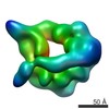
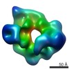
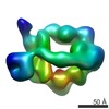
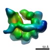
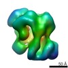
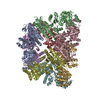
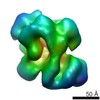
Dynein motor domain in the presence of ADP, with linker at position 1Motor protein
Map data
Reconstruction of dynein motor domain in 100 uM ADP
Sample
Sample: Dynein motor domain in 100 uM ADPMotor protein
Protein or peptide: Dynein heavy chain
Keywords
dynein / lis1 / regulation mechanism
Function / homology
Function and homology information
karyogamy / establishment of mitotic spindle localization / nuclear migration along microtubule / astral microtubule / minus-end-directed microtubule motor activity / cytoplasmic dynein complex / dynein light intermediate chain binding / spindle pole body / nuclear migration / dynein intermediate chain binding ...karyogamy / establishment of mitotic spindle localization / nuclear migration along microtubule / astral microtubule / minus-end-directed microtubule motor activity / cytoplasmic dynein complex / dynein light intermediate chain binding / spindle pole body / nuclear migration / dynein intermediate chain binding / mitotic sister chromatid segregation / establishment of mitotic spindle orientation / cytoplasmic microtubule / cytoplasmic microtubule organization / Neutrophil degranulation / mitotic spindle organization / cell cortex / ATP hydrolysis activity / ATP binding / cytoplasm Similarity search - Function
: / DYN1, AAA+ ATPase lid domain / Dynein heavy chain 3, AAA+ lid domain / AAA+ lid domain / P-loop containing dynein motor region / Dynein heavy chain, tail / Dynein heavy chain, N-terminal region 1 / Dynein heavy chain / Dynein heavy chain region D6 P-loop domain / Dynein heavy chain, linker ...: / DYN1, AAA+ ATPase lid domain / Dynein heavy chain 3, AAA+ lid domain / AAA+ lid domain / P-loop containing dynein motor region / Dynein heavy chain, tail / Dynein heavy chain, N-terminal region 1 / Dynein heavy chain / Dynein heavy chain region D6 P-loop domain / Dynein heavy chain, linker / Dynein heavy chain, AAA module D4 / Dynein heavy chain, coiled coil stalk / Dynein heavy chain, hydrolytic ATP-binding dynein motor region / Dynein heavy chain, ATP-binding dynein motor region / Dynein heavy chain AAA lid domain / Dynein heavy chain AAA lid domain superfamily / Dynein heavy chain, domain 2, N-terminal / Dynein heavy chain, linker, subdomain 3 / Dynein heavy chain, AAA1 domain, small subdomain / Dynein heavy chain region D6 P-loop domain / Dynein heavy chain, N-terminal region 2 / Hydrolytic ATP binding site of dynein motor region / Microtubule-binding stalk of dynein motor / P-loop containing dynein motor region D4 / ATP-binding dynein motor region / Dynein heavy chain AAA lid domain / ATPases associated with a variety of cellular activities / AAA+ ATPase domain / P-loop containing nucleoside triphosphate hydrolase Similarity search - Domain/homology
Journal: Elife / Year: 2014 Title: Lis1 regulates dynein by sterically blocking its mechanochemical cycle. Authors: Katerina Toropova / Sirui Zou / Anthony J Roberts / William B Redwine / Brian S Goodman / Samara L Reck-Peterson / Andres E Leschziner / Abstract: Regulation of cytoplasmic dynein's motor activity is essential for diverse eukaryotic functions, including cell division, intracellular transport, and brain development. The dynein regulator Lis1 is ...Regulation of cytoplasmic dynein's motor activity is essential for diverse eukaryotic functions, including cell division, intracellular transport, and brain development. The dynein regulator Lis1 is known to keep dynein bound to microtubules; however, how this is accomplished mechanistically remains unknown. We have used three-dimensional electron microscopy, single-molecule imaging, biochemistry, and in vivo assays to help establish this mechanism. The three-dimensional structure of the dynein-Lis1 complex shows that binding of Lis1 to dynein's AAA+ ring sterically prevents dynein's main mechanical element, the 'linker', from completing its normal conformational cycle. Single-molecule experiments show that eliminating this block by shortening the linker to a point where it can physically bypass Lis1 renders single dynein motors insensitive to regulation by Lis1. Our data reveal that Lis1 keeps dynein in a persistent microtubule-bound state by directly blocking the progression of its mechanochemical cycle.
History
Deposition
Aug 3, 2014
-
Header (metadata) release
Sep 24, 2014
-
Map release
Nov 19, 2014
-
Update
Nov 19, 2014
-
Current status
Nov 19, 2014
Processing site: RCSB / Status: Released
-
Structure visualization
Movie
Surface view with section colored by density value
pH: 8 Details: 50 mM Tris-HCl, 150 mM potassium acetate, 2 mM magnesium acetate, 1 mM EGTA, 1 mM DTT, 100 uM ADP
Staining
Type: NEGATIVE Details: Grids with adsorbed protein were floated on 2% w/v uranyl formate, then sandwiched with a thin layer of carbon, blotted, and frozen in liquid nitrogen.
Grid
Details: 200 mesh C-flat grid with thin carbon support
Vitrification
Cryogen name: NITROGEN / Instrument: OTHER Method: Manually blot, wait 10-20 seconds, then manually plunge into liquid nitrogen.
-
Electron microscopy
Microscope
FEI TECNAI 20
Electron beam
Acceleration voltage: 120 kV / Electron source: FIELD EMISSION GUN
Category: CCD / Film or detector model: GATAN ULTRASCAN 4000 (4k x 4k) / Digitization - Sampling interval: 15 µm / Number real images: 200 / Average electron dose: 25 e/Å2
-
Image processing
CTF correction
Details: phase flip
Final reconstruction
Resolution.type: BY AUTHOR / Resolution: 18.3 Å / Resolution method: OTHER / Software - Name: RELION Details: The dataset was first 3D-classified in RELION to sort different linker positions. Particles in this map displayed a linker unshifted relative to its position in the absence of nucleotide. Number images used: 7630
In the structure databanks used in Yorodumi, some data are registered as the other names, "COVID-19 virus" and "2019-nCoV". Here are the details of the virus and the list of structure data.
Jan 31, 2019. EMDB accession codes are about to change! (news from PDBe EMDB page)
EMDB accession codes are about to change! (news from PDBe EMDB page)
The allocation of 4 digits for EMDB accession codes will soon come to an end. Whilst these codes will remain in use, new EMDB accession codes will include an additional digit and will expand incrementally as the available range of codes is exhausted. The current 4-digit format prefixed with “EMD-” (i.e. EMD-XXXX) will advance to a 5-digit format (i.e. EMD-XXXXX), and so on. It is currently estimated that the 4-digit codes will be depleted around Spring 2019, at which point the 5-digit format will come into force.
The EM Navigator/Yorodumi systems omit the EMD- prefix.
Related info.:Q: What is EMD? / ID/Accession-code notation in Yorodumi/EM Navigator
Yorodumi is a browser for structure data from EMDB, PDB, SASBDB, etc.
This page is also the successor to EM Navigator detail page, and also detail information page/front-end page for Omokage search.
The word "yorodu" (or yorozu) is an old Japanese word meaning "ten thousand". "mi" (miru) is to see.
Related info.:EMDB / PDB / SASBDB / Comparison of 3 databanks / Yorodumi Search / Aug 31, 2016. New EM Navigator & Yorodumi / Yorodumi Papers / Jmol/JSmol / Function and homology information / Changes in new EM Navigator and Yorodumi
 Movie
Movie Controller

 Yorodumi
 Open data
 Basic information
 Motor protein
 Map data
 Sample
 Keywords
 Function and homology information
 Authors
 Citation
 Structure visualization
 Downloads & links
 400_6015.gif
 80_6015.gif
 http://ftp.pdbj.org/pub/emdb/structures/EMD-6015
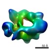


 Z
Z Y
Y X
X


 Sample components
 Processing
 Electron microscopy
