 Movie
Movie Controller

[English] 日本語
 Yorodumi
- PDB-5foj: Cryo electron microscopy structure of Grapevine Fanleaf Virus com... -
+ Open data

- Basic information
| Entry | Database: PDB / ID: 5foj | ||||||||||||
|---|---|---|---|---|---|---|---|---|---|---|---|---|---|
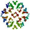
| Title | Cryo electron microscopy structure of Grapevine Fanleaf Virus complex with Nanobody | ||||||||||||
 Components Components |
| ||||||||||||
 Keywords Keywords | VIRUS / NANOBODY / COMPLEX | ||||||||||||
| Function / homology |  Function and homology information Function and homology informationtransport of virus in host, cell to cell / host cell plasmodesma / viral capsid / host cell cytoplasm / host cell nucleus / structural molecule activity Similarity search - Function | ||||||||||||
| Biological species |   Grapevine fanleaf virus Grapevine fanleaf virus | ||||||||||||
| Method | ELECTRON MICROSCOPY / single particle reconstruction / cryo EM / Resolution: 2.8 Å | ||||||||||||
 Authors Authors | Orlov, I. / Hemmer, C. / Ackerer, L. / Lorber, B. / Ghannam, A. / Poignavent, V. / Hleibieh, K. / Sauter, C. / Schmitt-Keichinger, C. / Belval, L. ...Orlov, I. / Hemmer, C. / Ackerer, L. / Lorber, B. / Ghannam, A. / Poignavent, V. / Hleibieh, K. / Sauter, C. / Schmitt-Keichinger, C. / Belval, L. / Hily, J.M. / Marmonier, A. / Komar, V. / Gersch, S. / Schellenberger, P. / Bron, P. / Vigne, E. / Muyldermans, S. / Lemaire, O. / Demangeat, G. / Ritzenthaler, C. / Klaholz, B.P. | ||||||||||||
| Funding support | 1items
| ||||||||||||
 Citation Citation | Journal: Proc Natl Acad Sci U S A / Year: 2020 Title: Structural basis of nanobody recognition of grapevine fanleaf virus and of virus resistance loss. Authors: Igor Orlov / Caroline Hemmer / Léa Ackerer / Bernard Lorber / Ahmed Ghannam / Vianney Poignavent / Kamal Hleibieh / Claude Sauter / Corinne Schmitt-Keichinger / Lorène Belval / Jean-Michel ...Authors: Igor Orlov / Caroline Hemmer / Léa Ackerer / Bernard Lorber / Ahmed Ghannam / Vianney Poignavent / Kamal Hleibieh / Claude Sauter / Corinne Schmitt-Keichinger / Lorène Belval / Jean-Michel Hily / Aurélie Marmonier / Véronique Komar / Sophie Gersch / Pascale Schellenberger / Patrick Bron / Emmanuelle Vigne / Serge Muyldermans / Olivier Lemaire / Gérard Demangeat / Christophe Ritzenthaler / Bruno P Klaholz /   Abstract: Grapevine fanleaf virus (GFLV) is a picorna-like plant virus transmitted by nematodes that affects vineyards worldwide. Nanobody (Nb)-mediated resistance against GFLV has been created recently, and ...Grapevine fanleaf virus (GFLV) is a picorna-like plant virus transmitted by nematodes that affects vineyards worldwide. Nanobody (Nb)-mediated resistance against GFLV has been created recently, and shown to be highly effective in plants, including grapevine, but the underlying mechanism is unknown. Here we present the high-resolution cryo electron microscopy structure of the GFLV-Nb23 complex, which provides the basis for molecular recognition by the Nb. The structure reveals a composite binding site bridging over three domains of one capsid protein (CP) monomer. The structure provides a precise mapping of the Nb23 epitope on the GFLV capsid in which the antigen loop is accommodated through an induced-fit mechanism. Moreover, we uncover and characterize several resistance-breaking GFLV isolates with amino acids mapping within this epitope, including C-terminal extensions of the CP, which would sterically interfere with Nb binding. Escape variants with such extended CP fail to be transmitted by nematodes linking Nb-mediated resistance to vector transmission. Together, these data provide insights into the molecular mechanism of Nb23-mediated recognition of GFLV and of virus resistance loss. | ||||||||||||
| History |
|
- Structure visualization
| Movie |
Movie viewer |
|---|---|
| Structure viewer | Molecule: MolmilJmol/JSmol |
- Downloads & links
-Download
| PDBx/mmCIF format | 5foj.cif.gz | 119.4 KB | Display | PDBx/mmCIF format |
|---|---|---|---|---|
| PDB format | pdb5foj.ent.gz | 93.6 KB | Display | PDB format |
| PDBx/mmJSON format | 5foj.json.gz | Tree view | PDBx/mmJSON format | |
| Others |  Other downloads Other downloads |
-Validation report
| Summary document | 5foj_validation.pdf.gz | 1.1 MB | Display | wwPDB validaton report |
|---|---|---|---|---|
| Full document | 5foj_full_validation.pdf.gz | 1.1 MB | Display | |
| Data in XML | 5foj_validation.xml.gz | 25.8 KB | Display | |
| Data in CIF | 5foj_validation.cif.gz | 36.6 KB | Display | |
| Arichive directory | https://data.pdbj.org/pub/pdb/validation_reports/fo/5fojftp://data.pdbj.org/pub/pdb/validation_reports/fo/5foj | HTTPS FTP |
-Related structure data
| Related structure data | 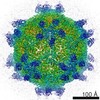 3246M M: map data used to model this data |
|---|---|
| Similar structure data |
-Links
 PDBj
PDBj


- Assembly
| Deposited unit | 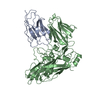
|
|---|---|
| 1 | x 60
|
| Symmetry | Point symmetry: (Schoenflies symbol: I (icosahedral)) |
-Components
| #1: Antibody | Mass: 14816.341 Da / Num. of mol.: 1 Source method: isolated from a genetically manipulated source Source: (gene. exp.)  |
|---|---|
| #2: Protein | Mass: 56073.141 Da / Num. of mol.: 1 / Fragment: UNP RESIDUES 606-1109 / Source method: isolated from a natural source / Source: (natural) Grapevine fanleaf virus / References: UniProt: P18474 |
-Experimental details
-Experiment
| Experiment | Method: ELECTRON MICROSCOPY |
|---|---|
| EM experiment | Aggregation state: PARTICLE / 3D reconstruction method: single particle reconstruction |
- Sample preparation
| Component | Name: Grapevine fanleaf virus / Type: VIRUS / Entity ID: all / Source: MULTIPLE SOURCES |
|---|---|
| Source (natural) | Organism: Grapevine fanleaf virus |
| Details of virus | Empty: NO / Enveloped: NO / Isolate: OTHER / Type: VIRION |
| Virus shell | Name: pseudo-T = 3 / Triangulation number (T number): 3 |
| Buffer solution | pH: 8.3 / Details: 100 mM TRIS pH 8.3, 50 MM NaCl. |
| Specimen | Conc.: 0.5 mg/ml / Embedding applied: NO / Shadowing applied: NO / Staining applied: NO / Vitrification applied: YES |
| Specimen support | Details: HOLEY CARBON |
| Vitrification | Instrument: FEI VITROBOT MARK III / Cryogen name: ETHANE Details: VITRIFICATION 1 -- CRYOGEN- ETHANE, HUMIDITY- 95, INSTRUMENT- FEI VITROBOT MARK III, METHOD- BLOT FOR 2 SECONDS BEFORE PLUNGING |
- Electron microscopy imaging
| Experimental equipment |  Model: Titan Krios / Image courtesy: FEI Company |
|---|---|
| Microscopy | Model: FEI TITAN KRIOS / Details: Cs-corrected microscope |
| Electron gun | Electron source:  FIELD EMISSION GUN / Accelerating voltage: 300 kV / Illumination mode: OTHER FIELD EMISSION GUN / Accelerating voltage: 300 kV / Illumination mode: OTHER |
| Electron lens | Mode: BRIGHT FIELD / Nominal magnification: 120000 X / Calibrated magnification: 127272 X / Nominal defocus max: 6000 nm / Nominal defocus min: 600 nm / Alignment procedure: ZEMLIN TABLEAU |
| Specimen holder | Cryogen: NITROGEN / Specimen holder model: FEI TITAN KRIOS AUTOGRID HOLDER |
| Image recording | Average exposure time: 1 sec. / Electron dose: 50 e/Å2 / Detector mode: COUNTING / Film or detector model: FEI FALCON II (4k x 4k) |
| EM imaging optics | Spherical aberration corrector: Cs-corrected microscope |
| Image scans | Width: 4096 / Height: 4096 / Movie frames/image: 7 |
- Processing
| Software | Name: PHENIX / Version: 1.17.1_3660: / Classification: refinement | ||||||||||||||||||||||||
|---|---|---|---|---|---|---|---|---|---|---|---|---|---|---|---|---|---|---|---|---|---|---|---|---|---|
| EM software |
| ||||||||||||||||||||||||
| CTF correction | Type: PHASE FLIPPING ONLY | ||||||||||||||||||||||||
| Symmetry | Point symmetry: I (icosahedral) | ||||||||||||||||||||||||
| 3D reconstruction | Resolution: 2.8 Å / Resolution method: FSC 0.143 CUT-OFF / Algorithm: BACK PROJECTION Details: SUBMISSION BASED ON EXPERIMENTAL DATA FROM EMDB EMD-3246. (DEPOSITION ID: 14064). | ||||||||||||||||||||||||
| Atomic model building | Protocol: FLEXIBLE FIT / Space: REAL Details: Initial atomic model building was done by a rigid body fitting, followed by extensive manual model building in Coot and real space refinement of the atomic model against the experimental map ...Details: Initial atomic model building was done by a rigid body fitting, followed by extensive manual model building in Coot and real space refinement of the atomic model against the experimental map using Phenix including restrained temperature factor refinement. | ||||||||||||||||||||||||
| Atomic model building | PDB-ID: 4V5T | ||||||||||||||||||||||||
| Refinement | Highest resolution: 2.8 Å | ||||||||||||||||||||||||
| Refinement step | Cycle: LAST / Highest resolution: 2.8 Å
| ||||||||||||||||||||||||
| Refine LS restraints |
|

