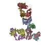
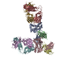
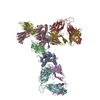
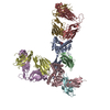
Movie
Movie Controller

[English] 日本語
 Yorodumi
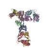
- PDB-5a8l: Human eRF1 and the hCMV nascent peptide in the translation termin... -
+ Open data

- Basic information
| Entry | Database: PDB / ID: 5a8l | |||||||||
|---|---|---|---|---|---|---|---|---|---|---|
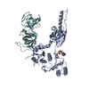
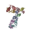
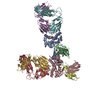
| Title | Human eRF1 and the hCMV nascent peptide in the translation termination complex | |||||||||
 Components Components |
| |||||||||
 Keywords Keywords | TRANSLATION / HUMAN / 80S / RIBOSOME / ERF1 | |||||||||
| Function / homology |  Function and homology information Function and homology informationtranslation termination factor activity / translation release factor complex / cytoplasmic translational termination / translation release factor activity / regulation of translational termination / translation release factor activity, codon specific / protein methylation / sequence-specific mRNA binding / aminoacyl-tRNA hydrolase activity / nuclear-transcribed mRNA catabolic process, nonsense-mediated decay ...translation termination factor activity / translation release factor complex / cytoplasmic translational termination / translation release factor activity / regulation of translational termination / translation release factor activity, codon specific / protein methylation / sequence-specific mRNA binding / aminoacyl-tRNA hydrolase activity / nuclear-transcribed mRNA catabolic process, nonsense-mediated decay / Protein hydroxylation / Peptide chain elongation / Selenocysteine synthesis / Formation of a pool of free 40S subunits / Eukaryotic Translation Termination / Response of EIF2AK4 (GCN2) to amino acid deficiency / SRP-dependent cotranslational protein targeting to membrane / Viral mRNA Translation / Nonsense Mediated Decay (NMD) independent of the Exon Junction Complex (EJC) / GTP hydrolysis and joining of the 60S ribosomal subunit / L13a-mediated translational silencing of Ceruloplasmin expression / Major pathway of rRNA processing in the nucleolus and cytosol / Nonsense Mediated Decay (NMD) enhanced by the Exon Junction Complex (EJC) / translational termination / cytosolic ribosome / rough endoplasmic reticulum / Regulation of expression of SLITs and ROBOs / ribosome binding / large ribosomal subunit rRNA binding / cytosolic large ribosomal subunit / cytoplasmic translation / postsynaptic density / structural constituent of ribosome / translation / ribonucleoprotein complex / focal adhesion / nucleolus / RNA binding / extracellular exosome / membrane / nucleus / cytoplasm / cytosol Similarity search - Function | |||||||||
| Biological species |  HOMO SAPIENS (human) HOMO SAPIENS (human) | |||||||||
| Method | ELECTRON MICROSCOPY / single particle reconstruction / cryo EM / Resolution: 3.8 Å | |||||||||
 Authors Authors | Matheisl, S. / Berninghausen, O. / Becker, T. / Beckmann, R. | |||||||||
 Citation Citation | Journal: Nucleic Acids Res / Year: 2015 Title: Structure of a human translation termination complex. Authors: Sarah Matheisl / Otto Berninghausen / Thomas Becker / Roland Beckmann /  Abstract: In contrast to bacteria that have two release factors, RF1 and RF2, eukaryotes only possess one unrelated release factor eRF1, which recognizes all three stop codons of the mRNA and hydrolyses the ...In contrast to bacteria that have two release factors, RF1 and RF2, eukaryotes only possess one unrelated release factor eRF1, which recognizes all three stop codons of the mRNA and hydrolyses the peptidyl-tRNA bond. While the molecular basis for bacterial termination has been elucidated, high-resolution structures of eukaryotic termination complexes have been lacking. Here we present a 3.8 Å structure of a human translation termination complex with eRF1 decoding a UAA(A) stop codon. The complex was formed using the human cytomegalovirus (hCMV) stalling peptide, which perturbs the peptidyltransferase center (PTC) to silence the hydrolysis activity of eRF1. Moreover, unlike sense codons or bacterial stop codons, the UAA stop codon adopts a U-turn-like conformation within a pocket formed by eRF1 and the ribosome. Inducing the U-turn conformation for stop codon recognition rationalizes how decoding by eRF1 includes monitoring geometry in order to discriminate against sense codons. | |||||||||
| History |
|
- Structure visualization
| Movie |
Movie viewer |
|---|---|
| Structure viewer | Molecule: MolmilJmol/JSmol |
- Downloads & links
-Download
| PDBx/mmCIF format | 5a8l.cif.gz | 404.2 KB | Display | PDBx/mmCIF format |
|---|---|---|---|---|
| PDB format | pdb5a8l.ent.gz | 235.5 KB | Display | PDB format |
| PDBx/mmJSON format | 5a8l.json.gz | Tree view | PDBx/mmJSON format | |
| Others |  Other downloads Other downloads |
-Validation report
| Summary document | 5a8l_validation.pdf.gz | 1.2 MB | Display | wwPDB validaton report |
|---|---|---|---|---|
| Full document | 5a8l_full_validation.pdf.gz | 1.2 MB | Display | |
| Data in XML | 5a8l_validation.xml.gz | 31.5 KB | Display | |
| Data in CIF | 5a8l_validation.cif.gz | 47.7 KB | Display | |
| Arichive directory | https://data.pdbj.org/pub/pdb/validation_reports/a8/5a8lftp://data.pdbj.org/pub/pdb/validation_reports/a8/5a8l | HTTPS FTP |
-Related structure data
| Related structure data | 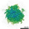 3099MC M: map data used to model this data C: citing same article ( |
|---|---|
| Similar structure data |

-Links
 PDBj
PDBj


- Assembly
| Deposited unit | 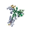
|
|---|---|
| 1 |
|
-Components
-RNA chain , 4 types, 4 molecules ABPR
| #1: RNA chain | Mass: 1625941.625 Da / Num. of mol.: 1 / Source method: isolated from a natural source / Details: HELA S3 / Source: (natural) HOMO SAPIENS (human) / References: GenBank: 337381 |
|---|---|
| #2: RNA chain | Mass: 602776.875 Da / Num. of mol.: 1 / Source method: isolated from a natural source / Details: HELA S3 / Source: (natural) HOMO SAPIENS (human) / References: GenBank: 36162 |
| #6: RNA chain | Mass: 5771.506 Da / Num. of mol.: 1 / Source method: isolated from a natural source / Details: HELA S3 / Source: (natural) HOMO SAPIENS (human) |
| #8: RNA chain | Mass: 2823.767 Da / Num. of mol.: 1 / Source method: isolated from a natural source / Details: HELA S3 / Source: (natural) HOMO SAPIENS (human) |
-60S RIBOSOMAL PROTEIN ... , 3 types, 3 molecules DGH
| #3: Protein | Mass: 21443.170 Da / Num. of mol.: 1 / Source method: isolated from a natural source / Details: HELA S3 / Source: (natural) HOMO SAPIENS (human) / References: UniProt: P18621 |
|---|---|
| #4: Protein | Mass: 17847.619 Da / Num. of mol.: 1 / Source method: isolated from a natural source / Details: HELA S3 / Source: (natural) HOMO SAPIENS (human) / References: UniProt: P30050 |
| #5: Protein | Mass: 47804.621 Da / Num. of mol.: 1 / Source method: isolated from a natural source / Details: HELA S3 / Source: (natural) HOMO SAPIENS (human) / References: UniProt: P36578 |
-Protein / Protein/peptide , 2 types, 2 molecules QZ
| #7: Protein | Mass: 48476.105 Da / Num. of mol.: 1 / Source method: isolated from a natural source / Details: HELA S3 / Source: (natural) HOMO SAPIENS (human) / References: UniProt: P62495 |
|---|---|
| #9: Protein/peptide | Mass: 2435.018 Da / Num. of mol.: 1 / Source method: isolated from a natural source / Details: HELA S3 / Source: (natural) HOMO SAPIENS (human) |
-Experimental details
-Experiment
| Experiment | Method: ELECTRON MICROSCOPY |
|---|---|
| EM experiment | Aggregation state: PARTICLE / 3D reconstruction method: single particle reconstruction |
- Sample preparation
| Component | Name: CMV STALLED HUMAN 80S RIBOSOME BOUND TO THE TRANSLATION TERMINATION FACTOR ERF1 Type: RIBOSOME |
|---|---|
| Buffer solution | Name: 20 MM HEPES, 100 MM KOAC, 2.5 MM MG(OAC)2, 0. 25 MM SPERMIDINE, 2 MM DTT, 0.06 U/UL RNASIN ( AMBION), 1/625 EDTA-FREE COMPLETE PROTEASE INHIBITOR (ROCHE) pH: 7.5 Details: 20 MM HEPES, 100 MM KOAC, 2.5 MM MG(OAC)2, 0. 25 MM SPERMIDINE, 2 MM DTT, 0.06 U/UL RNASIN ( AMBION), 1/625 EDTA-FREE COMPLETE PROTEASE INHIBITOR (ROCHE) |
| Specimen | Embedding applied: NO / Shadowing applied: NO / Staining applied: NO / Vitrification applied: YES |
| Specimen support | Details: CARBON |
| Vitrification | Instrument: FEI VITROBOT MARK IV / Cryogen name: ETHANE Details: VITRIFICATION 1 -- CRYOGEN- ETHANE, INSTRUMENT- FEI VITROBOT MARK IV, |
- Electron microscopy imaging
| Experimental equipment |  Model: Tecnai F20 / Image courtesy: FEI Company |
|---|---|
| Microscopy | Model: FEI TECNAI F20 / Date: Mar 11, 2015 |
| Electron gun | Electron source:  FIELD EMISSION GUN / Accelerating voltage: 300 kV / Illumination mode: FLOOD BEAM FIELD EMISSION GUN / Accelerating voltage: 300 kV / Illumination mode: FLOOD BEAM |
| Electron lens | Mode: BRIGHT FIELD / Nominal defocus max: 2700 nm / Nominal defocus min: 1000 nm / Cs: 2.7 mm |
| Image recording | Film or detector model: FEI FALCON II (4k x 4k) |
| Image scans | Num. digital images: 3 |
- Processing
| EM software |
| ||||||||||||
|---|---|---|---|---|---|---|---|---|---|---|---|---|---|
| CTF correction | Details: DEFOCUS GROUPS | ||||||||||||
| Symmetry | Point symmetry: C1 (asymmetric) | ||||||||||||
| 3D reconstruction | Resolution: 3.8 Å / Num. of particles: 33165 / Actual pixel size: 1.062 Å Details: SUBMISSION BASED ON EXPERIMENTAL DATA FROM EMDB EMD-3099. (DEPOSITION ID: 13610). Symmetry type: POINT | ||||||||||||
| Atomic model building | Protocol: FLEXIBLE FIT / Space: REAL / Details: METHOD--FLEXIBLE | ||||||||||||
| Atomic model building | PDB-ID: 3E1Y Accession code: 3E1Y / Source name: PDB / Type: experimental model | ||||||||||||
| Refinement | Highest resolution: 3.8 Å | ||||||||||||
| Refinement step | Cycle: LAST / Highest resolution: 3.8 Å
|