 Movie
Movie Controller

[English] 日本語
 Yorodumi
- EMDB-5982: Structure of 2G12 (Fab)2 in Complex with Soluble and Fully Glycos... -
+ Open data

- Basic information
| Entry | Database: EMDB / ID: EMD-5982 | |||||||||
|---|---|---|---|---|---|---|---|---|---|---|
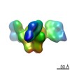
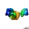
| Title | Structure of 2G12 (Fab)2 in Complex with Soluble and Fully Glycosylated HIV-1 Env by Negative-Stain Single Particle Electron Microscopy | |||||||||
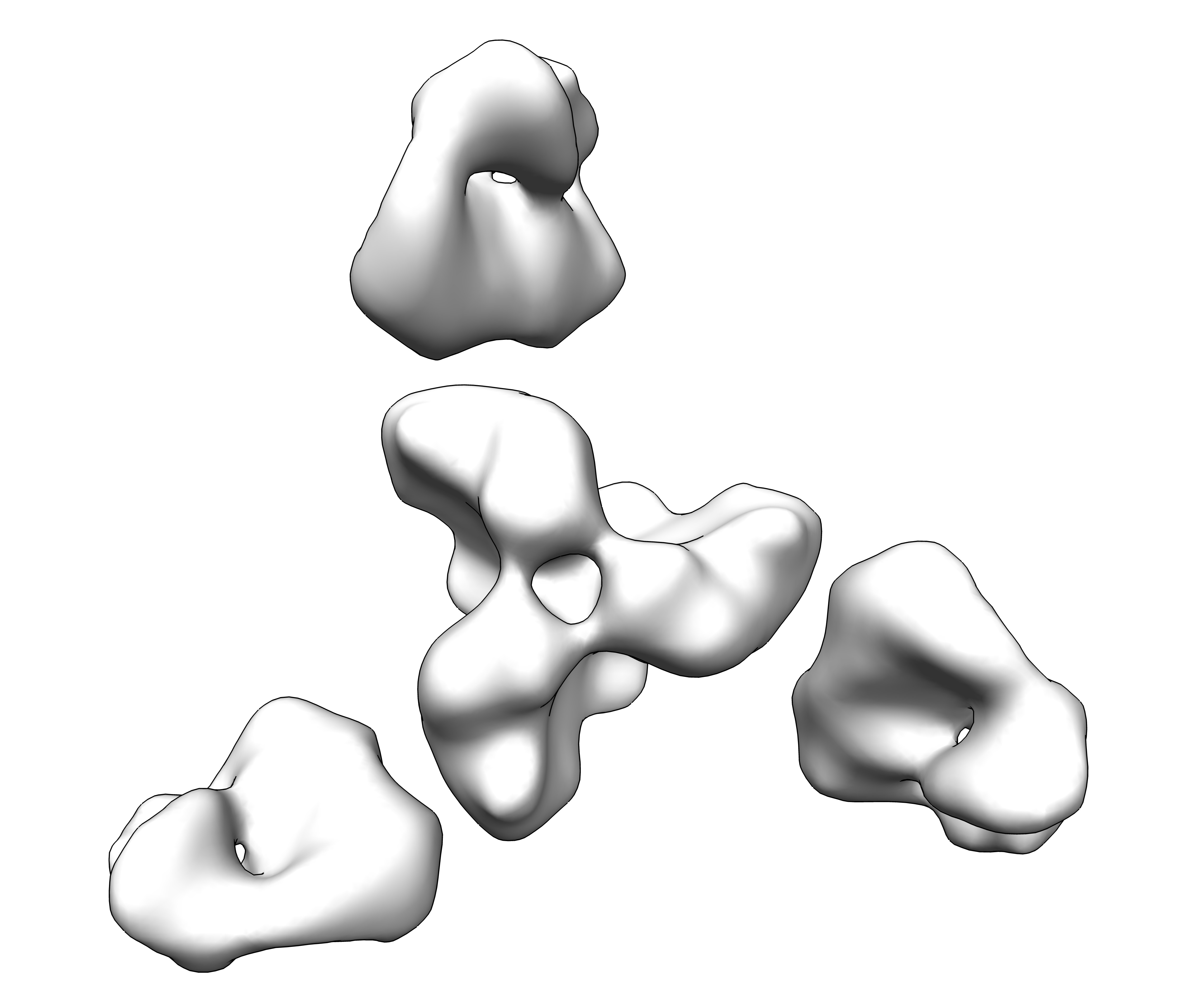
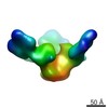
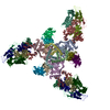
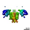
 Map data Map data | Reconstruction of HIV-1 Env SOSIP BG505.664 in complex with 2G12 Fab by negative stain EM | |||||||||
 Sample Sample |
| |||||||||
 Keywords Keywords |  HIV-1 / 2G12 / monoclonal antibodies / Envelope / CD4-bound Env HIV-1 / 2G12 / monoclonal antibodies / Envelope / CD4-bound Env | |||||||||
| Biological species |   Human immunodeficiency virus 1 / Human immunodeficiency virus 1 /  Homo sapiens (human) Homo sapiens (human) | |||||||||
| Method | single particle reconstruction / negative staining / Resolution: 17.0 Å | |||||||||
 Authors Authors | Murin CD / Julien JP / Stanfield R / Khayat R / Sok D / Burton DR / Wilson IA / Ward AB | |||||||||
 Citation Citation | Journal: J Virol / Year: 2014 Title: Structure of 2G12 Fab2 in complex with soluble and fully glycosylated HIV-1 Env by negative-stain single-particle electron microscopy. Authors: Charles D Murin / Jean-Philippe Julien / Devin Sok / Robyn L Stanfield / Reza Khayat / Albert Cupo / John P Moore / Dennis R Burton / Ian A Wilson / Andrew B Ward /  Abstract: The neutralizing anti-HIV-1 antibody 2G12 is of particular interest due to the sterilizing protection it provides from viral challenge in animal models. 2G12 is a unique, domain-exchanged antibody ...The neutralizing anti-HIV-1 antibody 2G12 is of particular interest due to the sterilizing protection it provides from viral challenge in animal models. 2G12 is a unique, domain-exchanged antibody that binds exclusively to conserved N-linked glycans that form the high-mannose patch on the gp120 outer domain centered on a glycan at position N332. Several glycans in and around the 2G12 epitope have been shown to interact with other potent, broadly neutralizing antibodies; therefore, this region constitutes a supersite of vulnerability on gp120. While crystal structures of 2G12 and 2G12 bound to high-mannose glycans have been solved, no structural information that describes the interaction of 2G12 with gp120 or the Env trimer is available. Here, we present a negative-stain single-particle electron microscopy reconstruction of 2G12 Fab2 in complex with a soluble, trimeric Env at ∼17-Å resolution that reveals the antibody's interaction with its native and fully glycosylated epitope. We also mapped relevant glycans in this epitope by fitting high-resolution crystal structures and by performing neutralization assays of glycan knockouts. In addition, a reconstruction at ∼26 Å of the ternary complex formed by 2G12 Fab2, soluble CD4, and Env indicates that 2G12 may block membrane fusion by induced steric hindrance upon primary receptor binding, thereby abrogating Env's interaction with coreceptor(s). These structures provide a basis for understanding 2G12 binding and neutralization, and our low-resolution model and glycan assignments provide a basis for higher-resolution studies to determine the molecular nature of the 2G12 epitope. IMPORTANCE: HIV-1 is a human virus that results in the deaths of millions of people around the world each year. While there are several effective therapeutics available to prolong life, a vaccine is ...IMPORTANCE: HIV-1 is a human virus that results in the deaths of millions of people around the world each year. While there are several effective therapeutics available to prolong life, a vaccine is the best long-term solution for curbing this global epidemic. Here, we present structural data that reveal the viral binding site of one of the first HIV-1-neutralizing antibodies isolated, 2G12, and provide a rationale for its effectiveness. These structures provide a basis for higher-resolution studies to determine the molecular nature of the 2G12 epitope, which will aid in vaccine design and antibody-based therapies. | |||||||||
| History |
|
- Structure visualization
| Movie |
 Movie viewer Movie viewer |
|---|---|
| Structure viewer | EM map: SurfViewMolmilJmol/JSmol |
| Supplemental images |
- Downloads & links
-EMDB archive
| Map data | emd_5982.map.gz | 1.7 MB | EMDB map data format | |
|---|---|---|---|---|
| Header (meta data) | emd-5982-v30.xmlemd-5982.xml | 13.4 KB 13.4 KB | Display Display | EMDB header |
| Images | emd_5982.tif | 2.2 MB | ||
| Archive directory |  http://ftp.pdbj.org/pub/emdb/structures/EMD-5982ftp://ftp.pdbj.org/pub/emdb/structures/EMD-5982 http://ftp.pdbj.org/pub/emdb/structures/EMD-5982ftp://ftp.pdbj.org/pub/emdb/structures/EMD-5982 | HTTPS FTP |
-Related structure data
-Links
| EMDB pages | EMDB (EBI/PDBe) / EMDataResource |
|---|
-Map
| File | Download / File: emd_5982.map.gz / Format: CCP4 / Size: 1.9 MB / Type: IMAGE STORED AS FLOATING POINT NUMBER (4 BYTES) | ||||||||||||||||||||||||||||||||||||||||||||||||||||||||||||||||||||
|---|---|---|---|---|---|---|---|---|---|---|---|---|---|---|---|---|---|---|---|---|---|---|---|---|---|---|---|---|---|---|---|---|---|---|---|---|---|---|---|---|---|---|---|---|---|---|---|---|---|---|---|---|---|---|---|---|---|---|---|---|---|---|---|---|---|---|---|---|---|
| Annotation | Reconstruction of HIV-1 Env SOSIP BG505.664 in complex with 2G12 Fab by negative stain EM | ||||||||||||||||||||||||||||||||||||||||||||||||||||||||||||||||||||
| Voxel size | X=Y=Z: 4.35 Å | ||||||||||||||||||||||||||||||||||||||||||||||||||||||||||||||||||||
| Density |
| ||||||||||||||||||||||||||||||||||||||||||||||||||||||||||||||||||||
| Symmetry | Space group: 1 | ||||||||||||||||||||||||||||||||||||||||||||||||||||||||||||||||||||
| Details | EMDB XML:
CCP4 map header:
| ||||||||||||||||||||||||||||||||||||||||||||||||||||||||||||||||||||
-Supplemental data
- Sample components
-Entire : Fab fragment of 2G12 monoclonal antibody bound to HIV-1 Env BG505.664
| Entire | Name: Fab fragment of 2G12 monoclonal antibody bound to HIV-1 Env BG505.664 |
|---|---|
| Components |
|
-Supramolecule #1000: Fab fragment of 2G12 monoclonal antibody bound to HIV-1 Env BG505.664
| Supramolecule | Name: Fab fragment of 2G12 monoclonal antibody bound to HIV-1 Env BG505.664 type: sample / ID: 1000 Oligomeric state: Env trimer bound to three 2G12 domain-swapped Fabs Number unique components: 2 |
|---|---|
| Molecular weight | Experimental: 660 KDa |
-Macromolecule #1: HIV-1 Env
| Macromolecule | Name: HIV-1 Env / type: protein_or_peptide / ID: 1 / Name.synonym: SOSIP BG505.664 / Number of copies: 1 / Oligomeric state: Trimer / Recombinant expression: Yes |
|---|---|
| Source (natural) | Organism: Human immunodeficiency virus 1 / synonym: HIV-1 |
| Molecular weight | Experimental: 360 KDa |
| Recombinant expression | Organism: Homo sapiens (human) / Recombinant cell: HEK 293F |
-Macromolecule #2: Human Monoclonal Antibody 2G12 IgG1 Fab Fragment
| Macromolecule | Name: Human Monoclonal Antibody 2G12 IgG1 Fab Fragment / type: protein_or_peptide / ID: 2 / Name.synonym: 2G12 Fab / Oligomeric state: Dimer of Fabs / Recombinant expression: Yes |
|---|---|
| Source (natural) | Organism: Homo sapiens (human) / synonym: Human |
| Molecular weight | Experimental: 100 KDa |
| Recombinant expression | Organism: Homo sapiens (human) / Recombinant cell: HEK 293F / Recombinant plasmid: phCMV3 |
-Experimental details
-Structure determination
| Method | negative staining |
|---|---|
 Processing Processing | single particle reconstruction |
| Aggregation state | particle |
-Sample preparation
| Concentration | 0.03 mg/mL |
|---|---|
| Buffer | pH: 7.4 / Details: 150 mM NaCl, 10 mM Tris |
| Staining | Type: NEGATIVE Details: Grids with adsorbed protein were floated on 2% w/v uranyl formate for 30 seconds. |
| Grid | Details: 400 mesh copper grid with thin carbon support, Gatan plasma cleaned |
| Vitrification | Cryogen name: NONE / Instrument: OTHER |
- Electron microscopy
| Microscope | FEI TECNAI F20 |
|---|---|
| Electron beam | Acceleration voltage: 120 kV / Electron source: TUNGSTEN HAIRPIN |
| Electron optics | Calibrated magnification: 100000 / Illumination mode: FLOOD BEAM / Imaging mode: BRIGHT FIELDBright-field microscopy / Nominal defocus max: 0.8 µm / Nominal defocus min: 0.5 µm / Nominal magnification: 100000 |
| Specialist optics | Energy filter - Name: FEI |
| Sample stage | Specimen holder model: HOME BUILD / Tilt angle max: 50 |
| Alignment procedure | Legacy - Astigmatism: Objective lens astigmatism was corrected at 100,000 times magnification. |
| Date | Jul 12, 2012 |
| Image recording | Category: CCD / Film or detector model: GENERIC GATAN (4k x 4k) / Number real images: 248 / Average electron dose: 30 e/Å2 |
| Tilt angle min | 0 |
| Experimental equipment |  Model: Tecnai F20 / Image courtesy: FEI Company |
-Image processing
| Final two d classification | Number classes: 130 |
|---|---|
| Final reconstruction | Algorithm: OTHER / Resolution.type: BY AUTHOR / Resolution: 17.0 Å / Resolution method: OTHER / Software - Name: EMAN2 / Details: Final map was low pass filtered to 20 Angstrom. / Number images used: 13143 |
| Details | The particles were selected using automatic selection program Leginon and processed using the Appion system. |
-Atomic model buiding 1
| Initial model | PDB ID: Chain - #0 - Chain ID: C / Chain - #1 - Chain ID: D / Chain - #2 - Chain ID: G / Chain - #3 - Chain ID: H / Chain - #4 - Chain ID: K / Chain - #5 - Chain ID: L |
|---|---|
| Software | Name: Chimera |
| Details | The HIV Env structure was fit by rigid body fitting using the UCSF Chimera volume fit option, simulating a map at an estimated resolution of 17 Angstrom. |
| Refinement | Space: REAL / Protocol: RIGID BODY FIT |
-Atomic model buiding 2
| Initial model | PDB ID:  1op5 |
|---|---|
| Software | Name: Chimera |
| Details | 2G12 Fab structures were fit by rigid body fitting using the UCSF Chimera volume fit option, simulating a map at an estimated resolution of 17 Angstrom. |
| Refinement | Space: REAL / Protocol: RIGID BODY FIT |
