Movie
Movie Controller

 Yorodumi
+ Open data

- Basic information
| Entry | Database: EMDB / ID: EMD-5927 | |||||||||
|---|---|---|---|---|---|---|---|---|---|---|
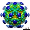
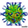
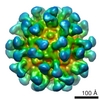
| Title | Kinetic and Structural Analysis of Coxsackievirus B3 Receptor Interactions and Formation of the A-particle | |||||||||
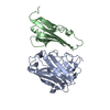
 Map data Map data | Reconstruction of CVB3 complexed with CAR | |||||||||
 Sample Sample |
| |||||||||
 Keywords Keywords | coxsackievirus b3 / cvb3 /  CAR / cryoEM / A-particle CAR / cryoEM / A-particle | |||||||||
| Function / homology |  Function and homology information Function and homology informationAV node cell-bundle of His cell adhesion involved in cell communication / cell adhesive protein binding involved in AV node cell-bundle of His cell communication / homotypic cell-cell adhesion / AV node cell to bundle of His cell communication / epithelial structure maintenance / gamma-delta T cell activation / regulation of AV node cell action potential / germ cell migration / apicolateral plasma membrane / cell-cell junction organization ...AV node cell-bundle of His cell adhesion involved in cell communication / cell adhesive protein binding involved in AV node cell-bundle of His cell communication / homotypic cell-cell adhesion / AV node cell to bundle of His cell communication / epithelial structure maintenance / gamma-delta T cell activation / regulation of AV node cell action potential / germ cell migration / apicolateral plasma membrane / cell-cell junction organization / transepithelial transport / connexin binding / cardiac muscle cell development / heterophilic cell-cell adhesion via plasma membrane cell adhesion molecules / bicellular tight junction / intercalated disc / mitochondrion organization / cell adhesion molecule binding / neutrophil chemotaxis / acrosomal vesicle / filopodium / PDZ domain binding / Cell surface interactions at the vascular wall / adherens junction / neuromuscular junction / beta-catenin binding / Immunoregulatory interactions between a Lymphoid and a non-Lymphoid cell / cell-cell junction / integrin binding / virus receptor activity / cell junction / cell body / heart development / growth cone / actin cytoskeleton organization / basolateral plasma membrane / defense response to virus / neuron projection / membrane raft / signaling receptor binding / protein-containing complex / extracellular space / extracellular region / nucleoplasm / identical protein binding / plasma membrane / cytoplasmSimilarity search - Function | |||||||||
| Biological species |  Homo sapiens (human) / Homo sapiens (human) /   Human coxsackievirus B3 Human coxsackievirus B3 | |||||||||
| Method | single particle reconstruction / cryo EM / Resolution: 9.0 Å | |||||||||
 Authors Authors | Organtini LJ / Makhov AM / Conway JF / Hafenstein S / Carson SD | |||||||||
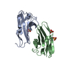
 Citation Citation | Journal: J Virol / Year: 2014 Title: Kinetic and structural analysis of coxsackievirus B3 receptor interactions and formation of the A-particle. Authors: Lindsey J Organtini / Alexander M Makhov / James F Conway / Susan Hafenstein / Steven D Carson /  Abstract: The coxsackievirus and adenovirus receptor (CAR) has been identified as the cellular receptor for group B coxsackieviruses, including serotype 3 (CVB3). CAR mediates infection by binding to CVB3 and ...The coxsackievirus and adenovirus receptor (CAR) has been identified as the cellular receptor for group B coxsackieviruses, including serotype 3 (CVB3). CAR mediates infection by binding to CVB3 and catalyzing conformational changes in the virus that result in formation of the altered, noninfectious A-particle. Kinetic analyses show that the apparent first-order rate constant for the inactivation of CVB3 by soluble CAR (sCAR) at physiological temperatures varies nonlinearly with sCAR concentration. Cryo-electron microscopy (cryo-EM) reconstruction of the CVB3-CAR complex resulted in a 9.0-Å resolution map that was interpreted with the four available crystal structures of CAR, providing a consensus footprint for the receptor binding site. The analysis of the cryo-EM structure identifies important virus-receptor interactions that are conserved across picornavirus species. These conserved interactions map to variable antigenic sites or structurally conserved regions, suggesting a combination of evolutionary mechanisms for receptor site preservation. The CAR-catalyzed A-particle structure was solved to a 6.6-Å resolution and shows significant rearrangement of internal features and symmetric interactions with the RNA genome. IMPORTANCE: This report presents new information about receptor use by picornaviruses and highlights the importance of attaining at least an ∼9-Å resolution for the interpretation of cryo-EM ...IMPORTANCE: This report presents new information about receptor use by picornaviruses and highlights the importance of attaining at least an ∼9-Å resolution for the interpretation of cryo-EM complex maps. The analysis of receptor binding elucidates two complementary mechanisms for preservation of the low-affinity (initial) interaction of the receptor and defines the kinetics of receptor-catalyzed conformational change to the A-particle. | |||||||||
| History |
|
- Structure visualization
| Movie |
Movie viewer |
|---|---|
| Structure viewer | EM map: SurfViewMolmilJmol/JSmol |
| Supplemental images |

- Downloads & links
-EMDB archive
| Map data | emd_5927.map.gz | 85.6 MB | EMDB map data format | |
|---|---|---|---|---|
| Header (meta data) | emd-5927-v30.xmlemd-5927.xml | 14.5 KB 14.5 KB | Display Display | EMDB header |
| Images | 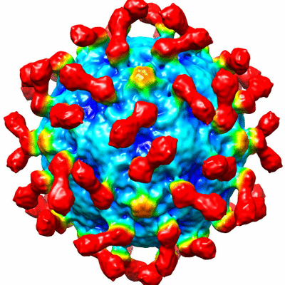 400_5927.gif 400_5927.gif 80_5927.gif 80_5927.gif | 84.9 KB 6.1 KB | ||
| Archive directory |  http://ftp.pdbj.org/pub/emdb/structures/EMD-5927ftp://ftp.pdbj.org/pub/emdb/structures/EMD-5927 http://ftp.pdbj.org/pub/emdb/structures/EMD-5927ftp://ftp.pdbj.org/pub/emdb/structures/EMD-5927 | HTTPS FTP |
-Related structure data
| Related structure data | 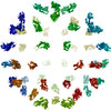 3j6lMC 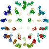 3j6mMC  3j6nMC  3j6oMC  5928C M: atomic model generated by this map C: citing same article ( |
|---|---|
| Similar structure data |

-Links
| EMDB pages | EMDB (EBI/PDBe) / EMDataResource |
|---|---|
| Related items in Molecule of the Month |


-Map
| File | Download / File: emd_5927.map.gz / Format: CCP4 / Size: 206 MB / Type: IMAGE STORED AS FLOATING POINT NUMBER (4 BYTES) | ||||||||||||||||||||||||||||||||||||||||||||||||||||||||||||||||||||
|---|---|---|---|---|---|---|---|---|---|---|---|---|---|---|---|---|---|---|---|---|---|---|---|---|---|---|---|---|---|---|---|---|---|---|---|---|---|---|---|---|---|---|---|---|---|---|---|---|---|---|---|---|---|---|---|---|---|---|---|---|---|---|---|---|---|---|---|---|---|
| Annotation | Reconstruction of CVB3 complexed with CAR | ||||||||||||||||||||||||||||||||||||||||||||||||||||||||||||||||||||
| Voxel size | X=Y=Z: 1.25 Å | ||||||||||||||||||||||||||||||||||||||||||||||||||||||||||||||||||||
| Density |
| ||||||||||||||||||||||||||||||||||||||||||||||||||||||||||||||||||||
| Symmetry | Space group: 1 | ||||||||||||||||||||||||||||||||||||||||||||||||||||||||||||||||||||
| Details | EMDB XML:
CCP4 map header:
| ||||||||||||||||||||||||||||||||||||||||||||||||||||||||||||||||||||
-Supplemental data
- Sample components
-Entire : Coxsackievirus B3 complexed with CAR
| Entire | Name: Coxsackievirus B3 complexed with CAR |
|---|---|
| Components |
|
-Supramolecule #1000: Coxsackievirus B3 complexed with CAR
| Supramolecule | Name: Coxsackievirus B3 complexed with CAR / type: sample / ID: 1000 / Details: purified virus and receptor complex in solution / Oligomeric state: icosahedral virus / Number unique components: 2 |
|---|---|
| Molecular weight | Experimental: 7 MDa |
-Supramolecule #1: Human coxsackievirus B3
| Supramolecule | Name: Human coxsackievirus B3 / type: virus / ID: 1 / Name.synonym: CVB3 Details: Virus was incubated with excess CAR at 4 degrees C. NCBI-ID: 12072 / Sci species name: Human coxsackievirus B3 / Sci species strain: CVB3/28 / Database: NCBI / Virus type: VIRION / Virus isolate: STRAIN / Virus enveloped: No / Virus empty: No / Syn species name: CVB3 |
|---|---|
| Host (natural) | Organism: Homo sapiens (human) / synonym: VERTEBRATES |
| Molecular weight | Experimental: 7 MDa |
| Virus shell | Shell ID: 1 / Name: VP1-4 / Diameter: 300 Å / T number (triangulation number): 1 |
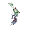
-Macromolecule #1: Coxsackievirus and adenovirus receptor
| Macromolecule | Name: Coxsackievirus and adenovirus receptor / type: protein_or_peptide / ID: 1 / Name.synonym: CAR / Recombinant expression: Yes |
|---|---|
| Source (natural) | Organism: Homo sapiens (human) / synonym: human |
| Recombinant expression | Organism:  Escherichia coli (E. coli) Escherichia coli (E. coli) |
| Sequence | UniProtKB: Coxsackievirus and adenovirus receptor |
-Experimental details
-Structure determination
| Method | cryo EM |
|---|---|
 Processing Processing | single particle reconstruction |
| Aggregation state | particle |
-Sample preparation
| Concentration | 0.1 mg/mL |
|---|---|
| Buffer | pH: 6 / Details: 50 mM MES, 100 mM NaCl |
| Grid | Details: glow-discharged holey carbon Quantifoil electron microscopy grids |
| Vitrification | Cryogen name: ETHANE-PROPANE MIXTURE / Chamber humidity: 95 % / Chamber temperature: 95 K / Instrument: FEI VITROBOT MARK III |
- Electron microscopy
| Microscope | FEI TECNAI F20 |
|---|---|
| Electron beam | Acceleration voltage: 200 kV / Electron source: FIELD EMISSION GUN |
| Electron optics | Calibrated magnification: 50000 / Illumination mode: SPOT SCAN / Imaging mode: BRIGHT FIELDBright-field microscopy / Cs: 2 mm / Nominal defocus max: 3.66 µm / Nominal defocus min: 1.98 µm / Nominal magnification: 50000 |
| Sample stage | Specimen holder model: GATAN LIQUID NITROGEN |
| Alignment procedure | Legacy - Astigmatism: CTFFIND3 |
| Date | Aug 1, 2012 |
| Image recording | Category: FILM / Film or detector model: KODAK SO-163 FILM / Digitization - Scanner: NIKON SUPER COOLSCAN 9000 / Digitization - Sampling interval: 6.35 µm / Number real images: 96 / Average electron dose: 15 e/Å2 |
| Experimental equipment |  Model: Tecnai F20 / Image courtesy: FEI Company |
-Image processing
| CTF correction | Details: AUTO3DEM |
|---|---|
| Final reconstruction | Algorithm: OTHER / Resolution.type: BY AUTHOR / Resolution: 9.0 Å / Resolution method: OTHER / Software - Name: EMAN, AUTO3DEM / Number images used: 9302 |
| Details | Particles were selected using EMAN. |
-Atomic model buiding 1
| Initial model | PDB ID: Chain - Chain ID: B |
|---|---|
| Software | Name: Situs |
| Details | The four available CAR structures were fit into the receptor density separately. |
| Refinement | Space: REAL / Protocol: RIGID BODY FIT / Target criteria: correlation coefficient |
| Output model | PDB-3j6l: PDB-3j6m: PDB-3j6n: PDB-3j6o: |
-Atomic model buiding 2
| Initial model | PDB ID: Chain - Chain ID: B |
|---|---|
| Software | Name: Situs |
| Details | The four available CAR structures were fit into the receptor density separately. |
| Refinement | Space: REAL / Protocol: RIGID BODY FIT / Target criteria: correlation coefficient |
| Output model | PDB-3j6l: PDB-3j6m: PDB-3j6n: PDB-3j6o: |
-Atomic model buiding 3
| Initial model | PDB ID: Chain - Chain ID: K |
|---|---|
| Software | Name: Situs |
| Details | The four available CAR structures were fit into the receptor density separately. |
| Refinement | Space: REAL / Protocol: RIGID BODY FIT / Target criteria: correlation coefficient |
| Output model | PDB-3j6l: PDB-3j6m: PDB-3j6n: PDB-3j6o: |

-Atomic model buiding 4
| Initial model | PDB ID: Chain - Chain ID: S |
|---|---|
| Software | Name: Situs |
| Details | The four available CAR structures were fit into the receptor density separately. |
| Refinement | Space: REAL / Protocol: RIGID BODY FIT / Target criteria: correlation coefficient |
| Output model | PDB-3j6l: PDB-3j6m: PDB-3j6n: PDB-3j6o: |