 Movie
Movie Controller


+ Open data

- Basic information
| Entry | Database: EMDB / ID: EMD-5915 | |||||||||
|---|---|---|---|---|---|---|---|---|---|---|
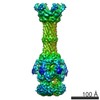
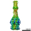
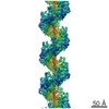
| Title | The structure of a complete multidrug efflux pump | |||||||||
 Map data Map data | Reconstruction of complete multidrug efflux pump | |||||||||
 Sample Sample |
| |||||||||
 Keywords Keywords | multidrug efflux pump | |||||||||
| Biological species |   Escherichia coli (E. coli) Escherichia coli (E. coli) | |||||||||
| Method | single particle reconstruction / cryo EM / Resolution: 15.0 Å | |||||||||
 Authors Authors | Du D / Wang Z / James NR / Voss JE / Klimont E / Ohene-Agyei T / Venter H / Chiu W / Luisi BF | |||||||||
 Citation Citation | Journal: Nature / Year: 2014 Title: Structure of the AcrAB-TolC multidrug efflux pump. Authors: Dijun Du / Zhao Wang / Nathan R James / Jarrod E Voss / Ewa Klimont / Thelma Ohene-Agyei / Henrietta Venter / Wah Chiu / Ben F Luisi /    Abstract: The capacity of numerous bacterial species to tolerate antibiotics and other toxic compounds arises in part from the activity of energy-dependent transporters. In Gram-negative bacteria, many of ...The capacity of numerous bacterial species to tolerate antibiotics and other toxic compounds arises in part from the activity of energy-dependent transporters. In Gram-negative bacteria, many of these transporters form multicomponent 'pumps' that span both inner and outer membranes and are driven energetically by a primary or secondary transporter component. A model system for such a pump is the acridine resistance complex of Escherichia coli. This pump assembly comprises the outer-membrane channel TolC, the secondary transporter AcrB located in the inner membrane, and the periplasmic AcrA, which bridges these two integral membrane proteins. The AcrAB-TolC efflux pump is able to transport vectorially a diverse array of compounds with little chemical similarity, thus conferring resistance to a broad spectrum of antibiotics. Homologous complexes are found in many Gram-negative species, including in animal and plant pathogens. Crystal structures are available for the individual components of the pump and have provided insights into substrate recognition, energy coupling and the transduction of conformational changes associated with the transport process. However, how the subunits are organized in the pump, their stoichiometry and the details of their interactions are not known. Here we present the pseudo-atomic structure of a complete multidrug efflux pump in complex with a modulatory protein partner from E. coli. The model defines the quaternary organization of the pump, identifies key domain interactions, and suggests a cooperative process for channel assembly and opening. These findings illuminate the basis for drug resistance in numerous pathogenic bacterial species. | |||||||||
| History |
|
- Structure visualization
| Movie |
 Movie viewer Movie viewer |
|---|---|
| Structure viewer | EM map: SurfViewMolmilJmol/JSmol |
| Supplemental images |

- Downloads & links
-EMDB archive
| Map data | emd_5915.map.gz | 59.9 MB | EMDB map data format | |
|---|---|---|---|---|
| Header (meta data) | emd-5915-v30.xmlemd-5915.xml | 10.4 KB 10.4 KB | Display Display | EMDB header |
| Images | 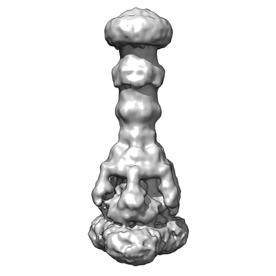 400_5915.gif 400_5915.gif 80_5915.gif 80_5915.gif | 26.2 KB 2.4 KB | ||
| Archive directory |  http://ftp.pdbj.org/pub/emdb/structures/EMD-5915ftp://ftp.pdbj.org/pub/emdb/structures/EMD-5915 http://ftp.pdbj.org/pub/emdb/structures/EMD-5915ftp://ftp.pdbj.org/pub/emdb/structures/EMD-5915 | HTTPS FTP |
-Related structure data


-Links
| EMDB pages | EMDB (EBI/PDBe) / EMDataResource |
|---|
-Map
| File | Download / File: emd_5915.map.gz / Format: CCP4 / Size: 62.5 MB / Type: IMAGE STORED AS FLOATING POINT NUMBER (4 BYTES) | ||||||||||||||||||||||||||||||||||||||||||||||||||||||||||||||||||||
|---|---|---|---|---|---|---|---|---|---|---|---|---|---|---|---|---|---|---|---|---|---|---|---|---|---|---|---|---|---|---|---|---|---|---|---|---|---|---|---|---|---|---|---|---|---|---|---|---|---|---|---|---|---|---|---|---|---|---|---|---|---|---|---|---|---|---|---|---|---|
| Annotation | Reconstruction of complete multidrug efflux pump | ||||||||||||||||||||||||||||||||||||||||||||||||||||||||||||||||||||
| Voxel size | X=Y=Z: 2.47 Å | ||||||||||||||||||||||||||||||||||||||||||||||||||||||||||||||||||||
| Density |
| ||||||||||||||||||||||||||||||||||||||||||||||||||||||||||||||||||||
| Symmetry | Space group: 1 | ||||||||||||||||||||||||||||||||||||||||||||||||||||||||||||||||||||
| Details | EMDB XML:
CCP4 map header:
| ||||||||||||||||||||||||||||||||||||||||||||||||||||||||||||||||||||
-Supplemental data
- Sample components
-Entire : AcrB-AcrA-TolC-AcrZ multidrug efflux pump
| Entire | Name: AcrB-AcrA-TolC-AcrZ multidrug efflux pump |
|---|---|
| Components |
|
-Supramolecule #1000: AcrB-AcrA-TolC-AcrZ multidrug efflux pump
| Supramolecule | Name: AcrB-AcrA-TolC-AcrZ multidrug efflux pump / type: sample / ID: 1000 Oligomeric state: One homotrimer of TolC and one homotrimer of AcrB bound to a homohexamer of AcrA; three AcrZ molecules are bound to the AcrB Number unique components: 1 |
|---|---|
| Molecular weight | Theoretical: 771 KDa |
-Macromolecule #1: AcrABZ-TolC complex
| Macromolecule | Name: AcrABZ-TolC complex / type: protein_or_peptide / ID: 1 / Name.synonym: acridine transport pump / Number of copies: 1 / Oligomeric state: hetero-pentadecamer (15-mer) / Recombinant expression: Yes |
|---|---|
| Source (natural) | Organism: Escherichia coli (E. coli) / Strain: W3110 |
| Molecular weight | Theoretical: 770 KDa |
| Recombinant expression | Organism: Escherichia coli. / Recombinant strain: C43 (DE3) / Recombinant plasmid: pET21a and pRSFDuet-1 |
-Experimental details
-Structure determination
| Method | cryo EM |
|---|---|
 Processing Processing | single particle reconstruction |
| Aggregation state | particle |
-Sample preparation
| Concentration | 2.0 mg/mL |
|---|---|
| Buffer | pH: 7.5 / Details: 50 mM HEPES pH 7.5, 400 mM NaCl, 0.03% DDM |
| Grid | Details: 200 mesh copper grid with holey carbon film |
| Vitrification | Cryogen name: ETHANE / Chamber humidity: 100 % / Instrument: FEI VITROBOT MARK IV / Method: Blot 1 second at force 0 |
- Electron microscopy
| Microscope | JEOL 3200FSC |
|---|---|
| Electron beam | Acceleration voltage: 300 kV / Electron source: FIELD EMISSION GUN |
| Electron optics | Illumination mode: FLOOD BEAM / Imaging mode: DARK FIELD / Nominal defocus max: 3.5 µm / Nominal defocus min: 0.5 µm / Nominal magnification: 20000 |
| Specialist optics | Energy filter - Name: JEOL |
| Sample stage | Specimen holder model: JEOL 3200FSC CRYOHOLDER |
| Temperature | Min: 78 K / Max: 80 K / Average: 79 K |
| Alignment procedure | Legacy - Astigmatism: Objective lens astigmatism was corrected at 100k magnification |
| Date | May 20, 2013 |
| Image recording | Category: CCD / Film or detector model: DIRECT ELECTRON DE-12 (4k x 3k) / Digitization - Sampling interval: 6 µm / Number real images: 1281 / Average electron dose: 25 e/Å2 |
-Image processing
| CTF correction | Details: per image |
|---|---|
| Final reconstruction | Resolution.type: BY AUTHOR / Resolution: 15.0 Å / Resolution method: OTHER / Software - Name: EMAN2, RELION Details: Resolution was estimated by gold standard eotest from two independent reconstructions. Number images used: 7000 |
| Details | Particle were selected by eman2 e2boxer. Initial model was generated by eman2 base on reference free 2d class averages. Further refinements were done by eman2.07, followed by Relion at the end. |
