 Movie
Movie Controller


+ Open data

- Basic information
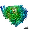
| Entry | Database: EMDB / ID: EMD-5903 | |||||||||
|---|---|---|---|---|---|---|---|---|---|---|
| Title | 3D Reconstruction of Membrane Protein Complex ExbB4-ExbD2 | |||||||||
 Map data Map data | ExbB4-ExbD2 complex with ExbD periplasmic domains dimerized in the membrane-parallel position | |||||||||
 Sample Sample |
| |||||||||
 Keywords Keywords | Membrane protein complex / coordinated rearrangement / flexible domain | |||||||||
| Function / homology |  Function and homology information Function and homology informationenergy transducer activity / bacteriocin transport / cobalamin transport / intracellular monoatomic cation homeostasis / plasma membrane protein complex / protein import / transmembrane transporter activity / transmembrane transporter complex / cell outer membrane / protein transport ...energy transducer activity / bacteriocin transport / cobalamin transport / intracellular monoatomic cation homeostasis / plasma membrane protein complex / protein import / transmembrane transporter activity / transmembrane transporter complex / cell outer membrane / protein transport / intracellular iron ion homeostasis / membrane => GO:0016020 / protein stabilization / protein homodimerization activity / identical protein binding / membrane / plasma membrane Similarity search - Function | |||||||||
| Biological species |  | |||||||||
| Method | single particle reconstruction / negative staining / Resolution: 24.0 Å | |||||||||
 Authors Authors | Sverzhinsky A / Fabre L / Cottreau AL / Biot-Pelletier DMP / Khalil S / Bostina M / Rouiller I / Coulton JW | |||||||||
 Citation Citation | Journal: Structure / Year: 2014 Title: Coordinated rearrangements between cytoplasmic and periplasmic domains of the membrane protein complex ExbB-ExbD of Escherichia coli. Authors: Aleksandr Sverzhinsky / Lucien Fabre / Andrew L Cottreau / Damien M P Biot-Pelletier / Sofia Khalil / Mihnea Bostina / Isabelle Rouiller / James W Coulton /  Abstract: Gram-negative bacteria rely on the ExbB-ExbD-TonB system for the import of essential nutrients. Despite decades of research, the stoichiometry, subunit organization, and mechanism of action of the ...Gram-negative bacteria rely on the ExbB-ExbD-TonB system for the import of essential nutrients. Despite decades of research, the stoichiometry, subunit organization, and mechanism of action of the membrane proteins of the Ton system remain unclear. We copurified ExbB with ExbD as an ∼240 kDa protein-detergent complex, measured by light scattering and by native gels. Quantitative Coomassie staining revealed a stoichiometry of ExbB4-ExbD2. Negative stain electron microscopy and 2D analysis showed particles of ∼10 nm diameter in multiple structural states. Nanogold labeling identified the position of the ExbD periplasmic domain. Random conical tilt was used to reconstruct the particles in three structural states followed by sorting of the single particles and refinement of each state. The different states are interpreted by coordinated structural rearrangements between the cytoplasmic domain and the periplasmic domain, concordant with in vivo predictions. | |||||||||
| History |
|
- Structure visualization
| Movie |
Movie viewer |
|---|---|
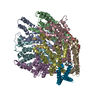
| Structure viewer | EM map: SurfViewMolmilJmol/JSmol |
| Supplemental images |

- Downloads & links
-EMDB archive
| Map data | emd_5903.map.gz | 7.4 MB | EMDB map data format | |
|---|---|---|---|---|
| Header (meta data) | emd-5903-v30.xmlemd-5903.xml | 12 KB 12 KB | Display Display | EMDB header |
| Images | 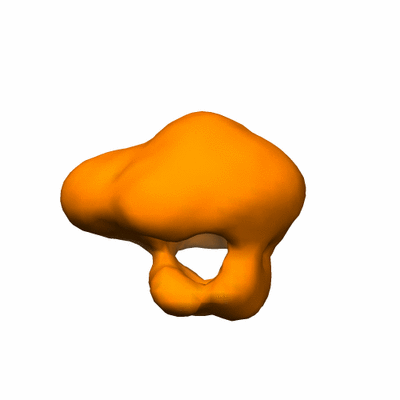 400_5903.gif 400_5903.gif 80_5903.gif 80_5903.gif | 16.6 KB 2.1 KB | ||
| Archive directory |  http://ftp.pdbj.org/pub/emdb/structures/EMD-5903ftp://ftp.pdbj.org/pub/emdb/structures/EMD-5903 http://ftp.pdbj.org/pub/emdb/structures/EMD-5903ftp://ftp.pdbj.org/pub/emdb/structures/EMD-5903 | HTTPS FTP |
-Validation report
| Summary document | emd_5903_validation.pdf.gz | 78.3 KB | Display | EMDB validaton report |
|---|---|---|---|---|
| Full document | emd_5903_full_validation.pdf.gz | 77.4 KB | Display | |
| Data in XML | emd_5903_validation.xml.gz | 493 B | Display | |
| Arichive directory | https://ftp.pdbj.org/pub/emdb/validation_reports/EMD-5903ftp://ftp.pdbj.org/pub/emdb/validation_reports/EMD-5903 | HTTPS FTP |
-Related structure data


-Links
| EMDB pages | EMDB (EBI/PDBe) / EMDataResource |
|---|
-Map
| File | Download / File: emd_5903.map.gz / Format: CCP4 / Size: 7.8 MB / Type: IMAGE STORED AS FLOATING POINT NUMBER (4 BYTES) | ||||||||||||||||||||||||||||||||||||||||||||||||||||||||||||
|---|---|---|---|---|---|---|---|---|---|---|---|---|---|---|---|---|---|---|---|---|---|---|---|---|---|---|---|---|---|---|---|---|---|---|---|---|---|---|---|---|---|---|---|---|---|---|---|---|---|---|---|---|---|---|---|---|---|---|---|---|---|
| Annotation | ExbB4-ExbD2 complex with ExbD periplasmic domains dimerized in the membrane-parallel position | ||||||||||||||||||||||||||||||||||||||||||||||||||||||||||||
| Voxel size | X=Y=Z: 2.2 Å | ||||||||||||||||||||||||||||||||||||||||||||||||||||||||||||
| Density |
| ||||||||||||||||||||||||||||||||||||||||||||||||||||||||||||
| Symmetry | Space group: 1 | ||||||||||||||||||||||||||||||||||||||||||||||||||||||||||||
| Details | EMDB XML:
CCP4 map header:
| ||||||||||||||||||||||||||||||||||||||||||||||||||||||||||||
-Supplemental data
- Sample components
-Entire : Membrane protein complex ExbB4-ExbD2 from Escherichia coli
| Entire | Name: Membrane protein complex ExbB4-ExbD2 from Escherichia coli |
|---|---|
| Components |
|
-Supramolecule #1000: Membrane protein complex ExbB4-ExbD2 from Escherichia coli
| Supramolecule | Name: Membrane protein complex ExbB4-ExbD2 from Escherichia coli type: sample / ID: 1000 / Oligomeric state: Four ExbB in complex with two ExbD / Number unique components: 2 |
|---|---|
| Molecular weight | Theoretical: 139 KDa |
-Macromolecule #1: Biopolymer Transport Protein ExbB
| Macromolecule | Name: Biopolymer Transport Protein ExbB / type: protein_or_peptide / ID: 1 / Name.synonym: ExbB / Number of copies: 4 / Oligomeric state: Tetramer / Recombinant expression: Yes |
|---|---|
| Source (natural) | Organism: |
| Molecular weight | Theoretical: 26 KDa |
| Recombinant expression | Organism: |
| Sequence | UniProtKB: Biopolymer transport protein ExbB / GO: membrane => GO:0016020 / InterPro: TonB-system energizer ExbB type-1 |
-Macromolecule #2: Biopolymer Transport Protein ExbD
| Macromolecule | Name: Biopolymer Transport Protein ExbD / type: protein_or_peptide / ID: 2 / Name.synonym: ExbD / Number of copies: 2 / Oligomeric state: Dimer / Recombinant expression: Yes |
|---|---|
| Source (natural) | Organism: |
| Molecular weight | Theoretical: 17 KDa |
| Recombinant expression | Organism: |
| Sequence | UniProtKB: Biopolymer transport protein ExbD / GO: membrane => GO:0016020 / InterPro: TonB system transport protein ExbD type-1 |
-Experimental details
-Structure determination
| Method | negative staining |
|---|---|
 Processing Processing | single particle reconstruction |
| Aggregation state | particle |
-Sample preparation
| Concentration | 0.02 mg/mL |
|---|---|
| Buffer | pH: 7.5 / Details: 25 mM Tris-HCl, 150 mM NaCl, 0.01% DDM |
| Staining | Type: NEGATIVE Details: Grids with adsorbed protein were stained with 1.5% uranyl formate for 1 minute. |
| Grid | Details: 400 mesh copper grid with thin carbon support, glow discharged |
| Vitrification | Cryogen name: NONE / Instrument: OTHER |
- Electron microscopy
| Microscope | FEI TECNAI F20 |
|---|---|
| Alignment procedure | Legacy - Astigmatism: Objective lens astigmatism was corrected at 50,000x magnification |
| Date | Mar 27, 2013 |
| Image recording | Category: CCD / Film or detector model: GATAN ULTRASCAN 4000 (4k x 4k) / Average electron dose: 20 e/Å2 |
| Electron beam | Acceleration voltage: 200 kV / Electron source:  FIELD EMISSION GUN FIELD EMISSION GUN |
| Electron optics | Calibrated magnification: 67147 / Illumination mode: FLOOD BEAM / Imaging mode: BRIGHT FIELD / Cs: 2.0 mm / Nominal defocus max: 3.5 µm / Nominal defocus min: 1.5 µm / Nominal magnification: 67000 |
| Sample stage | Specimen holder: Room temperature / Specimen holder model: SIDE ENTRY, EUCENTRIC |
| Experimental equipment |  Model: Tecnai F20 / Image courtesy: FEI Company |
-Image processing
| Details | Manual particle picking from random conical tilt pairs, 3D classification, projection matching angular refinement |
|---|---|
| CTF correction | Details: Each Micrograph |
| Final reconstruction | Algorithm: OTHER / Resolution.type: BY AUTHOR / Resolution: 24.0 Å / Resolution method: OTHER / Software - Name: Xmipp, EMAN2, SPARX / Number images used: 5562 |
