Movie
Movie Controller


+ Open data

- Basic information
| Entry | Database: EMDB / ID: EMD-5890 | |||||||||
|---|---|---|---|---|---|---|---|---|---|---|
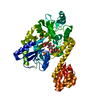
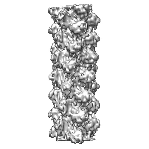
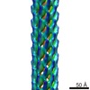
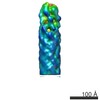
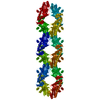
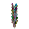
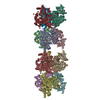
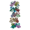
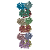
| Title | Cryo-EM structure of MAVS CARD filament | |||||||||
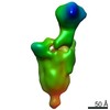
 Map data Map data | Cryo-EM map of MAVS CARD filament | |||||||||
 Sample Sample |
| |||||||||
 Keywords Keywords |  Innate immunity / helical filament Innate immunity / helical filament | |||||||||
| Function / homology |  Function and homology information Function and homology informationpositive regulation of IP-10 production / regulation of peroxisome organization / RIG-I binding / positive regulation of chemokine (C-C motif) ligand 5 production / positive regulation of myeloid dendritic cell cytokine production / CARD domain binding / NF-kB activation through FADD/RIP-1 pathway mediated by caspase-8 and -10 / positive regulation of response to cytokine stimulus / protein localization to mitochondrion / positive regulation of type I interferon-mediated signaling pathway ...positive regulation of IP-10 production / regulation of peroxisome organization / RIG-I binding / positive regulation of chemokine (C-C motif) ligand 5 production / positive regulation of myeloid dendritic cell cytokine production / CARD domain binding / NF-kB activation through FADD/RIP-1 pathway mediated by caspase-8 and -10 / positive regulation of response to cytokine stimulus / protein localization to mitochondrion / positive regulation of type I interferon-mediated signaling pathway / peroxisomal membrane / TRAF6 mediated IRF7 activation / negative regulation of type I interferon-mediated signaling pathway / positive regulation of NLRP3 inflammasome complex assembly / negative regulation of viral genome replication / cellular response to exogenous dsRNA / cytoplasmic pattern recognition receptor signaling pathway / type I interferon-mediated signaling pathway / positive regulation of interferon-alpha production / antiviral innate immune response / TRAF6 mediated NF-kB activation / positive regulation of type I interferon production / ubiquitin ligase complex / signaling adaptor activity / positive regulation of defense response to virus by host / molecular condensate scaffold activity / activation of innate immune response / positive regulation of interferon-beta production / Negative regulators of DDX58/IFIH1 signaling / positive regulation of interleukin-8 production / mitochondrial membrane / DDX58/IFIH1-mediated induction of interferon-alpha/beta / PKR-mediated signaling / positive regulation of interleukin-6 production / positive regulation of protein import into nucleus / SARS-CoV-1 activates/modulates innate immune responses / positive regulation of DNA-binding transcription factor activity / Ovarian tumor domain proteases / positive regulation of tumor necrosis factor production / TRAF3-dependent IRF activation pathway / defense response to virus / positive regulation of canonical NF-kappaB signal transduction / mitochondrial outer membrane / molecular adaptor activity / defense response to bacterium / positive regulation of protein phosphorylation / innate immune response / protein kinase binding / SARS-CoV-2 activates/modulates innate and adaptive immune responses / signal transduction / positive regulation of transcription by RNA polymerase II / mitochondrion / identical protein bindingSimilarity search - Function | |||||||||
| Biological species |  Homo sapiens (human) Homo sapiens (human) | |||||||||
| Method | helical reconstruction / cryo EM / Resolution: 9.6 Å | |||||||||
 Authors Authors | Xu H / He X / Zheng H / Huang LJ / Hou F / Yu Z / de la Cruz MJ / Borkowski B / Zhang X / Chen ZJ / Jiang Q-X | |||||||||
 Citation Citation | Journal: Elife / Year: 2014 Title: Structural basis for the prion-like MAVS filaments in antiviral innate immunity. Authors: Hui Xu / Xiaojing He / Hui Zheng / Lily J Huang / Fajian Hou / Zhiheng Yu / Michael Jason de la Cruz / Brian Borkowski / Xuewu Zhang / Zhijian J Chen / Qiu-Xing Jiang /  Abstract: Mitochondrial antiviral signaling (MAVS) protein is required for innate immune responses against RNA viruses. In virus-infected cells MAVS forms prion-like aggregates to activate antiviral signaling ...Mitochondrial antiviral signaling (MAVS) protein is required for innate immune responses against RNA viruses. In virus-infected cells MAVS forms prion-like aggregates to activate antiviral signaling cascades, but the underlying structural mechanism is unknown. Here we report cryo-electron microscopic structures of the helical filaments formed by both the N-terminal caspase activation and recruitment domain (CARD) of MAVS and a truncated MAVS lacking part of the proline-rich region and the C-terminal transmembrane domain. Both structures are left-handed three-stranded helical filaments, revealing specific interfaces between individual CARD subunits that are dictated by electrostatic interactions between neighboring strands and hydrophobic interactions within each strand. Point mutations at multiple locations of these two interfaces impaired filament formation and antiviral signaling. Super-resolution imaging of virus-infected cells revealed rod-shaped MAVS clusters on mitochondria. These results elucidate the structural mechanism of MAVS polymerization, and explain how an α-helical domain uses distinct chemical interactions to form self-perpetuating filaments. DOI: http://dx.doi.org/10.7554/eLife.01489.001. | |||||||||
| History |
|
- Structure visualization
| Movie |
Movie viewer |
|---|---|
| Structure viewer | EM map: SurfViewMolmilJmol/JSmol |
| Supplemental images |
- Downloads & links
-EMDB archive
| Map data | emd_5890.map.gz | 297.2 KB | EMDB map data format | |
|---|---|---|---|---|
| Header (meta data) | emd-5890-v30.xmlemd-5890.xml | 12 KB 12 KB | Display Display | EMDB header |
| Images | emd_5890.tif | 129.2 KB | ||
| Archive directory |  http://ftp.pdbj.org/pub/emdb/structures/EMD-5890ftp://ftp.pdbj.org/pub/emdb/structures/EMD-5890 http://ftp.pdbj.org/pub/emdb/structures/EMD-5890ftp://ftp.pdbj.org/pub/emdb/structures/EMD-5890 | HTTPS FTP |
-Related structure data
| Related structure data | 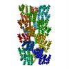 3j6cMC 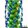 5891C 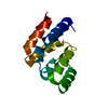 4o9fC 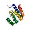 4o9lC M: atomic model generated by this map C: citing same article ( |
|---|---|
| Similar structure data | |
| EM raw data | EMPIAR-10014 (Title: MAVS CARD and DeltaProTM filaments / Data size: 35.3 / Data #1: CARD set1 micrographs [micrographs - single frame] / Data #2: CARD set2 micrographs [micrographs - single frame] / Data #3: CARD set3 micrographs [micrographs - single frame] / Data #4: CARD set4 micrographs [micrographs - single frame] Data #5: CARD set1 segments [picked particles - multiframe - unprocessed] Data #6: CARD set2 segments [picked particles - multiframe - unprocessed] Data #7: CARD set3 segments [picked particles - multiframe - unprocessed] Data #8: CARD set4 segments [picked particles - multiframe - unprocessed] Data #9: DeltProTM micrographs [micrographs - single frame] Data #10: DeltaProTM segments [picked particles - multiframe - unprocessed]) |
-Links
| EMDB pages | EMDB (EBI/PDBe) / EMDataResource |
|---|---|
| Related items in Molecule of the Month |


-Map
| File | Download / File: emd_5890.map.gz / Format: CCP4 / Size: 955.1 KB / Type: IMAGE STORED AS FLOATING POINT NUMBER (4 BYTES) | ||||||||||||||||||||||||||||||||||||||||||||||||||||||||||||||||||||
|---|---|---|---|---|---|---|---|---|---|---|---|---|---|---|---|---|---|---|---|---|---|---|---|---|---|---|---|---|---|---|---|---|---|---|---|---|---|---|---|---|---|---|---|---|---|---|---|---|---|---|---|---|---|---|---|---|---|---|---|---|---|---|---|---|---|---|---|---|---|
| Annotation | Cryo-EM map of MAVS CARD filament | ||||||||||||||||||||||||||||||||||||||||||||||||||||||||||||||||||||
| Voxel size | X=Y=Z: 2.333 Å | ||||||||||||||||||||||||||||||||||||||||||||||||||||||||||||||||||||
| Density |
| ||||||||||||||||||||||||||||||||||||||||||||||||||||||||||||||||||||
| Symmetry | Space group: 1 | ||||||||||||||||||||||||||||||||||||||||||||||||||||||||||||||||||||
| Details | EMDB XML:
CCP4 map header:
| ||||||||||||||||||||||||||||||||||||||||||||||||||||||||||||||||||||
-Supplemental data
- Sample components
-Entire : MAVS CARD filament
| Entire | Name: MAVS CARD filament |
|---|---|
| Components |
|
-Supramolecule #1000: MAVS CARD filament
| Supramolecule | Name: MAVS CARD filament / type: sample / ID: 1000 / Oligomeric state: polymer / Number unique components: 1 |
|---|---|
| Molecular weight | Theoretical: 546 KDa |
-Macromolecule #1: Mitochondrial antiviral-signaling protein (MAVS)
| Macromolecule | Name: Mitochondrial antiviral-signaling protein (MAVS) / type: protein_or_peptide / ID: 1 / Name.synonym: IPS1, KIAA1271, VISA / Oligomeric state: polymer / Recombinant expression: Yes |
|---|---|
| Source (natural) | Organism: Homo sapiens (human) / synonym: Human |
| Molecular weight | Theoretical: 13 KDa |
| Recombinant expression | Organism: Homo sapiens (human) / Recombinant cell: HEK293T / Recombinant plasmid: pcDNA3 |
| Sequence | UniProtKB: Mitochondrial antiviral-signaling protein |
-Experimental details
-Structure determination
| Method | cryo EM |
|---|---|
 Processing Processing | helical reconstruction |
| Aggregation state | filament |
-Sample preparation
| Buffer | pH: 7.5 / Details: 20mM Tris-HCl, 50mM NaCl, 1mM DTT |
|---|---|
| Grid | Details: Quantifoil grids coated with thin carbon on top |
| Vitrification | Cryogen name: ETHANE / Chamber humidity: 100 % / Instrument: FEI VITROBOT MARK III |
- Electron microscopy
| Microscope | JEOL 2200FS |
|---|---|
| Electron beam | Acceleration voltage: 200 kV / Electron source: FIELD EMISSION GUN |
| Electron optics | Calibrated magnification: 61950 / Illumination mode: FLOOD BEAM / Imaging mode: BRIGHT FIELDBright-field microscopy / Cs: 2.0 mm / Nominal defocus max: 2.0 µm / Nominal defocus min: 0.8 µm / Nominal magnification: 60000 |
| Specialist optics | Energy filter - Name: JEOL omega filter / Energy filter - Lower energy threshold: 0.0 eV / Energy filter - Upper energy threshold: 35.0 eV |
| Sample stage | Specimen holder model: GATAN LIQUID NITROGEN |
| Alignment procedure | Legacy - Astigmatism: Objective lens astigmatism was corrected at 100,000x magnification |
| Date | Oct 20, 2011 |
| Image recording | Category: FILM / Film or detector model: KODAK SO-163 FILM / Digitization - Scanner: ZEISS SCAI / Digitization - Sampling interval: 7 µm / Average electron dose: 20 e/Å2 |
-Image processing
| CTF correction | Details: Each filament segment |
|---|---|
| Final reconstruction | Applied symmetry - Helical parameters - Δz: 16.8 Å Applied symmetry - Helical parameters - Δ&Phi: 53.6 ° Applied symmetry - Helical parameters - Axial symmetry: C3 (3 fold cyclic )Algorithm: OTHER / Resolution.type: BY AUTHOR / Resolution: 9.6 Å / Resolution method: OTHER / Software - Name: Spider, MRC, EMAN, IMAGIC4D Details: Final data were calculated from three separate datasets from three sessions of data collection. The handedness of the map was determined by cryo-electron tomography. |
| Details | IHRSR |
-Atomic model buiding 1
| Initial model | PDB ID: Chain - Chain ID: A |
|---|---|
| Software | Name: Chimera, SITUS |
| Details | The docking of one X-ray model into a segmented map corresponding to one subunit was first done manually in Chimera, and then optimized using SITUS. |
| Refinement | Space: REAL / Protocol: RIGID BODY FIT |
| Output model | PDB-3j6c: |