 Movie
Movie Controller

[English] 日本語
 Yorodumi
- EMDB-5857: Architectures of Mcm2-7 Double Hexamer Assembly Intermediates Rev... -
+ Open data

- Basic information
| Entry | Database: EMDB / ID: EMD-5857 | |||||||||
|---|---|---|---|---|---|---|---|---|---|---|
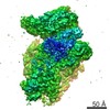
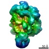
| Title | Architectures of Mcm2-7 Double Hexamer Assembly Intermediates Reveal Helicase Loading Mechanism | |||||||||
 Map data Map data | 3D reconstruction of Mcm2-7 double hexamer | |||||||||
 Sample Sample |
| |||||||||
 Keywords Keywords | Mcm2-7 double hexamer / MBP fusion / Helicase loading / 3DEM | |||||||||
| Biological species |   Saccharomyces cerevisiae (brewer's yeast) Saccharomyces cerevisiae (brewer's yeast) | |||||||||
| Method | single particle reconstruction / cryo EM / negative staining / Resolution: 15.0 Å | |||||||||
 Authors Authors | Sun J / Fernandez-Cid A / Riera A / Stillman B / Speck C / Li H | |||||||||
 Citation Citation | Journal: Genes Dev / Year: 2014 Title: Structural and mechanistic insights into Mcm2-7 double-hexamer assembly and function. Authors: Jingchuan Sun / Alejandra Fernandez-Cid / Alberto Riera / Silvia Tognetti / Zuanning Yuan / Bruce Stillman / Christian Speck / Huilin Li /   Abstract: Eukaryotic cells license each DNA replication origin during G1 phase by assembling a prereplication complex that contains a Mcm2-7 (minichromosome maintenance proteins 2-7) double hexamer. During S ...Eukaryotic cells license each DNA replication origin during G1 phase by assembling a prereplication complex that contains a Mcm2-7 (minichromosome maintenance proteins 2-7) double hexamer. During S phase, each Mcm2-7 hexamer forms the core of a replicative DNA helicase. However, the mechanisms of origin licensing and helicase activation are poorly understood. The helicase loaders ORC-Cdc6 function to recruit a single Cdt1-Mcm2-7 heptamer to replication origins prior to Cdt1 release and ORC-Cdc6-Mcm2-7 complex formation, but how the second Mcm2-7 hexamer is recruited to promote double-hexamer formation is not well understood. Here, structural evidence for intermediates consisting of an ORC-Cdc6-Mcm2-7 complex and an ORC-Cdc6-Mcm2-7-Mcm2-7 complex are reported, which together provide new insights into DNA licensing. Detailed structural analysis of the loaded Mcm2-7 double-hexamer complex demonstrates that the two hexamers are interlocked and misaligned along the DNA axis and lack ATP hydrolysis activity that is essential for DNA helicase activity. Moreover, we show that the head-to-head juxtaposition of the Mcm2-7 double hexamer generates a new protein interaction surface that creates a multisubunit-binding site for an S-phase protein kinase that is known to activate DNA replication. The data suggest how the double hexamer is assembled and how helicase activity is regulated during DNA licensing, with implications for cell cycle control of DNA replication and genome stability. | |||||||||
| History |
|
- Structure visualization
| Movie |
 Movie viewer Movie viewer |
|---|---|
| Structure viewer | EM map: SurfViewMolmilJmol/JSmol |
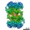
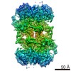
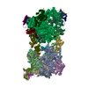
| Supplemental images |

- Downloads & links
-EMDB archive
| Map data | emd_5857.map.gz | 8 MB | EMDB map data format | |
|---|---|---|---|---|
| Header (meta data) | emd-5857-v30.xmlemd-5857.xml | 9.5 KB 9.5 KB | Display Display | EMDB header |
| Images | 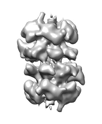 emd_5857.jpg emd_5857.jpg | 20.7 KB | ||
| Archive directory |  http://ftp.pdbj.org/pub/emdb/structures/EMD-5857ftp://ftp.pdbj.org/pub/emdb/structures/EMD-5857 http://ftp.pdbj.org/pub/emdb/structures/EMD-5857ftp://ftp.pdbj.org/pub/emdb/structures/EMD-5857 | HTTPS FTP |
-Related structure data
| Similar structure data |
|---|

-Links
| EMDB pages | EMDB (EBI/PDBe) / EMDataResource |
|---|
-Map
| File | Download / File: emd_5857.map.gz / Format: CCP4 / Size: 15.3 MB / Type: IMAGE STORED AS FLOATING POINT NUMBER (4 BYTES) | ||||||||||||||||||||||||||||||||||||||||||||||||||||||||||||||||||||
|---|---|---|---|---|---|---|---|---|---|---|---|---|---|---|---|---|---|---|---|---|---|---|---|---|---|---|---|---|---|---|---|---|---|---|---|---|---|---|---|---|---|---|---|---|---|---|---|---|---|---|---|---|---|---|---|---|---|---|---|---|---|---|---|---|---|---|---|---|---|
| Annotation | 3D reconstruction of Mcm2-7 double hexamer | ||||||||||||||||||||||||||||||||||||||||||||||||||||||||||||||||||||
| Voxel size | X=Y=Z: 2.12 Å | ||||||||||||||||||||||||||||||||||||||||||||||||||||||||||||||||||||
| Density |
| ||||||||||||||||||||||||||||||||||||||||||||||||||||||||||||||||||||
| Symmetry | Space group: 1 | ||||||||||||||||||||||||||||||||||||||||||||||||||||||||||||||||||||
| Details | EMDB XML:
CCP4 map header:
| ||||||||||||||||||||||||||||||||||||||||||||||||||||||||||||||||||||
-Supplemental data
- Sample components
-Entire : Mcm2-7 double hexamer from Saccharomyces cerevisiae
| Entire | Name: Mcm2-7 double hexamer from Saccharomyces cerevisiae |
|---|---|
| Components |
|
-Supramolecule #1000: Mcm2-7 double hexamer from Saccharomyces cerevisiae
| Supramolecule | Name: Mcm2-7 double hexamer from Saccharomyces cerevisiae / type: sample / ID: 1000 / Oligomeric state: dimer of hexamers / Number unique components: 1 |
|---|---|
| Molecular weight | Experimental: 1.2 MDa / Theoretical: 1.2 MDa / Method: Gel filtration |
-Macromolecule #1: Mini-Chromosome Maintenance
| Macromolecule | Name: Mini-Chromosome Maintenance / type: protein_or_peptide / ID: 1 / Name.synonym: Mcm / Number of copies: 2 / Oligomeric state: dimer of hexamers / Recombinant expression: Yes |
|---|---|
| Source (natural) | Organism: Saccharomyces cerevisiae (brewer's yeast) / synonym: Baker's yeast |
| Recombinant expression | Organism: Saccharomyces cerevisiae (brewer's yeast) |
-Experimental details
-Structure determination
| Method | negative staining, cryo EM |
|---|---|
 Processing Processing | single particle reconstruction |
| Aggregation state | particle |
-Sample preparation
| Staining | Type: NEGATIVE / Details: 1% uranyl acetate |
|---|---|
| Vitrification | Cryogen name: ETHANE / Chamber humidity: 70 % / Instrument: FEI VITROBOT MARK I / Method: Blot 5 seconds |
- Electron microscopy
| Microscope | JEOL 2010F |
|---|---|
| Electron beam | Acceleration voltage: 200 kV / Electron source: FIELD EMISSION GUN |
| Electron optics | Illumination mode: FLOOD BEAM / Imaging mode: BRIGHT FIELDBright-field microscopy / Cs: 2 mm / Nominal defocus max: 4.0 µm / Nominal defocus min: 1.0 µm / Nominal magnification: 50000 |
| Sample stage | Specimen holder model: GATAN LIQUID NITROGEN |
| Date | Oct 10, 2012 |
| Image recording | Category: CCD / Film or detector model: GATAN ULTRASCAN 4000 (4k x 4k) / Number real images: 100 / Average electron dose: 15 e/Å2 |
-Image processing
| CTF correction | Details: each particle set |
|---|---|
| Final reconstruction | Algorithm: OTHER / Resolution.type: BY AUTHOR / Resolution: 15.0 Å / Resolution method: OTHER / Software - Name: EMAN1.8 / Number images used: 22630 |
| Details | Particles were picked manually. |
-Atomic model buiding 1
| Initial model | PDB ID: |
|---|---|
| Software | Name: Chimera |
| Refinement | Space: REAL / Protocol: RIGID BODY FIT |

