 Movie
Movie Controller


+ Open data

- Basic information
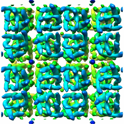
| Entry | Database: EMDB / ID: EMD-5802 | |||||||||
|---|---|---|---|---|---|---|---|---|---|---|
| Title | Electron crystallography of AQP0 with anionic phospholipids. | |||||||||
 Map data Map data | 3D reconstruction of AQP0_DMPG 2D crystal in p4212 form | |||||||||
 Sample Sample |
| |||||||||
 Keywords Keywords | aquaporin-0 / AQP0 / DMPG / phosphatidylglycerol / DMPS / phosphatidylserine / anionic phospholipids / electron crystallography | |||||||||
| Function / homology |  Function and homology information Function and homology informationgap junction-mediated intercellular transport / water transport / water channel activity / structural constituent of eye lens / gap junction / lens development in camera-type eye / positive regulation of cell adhesion / visual perception / protein homotetramerization / calmodulin binding ...gap junction-mediated intercellular transport / water transport / water channel activity / structural constituent of eye lens / gap junction / lens development in camera-type eye / positive regulation of cell adhesion / visual perception / protein homotetramerization / calmodulin binding / apical plasma membrane / endoplasmic reticulum / plasma membrane Similarity search - Function | |||||||||
| Biological species |  | |||||||||
| Method | electron crystallography / Resolution: 7.0 Å | |||||||||
 Authors Authors | Hite RK / Chiu P-L / Schuller J / Walz T | |||||||||
 Citation Citation | Journal: PLoS One / Year: 2015 Title: Effect of lipid head groups on double-layered two-dimensional crystals formed by aquaporin-0. Authors: Richard Kevin Hite / Po-Lin Chiu / Jan Michael Schuller / Thomas Walz /  Abstract: Aquaporin-0 (AQP0) is a lens-specific water channel that also forms membrane junctions. Reconstitution of AQP0 with dimyristoyl phosphatidylcholine (DMPC) and E. coli polar lipids (EPL) yielded well- ...Aquaporin-0 (AQP0) is a lens-specific water channel that also forms membrane junctions. Reconstitution of AQP0 with dimyristoyl phosphatidylcholine (DMPC) and E. coli polar lipids (EPL) yielded well-ordered, double-layered two-dimensional (2D) crystals that allowed electron crystallographic structure determination of the AQP0-mediated membrane junction. The interacting tetramers in the two crystalline layers are exactly in register, resulting in crystals with p422 symmetry. The high-resolution density maps also allowed modeling of the annular lipids surrounding the tetramers. Comparison of the DMPC and EPL bilayers suggested that the lipid head groups do not play an important role in the interaction of annular lipids with AQP0. We now reconstituted AQP0 with the anionic lipid dimyristoyl phosphatidylglycerol (DMPG), which yielded a mixture of 2D crystals with different symmetries. The different crystal symmetries result from shifts between the two crystalline layers, suggesting that the negatively charged PG head group destabilizes the interaction between the extracellular AQP0 surfaces. Reconstitution of AQP0 with dimyristoyl phosphatidylserine (DMPS), another anionic lipid, yielded crystals that had the usual p422 symmetry, but the crystals showed a pH-dependent tendency to stack through their cytoplasmic surfaces. Finally, AQP0 failed to reconstitute into membranes that were composed of more than 40% dimyristoyl phosphatidic acid (DMPA). Hence, although DMPG, DMPS, and DMPA are all negatively charged lipids, they have very different effects on AQP0 2D crystals, illustrating the importance of the specific lipid head group chemistry beyond its mere charge. | |||||||||
| History |
|
- Structure visualization
| Movie |
Movie viewer |
|---|---|
| Structure viewer | EM map: SurfViewMolmilJmol/JSmol |
| Supplemental images |
- Downloads & links
-EMDB archive
| Map data | emd_5802.map.gz | 57.4 MB | EMDB map data format | |
|---|---|---|---|---|
| Header (meta data) | emd-5802-v30.xmlemd-5802.xml | 10.6 KB 10.6 KB | Display Display | EMDB header |
| Images |  400_5802.gif 400_5802.gif 80_5802.gif 80_5802.gif | 117 KB 7.7 KB | ||
| Filedesc structureFactors | emd_5802_sf.cif.gz | 65.1 KB | ||
| Archive directory |  http://ftp.pdbj.org/pub/emdb/structures/EMD-5802ftp://ftp.pdbj.org/pub/emdb/structures/EMD-5802 http://ftp.pdbj.org/pub/emdb/structures/EMD-5802ftp://ftp.pdbj.org/pub/emdb/structures/EMD-5802 | HTTPS FTP |
-Validation report
| Summary document | emd_5802_validation.pdf.gz | 80.9 KB | Display | EMDB validaton report |
|---|---|---|---|---|
| Full document | emd_5802_full_validation.pdf.gz | 80 KB | Display | |
| Data in XML | emd_5802_validation.xml.gz | 494 B | Display | |
| Arichive directory | https://ftp.pdbj.org/pub/emdb/validation_reports/EMD-5802ftp://ftp.pdbj.org/pub/emdb/validation_reports/EMD-5802 | HTTPS FTP |
-Related structure data
-Links
| EMDB pages | EMDB (EBI/PDBe) / EMDataResource |
|---|---|
| Related items in Molecule of the Month |

-Map
| File | Download / File: emd_5802.map.gz / Format: CCP4 / Size: 120.4 MB / Type: IMAGE STORED AS FLOATING POINT NUMBER (4 BYTES) | ||||||||||||||||||||||||||||||||||||||||||||||||||||||||||||||||||||
|---|---|---|---|---|---|---|---|---|---|---|---|---|---|---|---|---|---|---|---|---|---|---|---|---|---|---|---|---|---|---|---|---|---|---|---|---|---|---|---|---|---|---|---|---|---|---|---|---|---|---|---|---|---|---|---|---|---|---|---|---|---|---|---|---|---|---|---|---|---|
| Annotation | 3D reconstruction of AQP0_DMPG 2D crystal in p4212 form | ||||||||||||||||||||||||||||||||||||||||||||||||||||||||||||||||||||
| Voxel size | X: 0.3275 Å / Y: 0.3275 Å / Z: 1 Å | ||||||||||||||||||||||||||||||||||||||||||||||||||||||||||||||||||||
| Density |
| ||||||||||||||||||||||||||||||||||||||||||||||||||||||||||||||||||||
| Symmetry | Space group: 90 | ||||||||||||||||||||||||||||||||||||||||||||||||||||||||||||||||||||
| Details | EMDB XML:
CCP4 map header:
| ||||||||||||||||||||||||||||||||||||||||||||||||||||||||||||||||||||
-Supplemental data
- Sample components
-Entire : 2D crystal structure of AQP0 (Ovis aries) with DMPG lipid in p421...
| Entire | Name: 2D crystal structure of AQP0 (Ovis aries) with DMPG lipid in p4212 form |
|---|---|
| Components |
|
-Supramolecule #1000: 2D crystal structure of AQP0 (Ovis aries) with DMPG lipid in p421...
| Supramolecule | Name: 2D crystal structure of AQP0 (Ovis aries) with DMPG lipid in p4212 form type: sample / ID: 1000 / Details: 2D crystals / Oligomeric state: Tetramer / Number unique components: 1 |
|---|---|
| Molecular weight | Theoretical: 113 KDa |
-Macromolecule #1: Aquaporin-0
| Macromolecule | Name: Aquaporin-0 / type: protein_or_peptide / ID: 1 / Name.synonym: AQP0 Details: Aquaporin-0 was crystallized with dimyristoylglycerol (DMPG) in p4212 symmetry. Number of copies: 32 / Oligomeric state: Tetramer / Recombinant expression: No / Database: NCBI |
|---|---|
| Source (natural) | Organism: |
| Sequence | UniProtKB: Lens fiber major intrinsic protein |
-Experimental details
-Structure determination
 Processing Processing | electron crystallography |
|---|---|
| Aggregation state | 2D array |
-Sample preparation
| Buffer | pH: 6 / Details: 10 mM MES, 50 mM Mg2Cl2, 150 mM NaCl, 0.05% NaN3 |
|---|---|
| Grid | Details: Carbon sandwich method combined with trehalose embedding |
| Vitrification | Cryogen name: NONE / Instrument: OTHER |
| Details | Dialysis |
| Crystal formation | Details: Dialysis |
- Electron microscopy
| Microscope | FEI POLARA 300 |
|---|---|
| Temperature | Min: 90 K / Max: 100 K / Average: 95 K |
| Date | Mar 31, 2012 |
| Image recording | Category: CCD / Film or detector model: GATAN ULTRASCAN 4000 (4k x 4k) / Digitization - Sampling interval: 15 µm / Number real images: 227 / Average electron dose: 20 e/Å2 |
| Electron beam | Acceleration voltage: 300 kV / Electron source:  FIELD EMISSION GUN FIELD EMISSION GUN |
| Electron optics | Calibrated magnification: 80000 / Illumination mode: FLOOD BEAM / Imaging mode: BRIGHT FIELD / Cs: 1.6 mm / Nominal defocus max: 0.65 µm / Nominal defocus min: 0.35 µm / Nominal magnification: 52000 |
| Sample stage | Specimen holder: liquid-nitrogen cooled / Specimen holder model: OTHER |
| Experimental equipment |  Model: Tecnai Polara / Image courtesy: FEI Company |
-Image processing
| Details | Crystal images were unbent and reconstructed using 2DX and MRC software. |
|---|---|
| Final reconstruction | Algorithm: OTHER / Resolution.type: BY AUTHOR / Resolution: 7.0 Å / Resolution method: DIFFRACTION PATTERN/LAYERLINES / Software - Name: 2DX, and, MRC / Details: The reconstruction map shows in 2x2 unit cells. |
| Crystal parameters | Unit cell - A: 65.5 Å / Unit cell - B: 65.5 Å / Unit cell - C: 200 Å / Unit cell - γ: 90.0 ° / Unit cell - α: 90.0 ° / Unit cell - β: 90.0 ° / Plane group: P 4 21 2 |
| CTF correction | Details: Phase flipping with each micrograph |
-Atomic model buiding 1
| Initial model | PDB ID: Chain - Chain ID: A |
|---|---|
| Software | Name: UCSF Chimera |
| Refinement | Space: REAL / Protocol: RIGID BODY FIT |

