 Movie
Movie Controller

[English] 日本語
 Yorodumi
- EMDB-5780: Cryo-EM structure of the BG505 SOSIP.664 HIV-1 Env trimer with 2 ... -
+ Open data

- Basic information
| Entry | Database: EMDB / ID: EMD-5780 | |||||||||
|---|---|---|---|---|---|---|---|---|---|---|
| Title | Cryo-EM structure of the BG505 SOSIP.664 HIV-1 Env trimer with 2 PGV04 Fabs | |||||||||
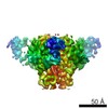
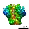
 Map data Map data | Reconstruction of the HIV-1 BG505 SOSIP.664 Envelope trimer with 2 broadly neutralizing PGV04 Fabs | |||||||||
 Sample Sample |
| |||||||||
 Keywords Keywords | HIV-1 trimeric spike / gp140 / SOSIP / broadly neutralizing antibody / PGV04 / Env / Envelope glycoprotein | |||||||||
| Biological species |   Human immunodeficiency virus 1 / Human immunodeficiency virus 1 /  Homo sapiens (human) Homo sapiens (human) | |||||||||
| Method | single particle reconstruction / cryo EM / Resolution: 7.9 Å | |||||||||
 Authors Authors | Lyumkis D / Julien JP / de Val N / Cupo A / Potter CS / Klasse PJ / Burton DR / Sanders RW / Moore JP / Carragher B ...Lyumkis D / Julien JP / de Val N / Cupo A / Potter CS / Klasse PJ / Burton DR / Sanders RW / Moore JP / Carragher B / Wilson IA / Ward AB | |||||||||
 Citation Citation | Journal: Science / Year: 2013 Title: Cryo-EM structure of a fully glycosylated soluble cleaved HIV-1 envelope trimer. Authors: Dmitry Lyumkis / Jean-Philippe Julien / Natalia de Val / Albert Cupo / Clinton S Potter / Per-Johan Klasse / Dennis R Burton / Rogier W Sanders / John P Moore / Bridget Carragher / Ian A ...Authors: Dmitry Lyumkis / Jean-Philippe Julien / Natalia de Val / Albert Cupo / Clinton S Potter / Per-Johan Klasse / Dennis R Burton / Rogier W Sanders / John P Moore / Bridget Carragher / Ian A Wilson / Andrew B Ward /  Abstract: The HIV-1 envelope glycoprotein (Env) trimer contains the receptor binding sites and membrane fusion machinery that introduce the viral genome into the host cell. As the only target for broadly ...The HIV-1 envelope glycoprotein (Env) trimer contains the receptor binding sites and membrane fusion machinery that introduce the viral genome into the host cell. As the only target for broadly neutralizing antibodies (bnAbs), Env is a focus for rational vaccine design. We present a cryo-electron microscopy reconstruction and structural model of a cleaved, soluble Env trimer (termed BG505 SOSIP.664 gp140) in complex with a CD4 binding site (CD4bs) bnAb, PGV04, at 5.8 angstrom resolution. The structure reveals the spatial arrangement of Env components, including the V1/V2, V3, HR1, and HR2 domains, as well as shielding glycans. The structure also provides insights into trimer assembly, gp120-gp41 interactions, and the CD4bs epitope cluster for bnAbs, which covers a more extensive area and defines a more complex site of vulnerability than previously described. | |||||||||
| History |
|
- Structure visualization
| Movie |
 Movie viewer Movie viewer |
|---|---|
| Structure viewer | EM map: SurfViewMolmilJmol/JSmol |
| Supplemental images |

- Downloads & links
-EMDB archive
| Map data | emd_5780.map.gz | 57.3 MB | EMDB map data format | |
|---|---|---|---|---|
| Header (meta data) | emd-5780-v30.xmlemd-5780.xml | 12.4 KB 12.4 KB | Display Display | EMDB header |
| Images | 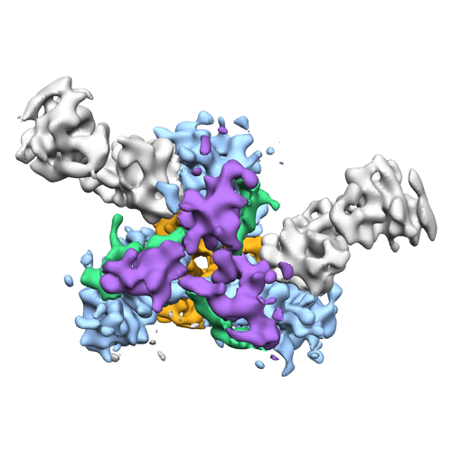 emd_5780_1.png emd_5780_1.png | 137.3 KB | ||
| Archive directory |  http://ftp.pdbj.org/pub/emdb/structures/EMD-5780ftp://ftp.pdbj.org/pub/emdb/structures/EMD-5780 http://ftp.pdbj.org/pub/emdb/structures/EMD-5780ftp://ftp.pdbj.org/pub/emdb/structures/EMD-5780 | HTTPS FTP |
-Validation report
| Summary document | emd_5780_validation.pdf.gz | 78.5 KB | Display | EMDB validaton report |
|---|---|---|---|---|
| Full document | emd_5780_full_validation.pdf.gz | 77.6 KB | Display | |
| Data in XML | emd_5780_validation.xml.gz | 494 B | Display | |
| Arichive directory | https://ftp.pdbj.org/pub/emdb/validation_reports/EMD-5780ftp://ftp.pdbj.org/pub/emdb/validation_reports/EMD-5780 | HTTPS FTP |
-Related structure data


-Links
| EMDB pages | EMDB (EBI/PDBe) / EMDataResource |
|---|
-Map
| File | Download / File: emd_5780.map.gz / Format: CCP4 / Size: 62.5 MB / Type: IMAGE STORED AS FLOATING POINT NUMBER (4 BYTES) | ||||||||||||||||||||||||||||||||||||||||||||||||||||||||||||||||||||
|---|---|---|---|---|---|---|---|---|---|---|---|---|---|---|---|---|---|---|---|---|---|---|---|---|---|---|---|---|---|---|---|---|---|---|---|---|---|---|---|---|---|---|---|---|---|---|---|---|---|---|---|---|---|---|---|---|---|---|---|---|---|---|---|---|---|---|---|---|---|
| Annotation | Reconstruction of the HIV-1 BG505 SOSIP.664 Envelope trimer with 2 broadly neutralizing PGV04 Fabs | ||||||||||||||||||||||||||||||||||||||||||||||||||||||||||||||||||||
| Voxel size | X=Y=Z: 1.21 Å | ||||||||||||||||||||||||||||||||||||||||||||||||||||||||||||||||||||
| Density |
| ||||||||||||||||||||||||||||||||||||||||||||||||||||||||||||||||||||
| Symmetry | Space group: 1 | ||||||||||||||||||||||||||||||||||||||||||||||||||||||||||||||||||||
| Details | EMDB XML:
CCP4 map header:
| ||||||||||||||||||||||||||||||||||||||||||||||||||||||||||||||||||||
-Supplemental data
- Sample components
-Entire : Fully glycosylated BG505 SOSIP.664 Envelope trimer with 2 broadly...
| Entire | Name: Fully glycosylated BG505 SOSIP.664 Envelope trimer with 2 broadly neutralizing PGV04 Fabs |
|---|---|
| Components |
|
-Supramolecule #1000: Fully glycosylated BG505 SOSIP.664 Envelope trimer with 2 broadly...
| Supramolecule | Name: Fully glycosylated BG505 SOSIP.664 Envelope trimer with 2 broadly neutralizing PGV04 Fabs type: sample / ID: 1000 Oligomeric state: three SOSIP.664 gp140 subunits (trimeric HIV-1 spike) with 2 PGV04 Fabs Number unique components: 2 |
|---|---|
| Molecular weight | Theoretical: 550 KDa |
-Macromolecule #1: BG505 SOSIP.664 HIV-1 Envelope glycoprotein gp140
| Macromolecule | Name: BG505 SOSIP.664 HIV-1 Envelope glycoprotein gp140 / type: protein_or_peptide / ID: 1 / Name.synonym: Env / Number of copies: 3 / Oligomeric state: trimer / Recombinant expression: Yes |
|---|---|
| Source (natural) | Organism: Human immunodeficiency virus 1 / Strain: BG505.W6M.ENV.A5 / synonym: human immunodeficiency virus type I |
| Molecular weight | Theoretical: 150 KDa |
| Recombinant expression | Organism: Homo sapiens (human) / Recombinant cell: HEK 293T |
-Macromolecule #2: Fragment antigen binding
| Macromolecule | Name: Fragment antigen binding / type: protein_or_peptide / ID: 2 / Name.synonym: Fab / Number of copies: 2 / Oligomeric state: heterodimer / Recombinant expression: Yes |
|---|---|
| Source (natural) | Organism: Homo sapiens (human) / synonym: Human |
| Molecular weight | Experimental: 50 KDa / Theoretical: 50 KDa |
| Recombinant expression | Organism: Homo sapiens (human) / Recombinant cell: HEK 293F |
-Experimental details
-Structure determination
| Method | cryo EM |
|---|---|
 Processing Processing | single particle reconstruction |
| Aggregation state | particle |
-Sample preparation
| Concentration | 0.72 mg/mL |
|---|---|
| Buffer | pH: 7.6 / Details: 20 mM Tris, 150 mM NaCl, 0.085 mM DDM |
| Grid | Details: 400 mesh C-Flat CF-22-4C, plasma treated for 5 seconds |
| Vitrification | Cryogen name: ETHANE / Instrument: HOMEMADE PLUNGER Method: Specimen was prepared for cryo-EM by applying 3 microliters of sample to a freshly plasma cleaned holey carbon C-flat grid (Protochips, Inc.), allowing the sample to adsorb to the grid for 30 ...Method: Specimen was prepared for cryo-EM by applying 3 microliters of sample to a freshly plasma cleaned holey carbon C-flat grid (Protochips, Inc.), allowing the sample to adsorb to the grid for 30 seconds, followed by blotting with a small piece of filter paper and plunge-freezing into liquid ethane using a manual cryo-plunger in an ambient environment (4 degrees C). |
- Electron microscopy
| Microscope | FEI TECNAI F20 |
|---|---|
| Alignment procedure | Legacy - Astigmatism: objective lens astigmatism was corrected by observing Thon rings with the Leginon software |
| Details | electron counting mode |
| Date | Feb 21, 2013 |
| Image recording | Category: CCD / Film or detector model: GATAN K2 (4k x 4k) / Digitization - Sampling interval: 5.0 µm / Number real images: 6355 / Average electron dose: 32 e/Å2 Details: The dose was fractionated over 20 raw frames collected over a 5-second exposure time (250 ms per frame) on the Gatan K2 Summit direct detection device, with each frame receiving a dose of ~9. ...Details: The dose was fractionated over 20 raw frames collected over a 5-second exposure time (250 ms per frame) on the Gatan K2 Summit direct detection device, with each frame receiving a dose of ~9.4 e-/pixel/sec. The individual frames were aligned using a GPU-enabled frame-alignment program that was generously provided by Yifan Cheng and Xueming Li. This program was used to track the global shifts between individual frames. |
| Tilt angle min | 0 |
| Electron beam | Acceleration voltage: 200 kV / Electron source:  FIELD EMISSION GUN FIELD EMISSION GUN |
| Electron optics | Calibrated magnification: 29000 / Illumination mode: FLOOD BEAM / Imaging mode: BRIGHT FIELD / Cs: 2 mm / Nominal defocus max: 5.0 µm / Nominal defocus min: 1.0 µm / Nominal magnification: 29000 |
| Sample stage | Specimen holder model: SIDE ENTRY, EUCENTRIC |
| Experimental equipment |  Model: Tecnai F20 / Image courtesy: FEI Company |
-Image processing
| Details | Reconstructed using resolution-limited refinement procedure implemented in Xmipp and Frealign. |
|---|---|
| CTF correction | Details: Frealign |
| Final reconstruction | Algorithm: OTHER / Resolution.type: BY AUTHOR / Resolution: 7.9 Å / Resolution method: FSC 0.143 CUT-OFF / Software - Name: Xmipp, Frealign Details: Final maps were calculated after sorting for the presence of sub-stoichiometrically labeled trimers. Number images used: 40817 |
