Movie
Movie Controller


+ Open data

- Basic information
| Entry | Database: EMDB / ID: EMD-5738 | |||||||||
|---|---|---|---|---|---|---|---|---|---|---|
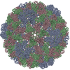
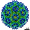
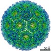
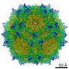
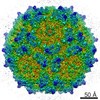
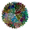
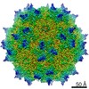
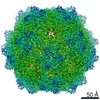
| Title | Electron cryo-microscopy of 2H15BMV | |||||||||
 Map data Map data | reconstruction of BMV 2H mutant | |||||||||
 Sample Sample |
| |||||||||
 Keywords Keywords |  Brome Mosaic Virus / protein / capsid / RNA encapsidation / electrostatic interaction Brome Mosaic Virus / protein / capsid / RNA encapsidation / electrostatic interaction | |||||||||
| Biological species |  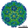 Brome mosaic virus Brome mosaic virus | |||||||||
| Method | single particle reconstruction / cryo EM / negative staining / Resolution: 5.0 Å | |||||||||
 Authors Authors | Ni P / Wang Z / Ma X / Das NC / Sokol P / Chiu W / Dragnea B / Hagan M / Kao CC | |||||||||
 Citation Citation | Journal: J Mol Biol / Year: 2012 Title: An examination of the electrostatic interactions between the N-terminal tail of the Brome Mosaic Virus coat protein and encapsidated RNAs. Authors: Peng Ni / Zhao Wang / Xiang Ma / Nayaran Chandra Das / Paul Sokol / Wah Chiu / Bogdan Dragnea / Michael Hagan / C Cheng Kao /  Abstract: The coat protein of positive-stranded RNA viruses often contains a positively charged tail that extends toward the center of the capsid and interacts with the viral genome. Electrostatic interaction ...The coat protein of positive-stranded RNA viruses often contains a positively charged tail that extends toward the center of the capsid and interacts with the viral genome. Electrostatic interaction between the tail and the RNA has been postulated as a major force in virus assembly and stabilization. The goal of this work is to examine the correlation between electrostatic interaction and amount of RNA packaged in the tripartite Brome Mosaic Virus (BMV). Nanoindentation experiment using atomic force microscopy showed that the stiffness of BMV virions with different RNAs varied by a range that is 10-fold higher than that would be predicted by electrostatics. BMV mutants with decreased positive charges encapsidated lower amounts of RNA while mutants with increased positive charges packaged additional RNAs up to ∼900 nt. However, the extra RNAs included truncated BMV RNAs, an additional copy of RNA4, potential cellular RNAs, or a combination of the three, indicating that change in the charge of the capsid could result in several different outcomes in RNA encapsidation. In addition, mutant with specific arginines changed to lysines in the capsid also exhibited defects in the specific encapsidation of BMV RNA4. The experimental results indicate that electrostatics is a major component in RNA encapsidation but was unable to account for all of the observed effects on RNA encapsidation. Thermodynamic modeling incorporating the electrostatics was able to predict the approximate length of the RNA to be encapsidated for the majority of mutant virions, but not for a mutant with extreme clustered positive charges. Cryo-electron microscopy of virions that encapsidated an additional copy of RNA4 revealed that, despite the increase in RNA encapsidated, the capsid structure was minimally changed. These results experimentally demonstrated the impact of electrostatics and additional restraints in the encapsidation of BMV RNAs, which could be applicable to other viruses. | |||||||||
| History |
|
- Structure visualization
| Movie |
 Movie viewer Movie viewer |
|---|---|
| Structure viewer | EM map: SurfViewMolmilJmol/JSmol |
| Supplemental images |

- Downloads & links
-EMDB archive
| Map data | emd_5738.map.gz | 58.3 MB | EMDB map data format | |
|---|---|---|---|---|
| Header (meta data) | emd-5738-v30.xmlemd-5738.xml | 9.4 KB 9.4 KB | Display Display | EMDB header |
| Images |  400_5738.gif 400_5738.gif 80_5738.gif 80_5738.gif | 59.9 KB 4.6 KB | ||
| Archive directory |  http://ftp.pdbj.org/pub/emdb/structures/EMD-5738ftp://ftp.pdbj.org/pub/emdb/structures/EMD-5738 http://ftp.pdbj.org/pub/emdb/structures/EMD-5738ftp://ftp.pdbj.org/pub/emdb/structures/EMD-5738 | HTTPS FTP |
-Related structure data
| Similar structure data |
|---|

-Links
| EMDB pages | EMDB (EBI/PDBe) / EMDataResource |
|---|
-Map
| File | Download / File: emd_5738.map.gz / Format: CCP4 / Size: 62.5 MB / Type: IMAGE STORED AS FLOATING POINT NUMBER (4 BYTES) | ||||||||||||||||||||||||||||||||||||||||||||||||||||||||||||||||||||
|---|---|---|---|---|---|---|---|---|---|---|---|---|---|---|---|---|---|---|---|---|---|---|---|---|---|---|---|---|---|---|---|---|---|---|---|---|---|---|---|---|---|---|---|---|---|---|---|---|---|---|---|---|---|---|---|---|---|---|---|---|---|---|---|---|---|---|---|---|---|
| Annotation | reconstruction of BMV 2H mutant | ||||||||||||||||||||||||||||||||||||||||||||||||||||||||||||||||||||
| Voxel size | X=Y=Z: 1.3 Å | ||||||||||||||||||||||||||||||||||||||||||||||||||||||||||||||||||||
| Density |
| ||||||||||||||||||||||||||||||||||||||||||||||||||||||||||||||||||||
| Symmetry | Space group: 1 | ||||||||||||||||||||||||||||||||||||||||||||||||||||||||||||||||||||
| Details | EMDB XML:
CCP4 map header:
| ||||||||||||||||||||||||||||||||||||||||||||||||||||||||||||||||||||
-Supplemental data
- Sample components
-Entire : 2H15 BMV
| Entire | Name: 2H15 BMV |
|---|---|
| Components |
|
-Supramolecule #1000: 2H15 BMV
| Supramolecule | Name: 2H15 BMV / type: sample / ID: 1000 / Number unique components: 1 |
|---|---|
| Molecular weight | Experimental: 4.6 MDa |
-Supramolecule #1: Brome mosaic virus
| Supramolecule | Name: Brome mosaic virus / type: virus / ID: 1 / NCBI-ID: 12302 / Sci species name: Brome mosaic virus / Database: NCBI / Virus type: VIRION / Virus isolate: SPECIES / Virus enveloped: No / Virus empty: No |
|---|---|
| Host (natural) | Organism:  Nicotiana benthamiana (plant) / synonym: PLANTAE(HIGHER PLANTS) Nicotiana benthamiana (plant) / synonym: PLANTAE(HIGHER PLANTS) |
| Molecular weight | Experimental: 4.6 MDa |
-Experimental details
-Structure determination
| Method | negative staining, cryo EM |
|---|---|
 Processing Processing | single particle reconstruction |
| Aggregation state | particle |
-Sample preparation
| Concentration | 1 mg/mL |
|---|---|
| Buffer | pH: 5.2 Details: virus 928 buffer II (50 mM NaOAc, 10 mM MgCl2, pH 5.2), 50% w/v CsCl |
| Staining | Type: NEGATIVE / Details: 1.2/1.3 Quantifoil grid |
| Vitrification | Cryogen name: ETHANE / Chamber humidity: 100 % / Chamber temperature: 20 K / Instrument: FEI VITROBOT MARK IV / Method: Blot for 2 seconds. |
- Electron microscopy
| Microscope | JEOL 3200FSC |
|---|---|
| Electron beam | Acceleration voltage: 300 kV / Electron source: FIELD EMISSION GUN |
| Electron optics | Illumination mode: OTHER / Imaging mode: BRIGHT FIELDBright-field microscopy / Nominal magnification: 80000 |
| Sample stage | Specimen holder model: JEOL 3200FSC CRYOHOLDER |
| Date | Sep 11, 2009 |
| Image recording | Category: CCD / Film or detector model: GENERIC GATAN (4k x 4k) / Number real images: 471 |
-Image processing
| CTF correction | Details: each image |
|---|---|
| Final reconstruction | Resolution.type: BY AUTHOR / Resolution: 5.0 Å / Resolution method: FSC 0.5 CUT-OFF / Software - Name: mpsa, EMAN1.8 / Number images used: 11576 |
| Details | Particles were selected from electron micrographs using the EMAN Boxer routine. The particles were translationally and rotationally aligned, classified, and averaged without applying symmetry using the EMAN Refine2d command. Initial model for reconstruction: approximately 3000 particles were selected by MPSA (Multi-Path Simulated Annealing) to build a subnanometer model of 2H15. The map was then refined using EMAN. |
-Atomic model buiding 1
| Initial model | PDB ID: |
|---|---|
| Refinement | Space: REAL / Protocol: RIGID BODY FIT |