 Movie
Movie Controller

[English] 日本語
 Yorodumi
- EMDB-5624: Broadly Neutralizing Antibody PGT121 Allosterically Modulates CD4... -
+ Open data

- Basic information
| Entry | Database: EMDB / ID: EMD-5624 | |||||||||
|---|---|---|---|---|---|---|---|---|---|---|
| Title | Broadly Neutralizing Antibody PGT121 Allosterically Modulates CD4 Binding via Recognition of the HIV-1 gp120 V3 Base and Multiple Surrounding Glycans | |||||||||
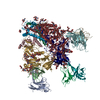
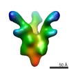
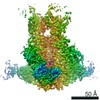
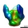
 Map data Map data | Reconstruction of SOSIP.664 HIV-1 envelope trimer in complex with broadly neutralizing PGT122 Fab | |||||||||
 Sample Sample |
| |||||||||
 Keywords Keywords | Single particle analysis / uranyl formate / tilt series / antibody / HIV gp120 trimer / Fab | |||||||||
| Biological species |   Human immunodeficiency virus 1 Human immunodeficiency virus 1 | |||||||||
| Method | single particle reconstruction / negative staining / Resolution: 14.0 Å | |||||||||
 Authors Authors | Khayat R / Lee JH / Julien JP / Wilson IA / Ward AB | |||||||||
 Citation Citation | Journal: PLoS Pathog / Year: 2013 Title: Broadly neutralizing antibody PGT121 allosterically modulates CD4 binding via recognition of the HIV-1 gp120 V3 base and multiple surrounding glycans. Authors: Jean-Philippe Julien / Devin Sok / Reza Khayat / Jeong Hyun Lee / Katie J Doores / Laura M Walker / Alejandra Ramos / Devan C Diwanji / Robert Pejchal / Albert Cupo / Umesh Katpally / Rafael ...Authors: Jean-Philippe Julien / Devin Sok / Reza Khayat / Jeong Hyun Lee / Katie J Doores / Laura M Walker / Alejandra Ramos / Devan C Diwanji / Robert Pejchal / Albert Cupo / Umesh Katpally / Rafael S Depetris / Robyn L Stanfield / Ryan McBride / Andre J Marozsan / James C Paulson / Rogier W Sanders / John P Moore / Dennis R Burton / Pascal Poignard / Andrew B Ward / Ian A Wilson /  Abstract: New broad and potent neutralizing HIV-1 antibodies have recently been described that are largely dependent on the gp120 N332 glycan for Env recognition. Members of the PGT121 family of antibodies, ...New broad and potent neutralizing HIV-1 antibodies have recently been described that are largely dependent on the gp120 N332 glycan for Env recognition. Members of the PGT121 family of antibodies, isolated from an African donor, neutralize ∼70% of circulating isolates with a median IC50 less than 0.05 µg ml(-1). Here, we show that three family members, PGT121, PGT122 and PGT123, have very similar crystal structures. A long 24-residue HCDR3 divides the antibody binding site into two functional surfaces, consisting of an open face, formed by the heavy chain CDRs, and an elongated face, formed by LCDR1, LCDR3 and the tip of the HCDR3. Alanine scanning mutagenesis of the antibody paratope reveals a crucial role in neutralization for residues on the elongated face, whereas the open face, which accommodates a complex biantennary glycan in the PGT121 structure, appears to play a more secondary role. Negative-stain EM reconstructions of an engineered recombinant Env gp140 trimer (SOSIP.664) reveal that PGT122 interacts with the gp120 outer domain at a more vertical angle with respect to the top surface of the spike than the previously characterized antibody PGT128, which is also dependent on the N332 glycan. We then used ITC and FACS to demonstrate that the PGT121 antibodies inhibit CD4 binding to gp120 despite the epitope being distal from the CD4 binding site. Together, these structural, functional and biophysical results suggest that the PGT121 antibodies may interfere with Env receptor engagement by an allosteric mechanism in which key structural elements, such as the V3 base, the N332 oligomannose glycan and surrounding glycans, including a putative V1/V2 complex biantennary glycan, are conformationally constrained. | |||||||||
| History |
|
- Structure visualization
| Movie |
 Movie viewer Movie viewer |
|---|---|
| Structure viewer | EM map: SurfViewMolmilJmol/JSmol |
| Supplemental images |

- Downloads & links
-EMDB archive
| Map data | emd_5624.map.gz | 11.5 MB | EMDB map data format | |
|---|---|---|---|---|
| Header (meta data) | emd-5624-v30.xmlemd-5624.xml | 12.7 KB 12.7 KB | Display Display | EMDB header |
| Images | 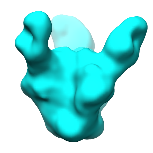 emd_5624_1.png emd_5624_1.png | 70.4 KB | ||
| Archive directory |  http://ftp.pdbj.org/pub/emdb/structures/EMD-5624ftp://ftp.pdbj.org/pub/emdb/structures/EMD-5624 http://ftp.pdbj.org/pub/emdb/structures/EMD-5624ftp://ftp.pdbj.org/pub/emdb/structures/EMD-5624 | HTTPS FTP |
-Validation report
| Summary document | emd_5624_validation.pdf.gz | 78.4 KB | Display | EMDB validaton report |
|---|---|---|---|---|
| Full document | emd_5624_full_validation.pdf.gz | 77.5 KB | Display | |
| Data in XML | emd_5624_validation.xml.gz | 494 B | Display | |
| Arichive directory | https://ftp.pdbj.org/pub/emdb/validation_reports/EMD-5624ftp://ftp.pdbj.org/pub/emdb/validation_reports/EMD-5624 | HTTPS FTP |
-Related structure data


-Links
| EMDB pages | EMDB (EBI/PDBe) / EMDataResource |
|---|
-Map
| File | Download / File: emd_5624.map.gz / Format: CCP4 / Size: 15.3 MB / Type: IMAGE STORED AS FLOATING POINT NUMBER (4 BYTES) | ||||||||||||||||||||||||||||||||||||||||||||||||||||||||||||||||||||
|---|---|---|---|---|---|---|---|---|---|---|---|---|---|---|---|---|---|---|---|---|---|---|---|---|---|---|---|---|---|---|---|---|---|---|---|---|---|---|---|---|---|---|---|---|---|---|---|---|---|---|---|---|---|---|---|---|---|---|---|---|---|---|---|---|---|---|---|---|---|
| Annotation | Reconstruction of SOSIP.664 HIV-1 envelope trimer in complex with broadly neutralizing PGT122 Fab | ||||||||||||||||||||||||||||||||||||||||||||||||||||||||||||||||||||
| Voxel size | X=Y=Z: 2.18 Å | ||||||||||||||||||||||||||||||||||||||||||||||||||||||||||||||||||||
| Density |
| ||||||||||||||||||||||||||||||||||||||||||||||||||||||||||||||||||||
| Symmetry | Space group: 1 | ||||||||||||||||||||||||||||||||||||||||||||||||||||||||||||||||||||
| Details | EMDB XML:
CCP4 map header:
| ||||||||||||||||||||||||||||||||||||||||||||||||||||||||||||||||||||
-Supplemental data
- Sample components
-Entire : Fab fragment of broadly neutralizing antibody PGT122 in complex w...
| Entire | Name: Fab fragment of broadly neutralizing antibody PGT122 in complex with HIV-1 SOSIP.664 from BG505 |
|---|---|
| Components |
|
-Supramolecule #1000: Fab fragment of broadly neutralizing antibody PGT122 in complex w...
| Supramolecule | Name: Fab fragment of broadly neutralizing antibody PGT122 in complex with HIV-1 SOSIP.664 from BG505 type: sample / ID: 1000 / Details: The sample was monodisperse / Oligomeric state: one SOSIP.664 trimer binds 3 PGT122 Fabs / Number unique components: 2 |
|---|---|
| Molecular weight | Experimental: 350 KDa / Theoretical: 350 KDa / Method: SDS-PAGE |
-Macromolecule #1: BG505 HIV-1 Env SOSIP.664
| Macromolecule | Name: BG505 HIV-1 Env SOSIP.664 / type: protein_or_peptide / ID: 1 / Details: Bound to Fab portion of PGT122 antibody / Number of copies: 3 / Oligomeric state: Trimer / Recombinant expression: Yes |
|---|---|
| Source (natural) | Organism: Human immunodeficiency virus 1 / Strain: BG505 / synonym: HIV-1 |
| Molecular weight | Experimental: 220 KDa / Theoretical: 220 KDa |
| Recombinant expression | Organism:  Homo sapiens (human) / Recombinant cell: HEK 293S GnT I-deficient cells Homo sapiens (human) / Recombinant cell: HEK 293S GnT I-deficient cells |
-Experimental details
-Structure determination
| Method | negative staining |
|---|---|
 Processing Processing | single particle reconstruction |
| Aggregation state | particle |
-Sample preparation
| Concentration | 0.1 mg/mL |
|---|---|
| Buffer | pH: 7 / Details: 20 mM Tris-HCl, pH 7.0, 50 mM NaCl |
| Staining | Type: NEGATIVE Details: Grids were briefly adsorbed with protein, wicked, and stained with 2% Nano-W for 30 seconds. |
| Grid | Details: 400 Cu mesh grid with thin carob support, glow discharged in natural atmosphere. |
| Vitrification | Cryogen name: NONE / Instrument: OTHER |
- Electron microscopy
| Microscope | FEI TECNAI F20 |
|---|---|
| Temperature | Min: 293 K / Max: 294 K / Average: 293 K |
| Alignment procedure | Legacy - Astigmatism: Astigmatism of objective lens was corrected at 100,000x Legacy - Electron beam tilt params: -2 |
| Date | Apr 30, 2012 |
| Image recording | Category: CCD / Film or detector model: GENERIC GATAN (4k x 4k) / Digitization - Sampling interval: 0.109 µm / Number real images: 340 / Average electron dose: 16 e/Å2 |
| Tilt angle min | 0 |
| Electron beam | Acceleration voltage: 120 kV / Electron source:  FIELD EMISSION GUN FIELD EMISSION GUN |
| Electron optics | Calibrated magnification: 100000 / Illumination mode: FLOOD BEAM / Imaging mode: BRIGHT FIELD / Nominal defocus max: 0.72 µm / Nominal defocus min: 0.6 µm / Nominal magnification: 100000 |
| Sample stage | Specimen holder model: SIDE ENTRY, EUCENTRIC / Tilt angle max: 55 |
| Experimental equipment |  Model: Tecnai F20 / Image courtesy: FEI Company |
