 Movie
Movie Controller

[English] 日本語
 Yorodumi
- EMDB-5560: CryoEM of Poliovirus Polymerase Tube, including Ghost Reflections -
+ Open data

- Basic information
| Entry | Database: EMDB / ID: EMD-5560 | |||||||||
|---|---|---|---|---|---|---|---|---|---|---|
| Title | CryoEM of Poliovirus Polymerase Tube, including Ghost Reflections | |||||||||
 Map data Map data | Helical reconstruction of a tube of poliovirus polymerase, including ghost reflections | |||||||||
 Sample Sample |
| |||||||||
 Keywords Keywords | poliovirus polymerase /  viral replication viral replication | |||||||||
| Biological species |   Human poliovirus 1 Mahoney Human poliovirus 1 Mahoney | |||||||||
| Method | helical reconstruction / cryo EM / Resolution: 16.0 Å | |||||||||
 Authors Authors | Wang J / Bullitt E | |||||||||
 Citation Citation | Journal: J Mol Biol / Year: 2013 Title: Surface for catalysis by poliovirus RNA-dependent RNA polymerase. Authors: Jing Wang / John M Lyle / Esther Bullitt /  Abstract: The poliovirus RNA-dependent RNA polymerase, 3Dpol, replicates the viral genomic RNA on the surface of virus-induced intracellular membranes. Macromolecular assemblies of 3Dpol form linear arrays of ...The poliovirus RNA-dependent RNA polymerase, 3Dpol, replicates the viral genomic RNA on the surface of virus-induced intracellular membranes. Macromolecular assemblies of 3Dpol form linear arrays of subunits that propagate along a strong protein-protein interaction called interface-I, as was observed in the crystal structure of wild-type poliovirus polymerase. These "filaments" recur with slight modifications in planar sheets and, with additional modifications that accommodate curvature, in helical tubes of the polymerase, by packing filaments together via a second set of interactions. Periodic variations of subunit orientations within 3Dpol tubes give rise to "ghost reflections" in diffraction patterns computed from electron cryomicrographs of helical arrays. The ghost reflections reveal that polymerase tubes are formed by bundles of four to five interface-I filaments, which are then connected to the next bundle of filaments with a perturbation of interface interactions between bundles. While enzymatically inactive polymerase is also capable of oligomerization, much thinner tubes that lack interface-I interactions between adjacent subunits are formed, suggesting that long-range allostery produces conformational changes that extend from the active site to the protein-protein interface. Macromolecular assemblies of poliovirus polymerase show repeated use of flexible interface interactions for polymerase lattice formation, suggesting that adaptability of polymerase-polymerase interactions facilitates RNA replication. In addition, the presence of a positively charged groove identified in polymerase arrays may help position and stabilize the RNA template during replication. | |||||||||
| History |
|
- Structure visualization
| Movie |
 Movie viewer Movie viewer |
|---|---|
| Structure viewer | EM map: SurfViewMolmilJmol/JSmol |
| Supplemental images |

- Downloads & links
-EMDB archive
| Map data | emd_5560.map.gz | 2.7 MB | EMDB map data format | |
|---|---|---|---|---|
| Header (meta data) | emd-5560-v30.xmlemd-5560.xml | 8.3 KB 8.3 KB | Display Display | EMDB header |
| Images | 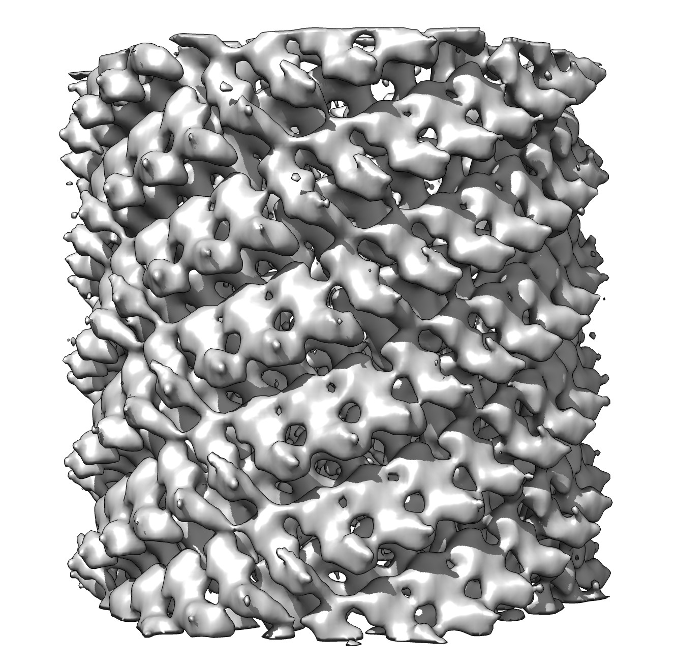 emd_5560.jpg emd_5560.jpg | 326.6 KB | ||
| Archive directory |  http://ftp.pdbj.org/pub/emdb/structures/EMD-5560ftp://ftp.pdbj.org/pub/emdb/structures/EMD-5560 http://ftp.pdbj.org/pub/emdb/structures/EMD-5560ftp://ftp.pdbj.org/pub/emdb/structures/EMD-5560 | HTTPS FTP |
-Related structure data
-Links
| EMDB pages | EMDB (EBI/PDBe) / EMDataResource |
|---|
-Map
| File | Download / File: emd_5560.map.gz / Format: CCP4 / Size: 13.9 MB / Type: IMAGE STORED AS FLOATING POINT NUMBER (4 BYTES) | ||||||||||||||||||||||||||||||||||||||||||||||||||||||||||||||||||||
|---|---|---|---|---|---|---|---|---|---|---|---|---|---|---|---|---|---|---|---|---|---|---|---|---|---|---|---|---|---|---|---|---|---|---|---|---|---|---|---|---|---|---|---|---|---|---|---|---|---|---|---|---|---|---|---|---|---|---|---|---|---|---|---|---|---|---|---|---|---|
| Annotation | Helical reconstruction of a tube of poliovirus polymerase, including ghost reflections | ||||||||||||||||||||||||||||||||||||||||||||||||||||||||||||||||||||
| Voxel size | X=Y=Z: 3.4 Å | ||||||||||||||||||||||||||||||||||||||||||||||||||||||||||||||||||||
| Density |
| ||||||||||||||||||||||||||||||||||||||||||||||||||||||||||||||||||||
| Symmetry | Space group: 1 | ||||||||||||||||||||||||||||||||||||||||||||||||||||||||||||||||||||
| Details | EMDB XML:
CCP4 map header:
| ||||||||||||||||||||||||||||||||||||||||||||||||||||||||||||||||||||
-Supplemental data
- Sample components
-Entire : Oligomer of poliovirus polymerase; helical structure with ghost r...
| Entire | Name: Oligomer of poliovirus polymerase; helical structure with ghost reflections included |
|---|---|
| Components |
|
-Supramolecule #1000: Oligomer of poliovirus polymerase; helical structure with ghost r...
| Supramolecule | Name: Oligomer of poliovirus polymerase; helical structure with ghost reflections included type: sample / ID: 1000 / Oligomeric state: oligomer of 331 components / Number unique components: 1 |
|---|---|
| Molecular weight | Theoretical: 17.4 MDa / Method: 52.5 kD monomer |
-Macromolecule #1: poliovirus polymerase
| Macromolecule | Name: poliovirus polymerase / type: protein_or_peptide / ID: 1 / Name.synonym: 3Dpol Details: polymerase was oligomerized by incubation at 30C followed by incubation at 4C. Number of copies: 331 / Oligomeric state: oligomer / Recombinant expression: Yes |
|---|---|
| Source (natural) | Organism: Human poliovirus 1 Mahoney / Strain: Mahoney / synonym: poliovirus |
| Molecular weight | Theoretical: 17.4 MDa |
| Recombinant expression | Organism:  Escherichia coli (E. coli) / Recombinant plasmid: pT5T-3D Escherichia coli (E. coli) / Recombinant plasmid: pT5T-3D |
-Experimental details
-Structure determination
| Method | cryo EM |
|---|---|
 Processing Processing | helical reconstruction |
| Aggregation state | filament |
-Sample preparation
| Grid | Details: Quantifoil grids |
|---|---|
| Vitrification | Cryogen name: ETHANE / Chamber humidity: 100 % / Chamber temperature: 77 K / Instrument: FEI VITROBOT MARK I / Method: Blot for 3 sec before plunging |
- Electron microscopy
| Microscope | FEI TECNAI 20 |
|---|---|
| Electron beam | Acceleration voltage: 200 kV / Electron source: FIELD EMISSION GUN |
| Electron optics | Illumination mode: FLOOD BEAM / Imaging mode: BRIGHT FIELDBright-field microscopy / Cs: 2 mm |
| Sample stage | Specimen holder model: OTHER |
| Date | Jan 1, 2011 |
| Image recording | Category: CCD / Film or detector model: TVIPS TEMCAM-F415 (4k x 4k) |
-Image processing
| Final reconstruction | Applied symmetry - Helical parameters - Δz: 3.040994 Å Applied symmetry - Helical parameters - Δ&Phi: 58.136646 ° Resolution.type: BY AUTHOR / Resolution: 16.0 Å / Software - Name: EMIP, Chimera |
|---|---|
| Details | n,l (1,0): -31, 8 n,l (0,1): 6, 40 t= -208, u= 1288 (l = -208n + 1288m) |
