 Movie
Movie Controller

[English] 日本語
 Yorodumi
- EMDB-5527: Reconstruction of the deltaGCC mutant HCV IRES bound to rabbit 40... -
+ Open data

- Basic information
| Entry | Database: EMDB / ID: EMD-5527 | |||||||||
|---|---|---|---|---|---|---|---|---|---|---|
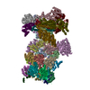
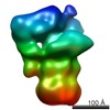
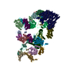
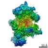
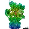
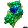
| Title | Reconstruction of the deltaGCC mutant HCV IRES bound to rabbit 40S ribosomal subunit | |||||||||
 Map data Map data | Reconstruction of mutant HCV IRES bound to 40S | |||||||||
 Sample Sample |
| |||||||||
 Keywords Keywords |  HCV IRES / 40S HCV IRES / 40S | |||||||||
| Biological species |  Oryctolagus cuniculus (rabbit) / Oryctolagus cuniculus (rabbit) /  Hepatitis C virus Hepatitis C virus | |||||||||
| Method | single particle reconstruction / cryo EM / Resolution: 17.5 Å | |||||||||
 Authors Authors | Vollmar BS / Shi D / Filbin ME / Kieft JS / Gonen T | |||||||||
 Citation Citation | Journal: Nat Struct Mol Biol / Year: 2013 Title: HCV IRES manipulates the ribosome to promote the switch from translation initiation to elongation. Authors: Megan E Filbin / Breanna S Vollmar / Dan Shi / Tamir Gonen / Jeffrey S Kieft /  Abstract: The internal ribosome entry site (IRES) of the hepatitis C virus (HCV) drives noncanonical initiation of protein synthesis necessary for viral replication. Functional studies of the HCV IRES have ...The internal ribosome entry site (IRES) of the hepatitis C virus (HCV) drives noncanonical initiation of protein synthesis necessary for viral replication. Functional studies of the HCV IRES have focused on 80S ribosome formation but have not explored its role after the 80S ribosome is poised at the start codon. Here, we report that mutations of an IRES domain that docks in the 40S subunit's decoding groove cause only a local perturbation in IRES structure and result in conformational changes in the IRES-rabbit 40S subunit complex. Functionally, the mutations decrease IRES activity by inhibiting the first ribosomal translocation event, and modeling results suggest that this effect occurs through an interaction with a single ribosomal protein. The ability of the HCV IRES to manipulate the ribosome provides insight into how the ribosome's structure and function can be altered by bound RNAs, including those derived from cellular invaders. | |||||||||
| History |
|
- Structure visualization
| Movie |
 Movie viewer Movie viewer |
|---|---|
| Structure viewer | EM map: SurfViewMolmilJmol/JSmol |
| Supplemental images |

- Downloads & links
-EMDB archive
| Map data | emd_5527.map.gz | 11.1 MB | EMDB map data format | |
|---|---|---|---|---|
| Header (meta data) | emd-5527-v30.xmlemd-5527.xml | 9.2 KB 9.2 KB | Display Display | EMDB header |
| Images | emd_5527_1.tif | 164.9 KB | ||
| Archive directory |  http://ftp.pdbj.org/pub/emdb/structures/EMD-5527ftp://ftp.pdbj.org/pub/emdb/structures/EMD-5527 http://ftp.pdbj.org/pub/emdb/structures/EMD-5527ftp://ftp.pdbj.org/pub/emdb/structures/EMD-5527 | HTTPS FTP |
-Related structure data
| Similar structure data |
|---|


-Links
| EMDB pages | EMDB (EBI/PDBe) / EMDataResource |
|---|---|
| Related items in Molecule of the Month |

-Map
| File | Download / File: emd_5527.map.gz / Format: CCP4 / Size: 12.6 MB / Type: IMAGE STORED AS FLOATING POINT NUMBER (4 BYTES) | ||||||||||||||||||||||||||||||||||||||||||||||||||||||||||||||||||||
|---|---|---|---|---|---|---|---|---|---|---|---|---|---|---|---|---|---|---|---|---|---|---|---|---|---|---|---|---|---|---|---|---|---|---|---|---|---|---|---|---|---|---|---|---|---|---|---|---|---|---|---|---|---|---|---|---|---|---|---|---|---|---|---|---|---|---|---|---|---|
| Annotation | Reconstruction of mutant HCV IRES bound to 40S | ||||||||||||||||||||||||||||||||||||||||||||||||||||||||||||||||||||
| Voxel size | X=Y=Z: 2.66 Å | ||||||||||||||||||||||||||||||||||||||||||||||||||||||||||||||||||||
| Density |
| ||||||||||||||||||||||||||||||||||||||||||||||||||||||||||||||||||||
| Symmetry | Space group: 1 | ||||||||||||||||||||||||||||||||||||||||||||||||||||||||||||||||||||
| Details | EMDB XML:
CCP4 map header:
| ||||||||||||||||||||||||||||||||||||||||||||||||||||||||||||||||||||
-Supplemental data
- Sample components
-Entire : mutant HCV IRES bound to 40S
| Entire | Name: mutant HCV IRES bound to 40S |
|---|---|
| Components |
|
-Supramolecule #1000: mutant HCV IRES bound to 40S
| Supramolecule | Name: mutant HCV IRES bound to 40S / type: sample / ID: 1000 / Number unique components: 2 |
|---|
-Supramolecule #1: Rabbit 40S ribosomal subunit
| Supramolecule | Name: Rabbit 40S ribosomal subunit / type: complex / ID: 1 / Name.synonym: 40S ribosome / Details: Mutant HCV IRES bound to 40S / Recombinant expression: No / Database: NCBI / Ribosome-details: ribosome-eukaryote: SSU 40S |
|---|---|
| Source (natural) | Organism: Oryctolagus cuniculus (rabbit) / synonym: rabbit / Cell: reticulocyte |
-Macromolecule #1: hepatitis C virus internal ribosome entry site
| Macromolecule | Name: hepatitis C virus internal ribosome entry site / type: rna / ID: 1 / Name.synonym: HCV IRES Details: Deletion of GCC (82-84) of HCV IRES (nucleotides 40-372) Classification: OTHER / Structure: DOUBLE HELIX / Synthetic?: Yes |
|---|---|
| Source (natural) | Organism: Hepatitis C virus / synonym: HCV |
-Experimental details
-Structure determination
| Method | cryo EM |
|---|---|
 Processing Processing | single particle reconstruction |
| Aggregation state | particle |
-Sample preparation
| Buffer | pH: 7.4 Details: 20 mM Tris-HCl, pH 7.4, 200 mM KOAc, 2.5 mM MgCl2, 2 mM DTT, 40 mM KCl |
|---|---|
| Grid | Details: Quantifoil R1.2/1.3 400 mesh copper grids with holey carbon |
| Vitrification | Cryogen name: ETHANE / Chamber humidity: 100 % / Instrument: FEI VITROBOT MARK IV / Method: Blot and plunge into liquid ethane |
- Electron microscopy
| Microscope | FEI TECNAI F20 |
|---|---|
| Electron beam | Acceleration voltage: 200 kV / Electron source: FIELD EMISSION GUN |
| Electron optics | Calibrated magnification: 117293 / Illumination mode: SPOT SCAN / Imaging mode: BRIGHT FIELDBright-field microscopy / Cs: 2.0 mm / Nominal defocus max: 5.0 µm / Nominal defocus min: 1.25 µm / Nominal magnification: 62000 |
| Sample stage | Specimen holder model: GATAN LIQUID NITROGEN |
| Date | Mar 19, 2012 |
| Image recording | Category: CCD / Film or detector model: GENERIC TVIPS (4k x 4k) / Number real images: 1790 / Average electron dose: 15 e/Å2 Details: Used 2x binned images yielding a pixel size of 2.66 A/pixel. |
| Tilt angle min | 0 |
| Tilt angle max | 0 |
| Experimental equipment |  Model: Tecnai F20 / Image courtesy: FEI Company |
-Image processing
| CTF correction | Details: CTFFIND3 |
|---|---|
| Final reconstruction | Resolution.type: BY AUTHOR / Resolution: 17.5 Å / Resolution method: OTHER / Software - Name: FREALIGN / Number images used: 29920 |
| Details | Particles selected manually using Electron Micrographe Utility (cryoem.ucsf.edu) |
