 Movie
Movie Controller


+ Open data

- Basic information
| Entry | Database: EMDB / ID: EMD-5352 | |||||||||
|---|---|---|---|---|---|---|---|---|---|---|
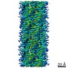
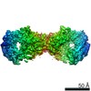
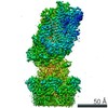
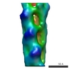
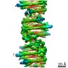
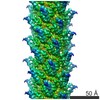
| Title | Structure of a type III secretion needle | |||||||||
 Map data Map data | This is an reconstruction of the type III secretion needle | |||||||||
 Sample Sample |
| |||||||||
 Keywords Keywords | type III secretion system / needle / helical filament | |||||||||
| Function / homology |  Function and homology information Function and homology informationtype III protein secretion system complex / protein secretion by the type III secretion system / : / cell surface / extracellular region / identical protein binding Similarity search - Function | |||||||||
| Biological species |  Shigella flexneri (bacteria) Shigella flexneri (bacteria) | |||||||||
| Method | helical reconstruction / cryo EM / Resolution: 7.7 Å | |||||||||
 Authors Authors | Fujii T / Cheung M / Blanco A / Kato T / Blocker AJ / Namba K | |||||||||
 Citation Citation | Journal: Proc Natl Acad Sci U S A / Year: 2012 Title: Structure of a type III secretion needle at 7-Å resolution provides insights into its assembly and signaling mechanisms. Authors: Takashi Fujii / Martin Cheung / Amandine Blanco / Takayuki Kato / Ariel J Blocker / Keiichi Namba /  Abstract: Type III secretion systems of Gram-negative bacteria form injection devices that deliver effector proteins into eukaryotic cells during infection. They span both bacterial membranes and the ...Type III secretion systems of Gram-negative bacteria form injection devices that deliver effector proteins into eukaryotic cells during infection. They span both bacterial membranes and the extracellular space to connect with the host cell plasma membrane. Their extracellular portion is a needle-like, hollow tube that serves as a secretion conduit for effector proteins. The needle of Shigella flexneri is approximately 50-nm long and 7-nm thick and is made by the helical assembly of one protein, MxiH. We provide a 7-Å resolution 3D image reconstruction of the Shigella needle by electron cryomicroscopy, which resolves α-helices and a β-hairpin that has never been observed in the crystal and solution structures of needle proteins, including MxiH. An atomic model of the needle based on the 3D-density map, in comparison with that of the bacterial-flagellar filament, provides insights into how such a thin tubular structure is stably assembled by intricate intermolecular interactions. The map also illuminates how the needle-length control protein functions as a ruler within the central channel during export of MxiH for assembly at the distal end of the needle, and how the secretion-activation signal may be transduced through a conformational change of the needle upon host-cell contact. | |||||||||
| History |
|
- Structure visualization
| Movie |
Movie viewer |
|---|---|
| Structure viewer | EM map: SurfViewMolmilJmol/JSmol |
| Supplemental images |

- Downloads & links
-EMDB archive
| Map data | emd_5352.map.gz | 3.6 MB | EMDB map data format | |
|---|---|---|---|---|
| Header (meta data) | emd-5352-v30.xmlemd-5352.xml | 9.5 KB 9.5 KB | Display Display | EMDB header |
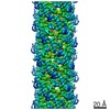
| Images | 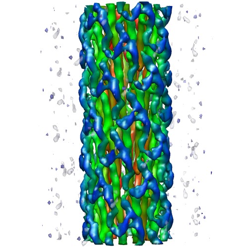 emd_5352_1.jpg emd_5352_1.jpg | 61.4 KB | ||
| Archive directory |  http://ftp.pdbj.org/pub/emdb/structures/EMD-5352ftp://ftp.pdbj.org/pub/emdb/structures/EMD-5352 http://ftp.pdbj.org/pub/emdb/structures/EMD-5352ftp://ftp.pdbj.org/pub/emdb/structures/EMD-5352 | HTTPS FTP |
-Validation report
| Summary document | emd_5352_validation.pdf.gz | 381.5 KB | Display | EMDB validaton report |
|---|---|---|---|---|
| Full document | emd_5352_full_validation.pdf.gz | 381 KB | Display | |
| Data in XML | emd_5352_validation.xml.gz | 5.4 KB | Display | |
| Arichive directory | https://ftp.pdbj.org/pub/emdb/validation_reports/EMD-5352ftp://ftp.pdbj.org/pub/emdb/validation_reports/EMD-5352 | HTTPS FTP |
-Related structure data
| Related structure data |  2mmeM  3j0rMC M: atomic model generated by this map C: citing same article ( |
|---|---|
| Similar structure data |
-Links
| EMDB pages | EMDB (EBI/PDBe) / EMDataResource |
|---|
-Map
| File | Download / File: emd_5352.map.gz / Format: CCP4 / Size: 3.7 MB / Type: IMAGE STORED AS FLOATING POINT NUMBER (4 BYTES) | ||||||||||||||||||||||||||||||||||||||||||||||||||||||||||||||||||||
|---|---|---|---|---|---|---|---|---|---|---|---|---|---|---|---|---|---|---|---|---|---|---|---|---|---|---|---|---|---|---|---|---|---|---|---|---|---|---|---|---|---|---|---|---|---|---|---|---|---|---|---|---|---|---|---|---|---|---|---|---|---|---|---|---|---|---|---|---|---|
| Annotation | This is an reconstruction of the type III secretion needle | ||||||||||||||||||||||||||||||||||||||||||||||||||||||||||||||||||||
| Voxel size | X=Y=Z: 1.68 Å | ||||||||||||||||||||||||||||||||||||||||||||||||||||||||||||||||||||
| Density |
| ||||||||||||||||||||||||||||||||||||||||||||||||||||||||||||||||||||
| Symmetry | Space group: 1 | ||||||||||||||||||||||||||||||||||||||||||||||||||||||||||||||||||||
| Details | EMDB XML:
CCP4 map header:
| ||||||||||||||||||||||||||||||||||||||||||||||||||||||||||||||||||||
-Supplemental data
- Sample components
-Entire : Shigella needle
| Entire | Name: Shigella needle |
|---|---|
| Components |
|
-Supramolecule #1000: Shigella needle
| Supramolecule | Name: Shigella needle / type: sample / ID: 1000 / Oligomeric state: helical assembly of MxiH / Number unique components: 1 |
|---|
-Macromolecule #1: MxiH
| Macromolecule | Name: MxiH / type: protein_or_peptide / ID: 1 / Name.synonym: MxiH / Recombinant expression: Yes / Database: NCBI |
|---|---|
| Source (natural) | Organism: Shigella flexneri (bacteria) |
-Experimental details
-Structure determination
| Method | cryo EM |
|---|---|
 Processing Processing | helical reconstruction |
| Aggregation state | filament |
-Sample preparation
| Buffer | pH: 7.4 / Details: 20 mM Tris, pH 7.4, 150 mM NaCl, 2mM MgSO4 |
|---|---|
| Vitrification | Cryogen name: ETHANE / Chamber humidity: 100 % / Instrument: OTHER / Details: Vitrification instrument: Vitrobot / Method: Blot for 3.5 seconds before plunging |
- Electron microscopy
| Microscope | JEOL 3200FSC |
|---|---|
| Temperature | Min: 50 K / Max: 60 K / Average: 50 K |
| Specialist optics | Energy filter - Name: JEOL Omega filter / Energy filter - Lower energy threshold: 0.0 eV / Energy filter - Upper energy threshold: 10.0 eV |
| Date | Jul 2, 2008 |
| Image recording | Category: CCD / Film or detector model: TVIPS TEMCAM-F415 (4k x 4k) / Digitization - Sampling interval: 15 µm / Number real images: 330 / Average electron dose: 20 e/Å2 / Bits/pixel: 16 |
| Electron beam | Acceleration voltage: 200 kV / Electron source:  FIELD EMISSION GUN FIELD EMISSION GUN |
| Electron optics | Calibrated magnification: 89285 / Illumination mode: FLOOD BEAM / Imaging mode: BRIGHT FIELD / Cs: 1.6 mm / Nominal defocus max: 2.0 µm / Nominal defocus min: 1.0 µm / Nominal magnification: 50000 |
| Sample stage | Specimen holder: top entry / Specimen holder model: OTHER |
-Image processing
| Final reconstruction | Applied symmetry - Helical parameters - Δz: 4.30 Å Applied symmetry - Helical parameters - Δ&Phi: 64.06 ° Algorithm: OTHER / Resolution.type: BY AUTHOR / Resolution: 7.7 Å / Resolution method: FSC 0.5 CUT-OFF / Software - Name: SPIDER |
|---|---|
| CTF correction | Details: CTFFIND3 Each particle |
