 Movie
Movie Controller

[English] 日本語
 Yorodumi
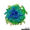
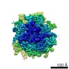
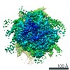
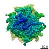
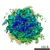
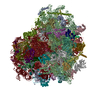
- EMDB-5329: Cryo EM reconstruction of mammalian 80S ribosome in rotated PRE s... -
+ Open data

- Basic information
| Entry | Database: EMDB / ID: EMD-5329 | |||||||||
|---|---|---|---|---|---|---|---|---|---|---|
| Title | Cryo EM reconstruction of mammalian 80S ribosome in rotated PRE state 2 | |||||||||
 Map data Map data | 80S mammalian ribosome | |||||||||
 Sample Sample |
| |||||||||
 Keywords Keywords |  80S ribosome / mammalian / elongation cycle / tRNA 80S ribosome / mammalian / elongation cycle / tRNA | |||||||||
| Function / homology |  Function and homology information Function and homology informationSRP-dependent cotranslational protein targeting to membrane / GTP hydrolysis and joining of the 60S ribosomal subunit / Formation of a pool of free 40S subunits / Nonsense Mediated Decay (NMD) independent of the Exon Junction Complex (EJC) / Nonsense Mediated Decay (NMD) enhanced by the Exon Junction Complex (EJC) / L13a-mediated translational silencing of Ceruloplasmin expression / ribosomal large subunit export from nucleus / maturation of LSU-rRNA / ribosomal large subunit assembly / cytoplasmic translation ...SRP-dependent cotranslational protein targeting to membrane / GTP hydrolysis and joining of the 60S ribosomal subunit / Formation of a pool of free 40S subunits / Nonsense Mediated Decay (NMD) independent of the Exon Junction Complex (EJC) / Nonsense Mediated Decay (NMD) enhanced by the Exon Junction Complex (EJC) / L13a-mediated translational silencing of Ceruloplasmin expression / ribosomal large subunit export from nucleus / maturation of LSU-rRNA / ribosomal large subunit assembly / cytoplasmic translation / cytosolic large ribosomal subunit / rRNA binding / structural constituent of ribosome / RNA binding / nucleus / cytosolSimilarity search - Function | |||||||||
| Biological species |  Oryctolagus cuniculus (rabbit) Oryctolagus cuniculus (rabbit) | |||||||||
| Method | single particle reconstruction / cryo EM / Resolution: 10.6 Å | |||||||||
 Authors Authors | Budkevich T / Giesebrecht J / Altman R / Munro J / Mielke T / Nierhaus K / Blanchard S / Spahn C | |||||||||
 Citation Citation | Journal: Mol Cell / Year: 2011 Title: Structure and dynamics of the mammalian ribosomal pretranslocation complex. Authors: Tatyana Budkevich / Jan Giesebrecht / Roger B Altman / James B Munro / Thorsten Mielke / Knud H Nierhaus / Scott C Blanchard / Christian M T Spahn /  Abstract: Although the structural core of the ribosome is conserved in all kingdoms of life, eukaryotic ribosomes are significantly larger and more complex than their bacterial counterparts. The extent to ...Although the structural core of the ribosome is conserved in all kingdoms of life, eukaryotic ribosomes are significantly larger and more complex than their bacterial counterparts. The extent to which these differences influence the molecular mechanism of translation remains elusive. Multiparticle cryo-electron microscopy and single-molecule FRET investigations of the mammalian pretranslocation complex reveal spontaneous, large-scale conformational changes, including an intersubunit rotation of the ribosomal subunits. Through structurally related processes, tRNA substrates oscillate between classical and at least two distinct hybrid configurations facilitated by localized changes in their L-shaped fold. Hybrid states are favored within the mammalian complex. However, classical tRNA positions can be restored by tRNA binding to the E site or by the eukaryotic-specific antibiotic and translocation inhibitor cycloheximide. These findings reveal critical distinctions in the structural and energetic features of bacterial and mammalian ribosomes, providing a mechanistic basis for divergent translation regulation strategies and species-specific antibiotic action. | |||||||||
| History |
|
- Structure visualization
| Movie |
Movie viewer |
|---|---|
| Structure viewer | EM map: SurfViewMolmilJmol/JSmol |
| Supplemental images |

- Downloads & links
-EMDB archive
| Map data | emd_5329.map.gz | 1.1 MB | EMDB map data format | |
|---|---|---|---|---|
| Header (meta data) | emd-5329-v30.xmlemd-5329.xml | 10.1 KB 10.1 KB | Display Display | EMDB header |
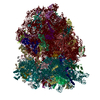
| Images | 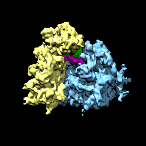 emd_5329_1.jpg emd_5329_1.jpg | 101.7 KB | ||
| Archive directory |  http://ftp.pdbj.org/pub/emdb/structures/EMD-5329ftp://ftp.pdbj.org/pub/emdb/structures/EMD-5329 http://ftp.pdbj.org/pub/emdb/structures/EMD-5329ftp://ftp.pdbj.org/pub/emdb/structures/EMD-5329 | HTTPS FTP |
-Related structure data
| Related structure data |  3j0qMC 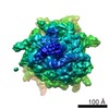 5326C 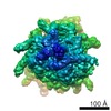 5327C  5328C 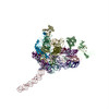 3j0lC 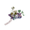 3j0oC 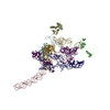 3j0pC M: atomic model generated by this map C: citing same article ( |
|---|---|
| Similar structure data |

-Links
| EMDB pages | EMDB (EBI/PDBe) / EMDataResource |
|---|---|
| Related items in Molecule of the Month |


-Map
| File | Download / File: emd_5329.map.gz / Format: CCP4 / Size: 21.7 MB / Type: IMAGE STORED AS FLOATING POINT NUMBER (4 BYTES) | ||||||||||||||||||||||||||||||||||||||||||||||||||||||||||||||||||||
|---|---|---|---|---|---|---|---|---|---|---|---|---|---|---|---|---|---|---|---|---|---|---|---|---|---|---|---|---|---|---|---|---|---|---|---|---|---|---|---|---|---|---|---|---|---|---|---|---|---|---|---|---|---|---|---|---|---|---|---|---|---|---|---|---|---|---|---|---|---|
| Annotation | 80S mammalian ribosome | ||||||||||||||||||||||||||||||||||||||||||||||||||||||||||||||||||||
| Voxel size | X=Y=Z: 2.52 Å | ||||||||||||||||||||||||||||||||||||||||||||||||||||||||||||||||||||
| Density |
| ||||||||||||||||||||||||||||||||||||||||||||||||||||||||||||||||||||
| Symmetry | Space group: 1 | ||||||||||||||||||||||||||||||||||||||||||||||||||||||||||||||||||||
| Details | EMDB XML:
CCP4 map header:
| ||||||||||||||||||||||||||||||||||||||||||||||||||||||||||||||||||||
-Supplemental data
- Sample components
-Entire : 80S-PRE-rotated-2 complex from rabbit (Oryctolagus cuniculus) liver
| Entire | Name: 80S-PRE-rotated-2 complex from rabbit (Oryctolagus cuniculus) liver |
|---|---|
| Components |
|
-Supramolecule #1000: 80S-PRE-rotated-2 complex from rabbit (Oryctolagus cuniculus) liver
| Supramolecule | Name: 80S-PRE-rotated-2 complex from rabbit (Oryctolagus cuniculus) liver type: sample / ID: 1000 / Details: Sample was re-associated from subunits / Oligomeric state: monomer / Number unique components: 5 |
|---|---|
| Molecular weight | Experimental: 4 MDa / Method: Sedimentation |
-Supramolecule #1: 80S ribosome
| Supramolecule | Name: 80S ribosome / type: complex / ID: 1 / Recombinant expression: No / Database: NCBI / Ribosome-details: ribosome-eukaryote: LSU 60S, SSU 40S |
|---|---|
| Source (natural) | Organism: Oryctolagus cuniculus (rabbit) / synonym: rabbit / Tissue: liver |
| Molecular weight | Theoretical: 4 MDa |
-Experimental details
-Structure determination
| Method | cryo EM |
|---|---|
 Processing Processing | single particle reconstruction |
| Aggregation state | particle |
-Sample preparation
| Buffer | pH: 7.5 |
|---|---|
| Grid | Details: Quantifoil grids |
| Vitrification | Cryogen name: ETHANE / Chamber humidity: 100 % / Chamber temperature: 77 K / Instrument: OTHER / Details: Vitrification instrument: VitroBot (FEI) Method: Flash-frozen in liquid ethane on carbon coated Quantifoil grids |
- Electron microscopy
| Microscope | FEI POLARA 300 |
|---|---|
| Electron beam | Acceleration voltage: 300 kV / Electron source: FIELD EMISSION GUN |
| Electron optics | Calibrated magnification: 65520 / Illumination mode: SPOT SCAN / Imaging mode: BRIGHT FIELDBright-field microscopy / Cs: 2 mm / Nominal defocus max: 4.0 µm / Nominal defocus min: 2.0 µm / Nominal magnification: 39000 |
| Sample stage | Specimen holder: Cartridge / Specimen holder model: GATAN HELIUM |
| Temperature | Average: 77 K |
| Alignment procedure | Legacy - Astigmatism: Objective lens astigmatism was corrected at 200,000 times magnification |
| Date | Oct 17, 2006 |
| Image recording | Category: FILM / Film or detector model: KODAK SO-163 FILM / Digitization - Scanner: PRIMESCAN / Digitization - Sampling interval: 4.7 µm / Number real images: 87 / Average electron dose: 20 e/Å2 |
| Experimental equipment |  Model: Tecnai Polara / Image courtesy: FEI Company |
-Image processing
| CTF correction | Details: By defocus groups |
|---|---|
| Final reconstruction | Algorithm: OTHER / Resolution.type: BY AUTHOR / Resolution: 10.6 Å / Resolution method: FSC 0.5 CUT-OFF / Software - Name: SPIDER Details: Several rounds of multireference 3D projection refinement Number images used: 23347 |
