 Movie
Movie Controller

[English] 日本語
 Yorodumi
- EMDB-5173: Cryo-EM structure of virion-sized hepatitis E virus-like particle -
+ Open data

- Basic information
| Entry | Database: EMDB / ID: EMD-5173 | |||||||||
|---|---|---|---|---|---|---|---|---|---|---|
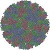
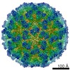
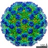
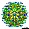
| Title | Cryo-EM structure of virion-sized hepatitis E virus-like particle | |||||||||
 Map data Map data | This is a map for the virion-sized hepatitis E virus-like particle | |||||||||
 Sample Sample |
| |||||||||
 Keywords Keywords | assembly pathway | |||||||||
| Function / homology |  Function and homology information Function and homology informationviral capsid / host cell surface / host cell cytoplasm / structural molecule activity / RNA binding / cytoplasm Similarity search - Function | |||||||||
| Biological species |  Hepatitis E virus Hepatitis E virus | |||||||||
| Method | single particle reconstruction / cryo EM / Resolution: 10.5 Å | |||||||||
 Authors Authors | Xing L / Mayazaki N / Li TC / Simons MN / Wall JS / Moore M / Wang CY / Takeda N / Wakita T / Miyamura T / Cheng RH | |||||||||
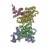
 Citation Citation | Journal: J Biol Chem / Year: 2010 Title: Structure of hepatitis E virion-sized particle reveals an RNA-dependent viral assembly pathway. Authors: Li Xing / Tian-Cheng Li / Naoyuki Mayazaki / Martha N Simon / Joseph S Wall / Mary Moore / Che-Yen Wang / Naokazu Takeda / Takaji Wakita / Tatsuo Miyamura / R Holland Cheng /    Abstract: Hepatitis E virus (HEV) induces acute hepatitis in humans with a high fatality rate in pregnant women. There is a need for anti-HEV research to understand the assembly process of HEV native capsid. ...Hepatitis E virus (HEV) induces acute hepatitis in humans with a high fatality rate in pregnant women. There is a need for anti-HEV research to understand the assembly process of HEV native capsid. Here, we produced a large virion-sized and a small T=1 capsid by expressing the HEV capsid protein in insect cells with and without the N-terminal 111 residues, respectively, for comparative structural analysis. The virion-sized capsid demonstrates a T=3 icosahedral lattice and contains RNA fragment in contrast to the RNA-free T=1 capsid. However, both capsids shared common decameric organization. The in vitro assembly further demonstrated that HEV capsid protein had the intrinsic ability to form decameric intermediate. Our data suggest that RNA binding is the extrinsic factor essential for the assembly of HEV native capsids. | |||||||||
| History |
|
- Structure visualization
| Movie |
Movie viewer |
|---|---|
| Structure viewer | EM map: SurfViewMolmilJmol/JSmol |
| Supplemental images |

- Downloads & links
-EMDB archive
| Map data | emd_5173.map.gz | 49.5 MB | EMDB map data format | |
|---|---|---|---|---|
| Header (meta data) | emd-5173-v30.xmlemd-5173.xml | 10.6 KB 10.6 KB | Display Display | EMDB header |
| Images | 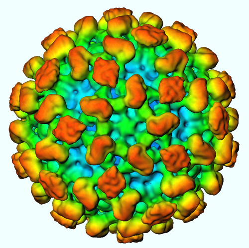 emd_5173_1.png emd_5173_1.png | 312.3 KB | ||
| Archive directory |  http://ftp.pdbj.org/pub/emdb/structures/EMD-5173ftp://ftp.pdbj.org/pub/emdb/structures/EMD-5173 http://ftp.pdbj.org/pub/emdb/structures/EMD-5173ftp://ftp.pdbj.org/pub/emdb/structures/EMD-5173 | HTTPS FTP |
-Validation report
| Summary document | emd_5173_validation.pdf.gz | 407.7 KB | Display | EMDB validaton report |
|---|---|---|---|---|
| Full document | emd_5173_full_validation.pdf.gz | 407.3 KB | Display | |
| Data in XML | emd_5173_validation.xml.gz | 6.4 KB | Display | |
| Arichive directory | https://ftp.pdbj.org/pub/emdb/validation_reports/EMD-5173ftp://ftp.pdbj.org/pub/emdb/validation_reports/EMD-5173 | HTTPS FTP |
-Related structure data
| Related structure data |  3iyoMC M: atomic model generated by this map C: citing same article ( |
|---|---|
| Similar structure data |

-Links
| EMDB pages | EMDB (EBI/PDBe) / EMDataResource |
|---|
-Map
| File | Download / File: emd_5173.map.gz / Format: CCP4 / Size: 118.7 MB / Type: IMAGE STORED AS FLOATING POINT NUMBER (4 BYTES) | ||||||||||||||||||||||||||||||||||||||||||||||||||||||||||||||||||||
|---|---|---|---|---|---|---|---|---|---|---|---|---|---|---|---|---|---|---|---|---|---|---|---|---|---|---|---|---|---|---|---|---|---|---|---|---|---|---|---|---|---|---|---|---|---|---|---|---|---|---|---|---|---|---|---|---|---|---|---|---|---|---|---|---|---|---|---|---|---|
| Annotation | This is a map for the virion-sized hepatitis E virus-like particle | ||||||||||||||||||||||||||||||||||||||||||||||||||||||||||||||||||||
| Voxel size | X=Y=Z: 2 Å | ||||||||||||||||||||||||||||||||||||||||||||||||||||||||||||||||||||
| Density |
| ||||||||||||||||||||||||||||||||||||||||||||||||||||||||||||||||||||
| Symmetry | Space group: 1 | ||||||||||||||||||||||||||||||||||||||||||||||||||||||||||||||||||||
| Details | EMDB XML:
CCP4 map header:
| ||||||||||||||||||||||||||||||||||||||||||||||||||||||||||||||||||||
-Supplemental data
- Sample components
-Entire : Hepatitis E virus like particle
| Entire | Name: Hepatitis E virus like particle |
|---|---|
| Components |
|
-Supramolecule #1000: Hepatitis E virus like particle
| Supramolecule | Name: Hepatitis E virus like particle / type: sample / ID: 1000 / Oligomeric state: 1 / Number unique components: 2 |
|---|---|
| Molecular weight | Experimental: 11.8 MDa / Theoretical: 12.2 MDa / Method: STEM |
-Supramolecule #1: Hepatitis E virus
| Supramolecule | Name: Hepatitis E virus / type: virus / ID: 1 / Name.synonym: recombinant virus-like particle / NCBI-ID: 12461 / Sci species name: Hepatitis E virus / Database: NCBI / Virus type: VIRUS-LIKE PARTICLE / Virus isolate: SEROTYPE / Virus enveloped: No / Virus empty: No / Syn species name: recombinant virus-like particle |
|---|---|
| Host (natural) | Organism:  Homo sapiens (human) / synonym: VERTEBRATES Homo sapiens (human) / synonym: VERTEBRATES |
| Molecular weight | Experimental: 11.8 MDa / Theoretical: 12.2 MDa |
| Virus shell | Shell ID: 1 / Name: ORF2 protein / Diameter: 410 Å / T number (triangulation number): 3 |
-Experimental details
-Structure determination
| Method | cryo EM |
|---|---|
 Processing Processing | single particle reconstruction |
| Aggregation state | particle |
-Sample preparation
| Concentration | 1 mg/mL |
|---|---|
| Buffer | pH: 6.2 / Details: 20mM MES-K |
| Grid | Details: 300 mesh holey carbon |
| Vitrification | Cryogen name: ETHANE / Chamber temperature: 93 K / Instrument: HOMEMADE PLUNGER / Details: Vitrification instrument: manual plunger |
- Electron microscopy
| Microscope | JEOL 2100F |
|---|---|
| Image recording | Category: CCD / Film or detector model: TVIPS TEMCAM-F415 (4k x 4k) / Average electron dose: 10 e/Å2 / Bits/pixel: 16 |
| Electron beam | Acceleration voltage: 200 kV / Electron source:  FIELD EMISSION GUN FIELD EMISSION GUN |
| Electron optics | Illumination mode: FLOOD BEAM / Imaging mode: BRIGHT FIELD / Cs: 2.0 mm / Nominal magnification: 50000 |
| Sample stage | Specimen holder: single-tilt / Specimen holder model: GATAN LIQUID NITROGEN |
-Image processing
| CTF correction | Details: each micrograph |
|---|---|
| Final reconstruction | Algorithm: OTHER / Resolution.type: BY AUTHOR / Resolution: 10.5 Å / Resolution method: FSC 0.5 CUT-OFF / Software - Name: P3DR / Number images used: 4348 |
-Atomic model buiding 1
| Initial model | PDB ID: Chain - Chain ID: A |
|---|---|
| Software | Name: Situs |
| Details | PDBEntryID_givenInChain. Protocol: Rigid Body. Monomer C were divided into Shell and Spike domains and separately fitted by manual docking using program O and followed by Situs |
| Refinement | Space: REAL / Protocol: RIGID BODY FIT / Target criteria: cross-correlation |
| Output model | PDB-3iyo: |

