Movie
Movie Controller


+ Open data

- Basic information
| Entry | Database: EMDB / ID: EMD-5119 | |||||||||
|---|---|---|---|---|---|---|---|---|---|---|
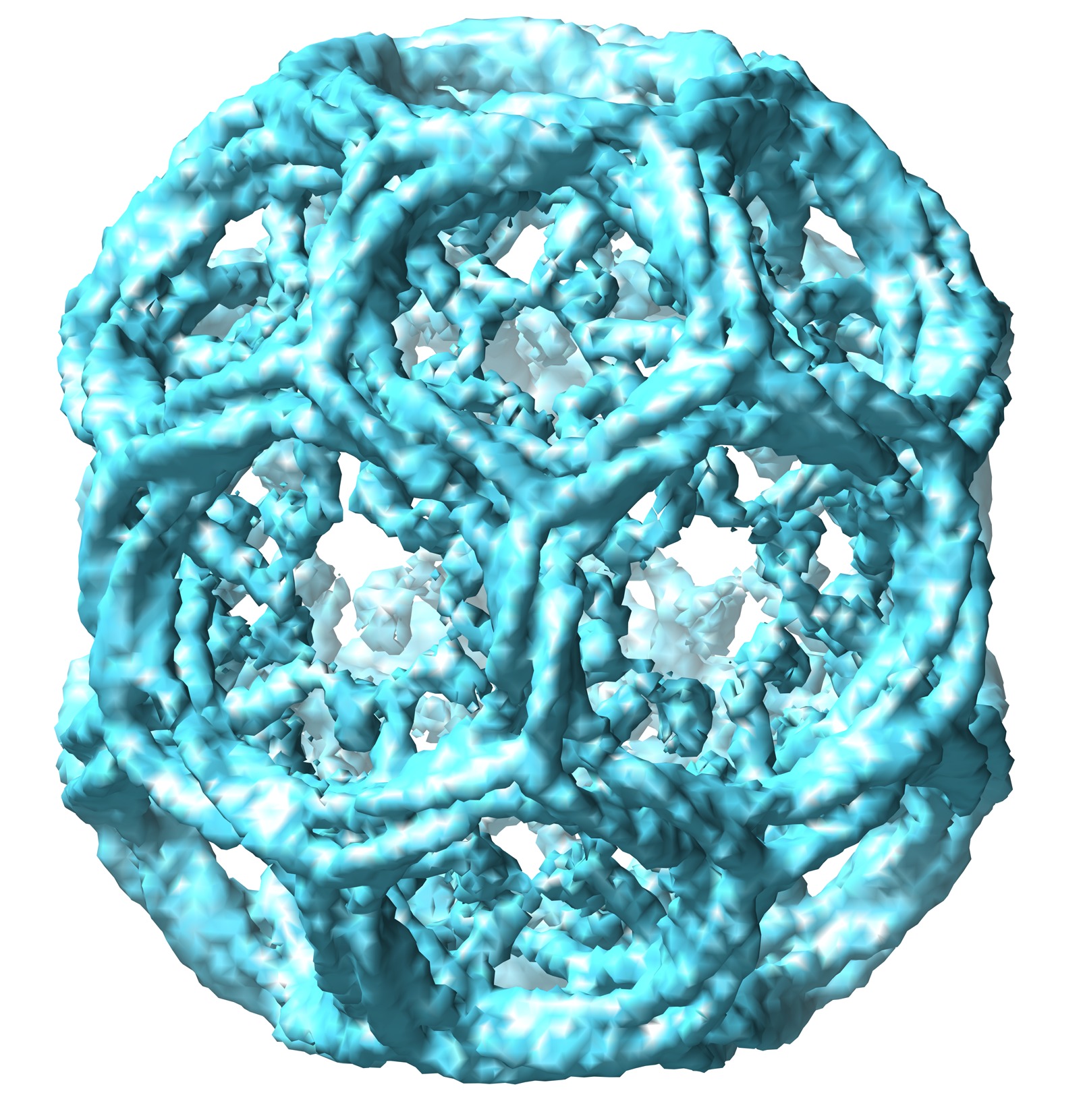
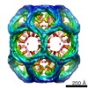
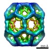
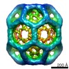
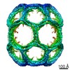
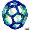
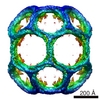
| Title | Clathrin D6 coat | |||||||||
 Map data Map data | This is an averaged map of clathrin D6 coat | |||||||||
 Sample Sample |
| |||||||||
 Keywords Keywords |  CLATHRIN / ALPHA-ZIG-ZAG / BETA-PROPELLER / ENDOCYTOSIS/EXOCYTOSIS COMPLEX CLATHRIN / ALPHA-ZIG-ZAG / BETA-PROPELLER / ENDOCYTOSIS/EXOCYTOSIS COMPLEX | |||||||||
| Function / homology |  Function and homology information Function and homology informationpostsynaptic endocytic zone cytoplasmic component / Retrograde neurotrophin signalling / Recycling pathway of L1 / WNT5A-dependent internalization of FZD4 / WNT5A-dependent internalization of FZD2, FZD5 and ROR2 / LDL clearance / Gap junction degradation / Formation of annular gap junctions / Golgi Associated Vesicle Biogenesis / RHOU GTPase cycle ...postsynaptic endocytic zone cytoplasmic component / Retrograde neurotrophin signalling / Recycling pathway of L1 / WNT5A-dependent internalization of FZD4 / WNT5A-dependent internalization of FZD2, FZD5 and ROR2 / LDL clearance / Gap junction degradation / Formation of annular gap junctions / Golgi Associated Vesicle Biogenesis / RHOU GTPase cycle / RHOV GTPase cycle / clathrin coat of trans-Golgi network vesicle / clathrin vesicle coat / Lysosome Vesicle Biogenesis / negative regulation of hyaluronan biosynthetic process / clathrin light chain binding / clathrin complex / MHC class II antigen presentation / VLDLR internalisation and degradation / clathrin heavy chain binding / clathrin coat of coated pit / clathrin coat disassembly / Cargo recognition for clathrin-mediated endocytosis / clathrin coat assembly / clathrin-coated endocytic vesicle / membrane coat / Clathrin-mediated endocytosis / clathrin-dependent endocytosis / arrestin family protein binding / receptor-mediated endocytosis / intracellular protein transport / synaptic vesicle membrane / spindle / autophagy / disordered domain specific binding / melanosome / mitotic cell cycle / cell cycle / cell division / protein domain specific binding / structural molecule activity / mitochondrion / identical protein binding / plasma membraneSimilarity search - Function | |||||||||
| Biological species |  Bos taurus (cattle) Bos taurus (cattle) | |||||||||
| Method | single particle reconstruction / cryo EM / Resolution: 7.9 Å | |||||||||
 Authors Authors | Fotin A / Cheng Y / Sliz P / Grigorieff N / Harrison SC / Kirchhausen T / Walz T | |||||||||
 Citation Citation | Journal: Nature / Year: 2004 Title: Molecular model for a complete clathrin lattice from electron cryomicroscopy. Authors: Alexander Fotin / Yifan Cheng / Piotr Sliz / Nikolaus Grigorieff / Stephen C Harrison / Tomas Kirchhausen / Thomas Walz /  Abstract: Clathrin-coated vesicles are important vehicles of membrane traffic in cells. We report the structure of a clathrin lattice at subnanometre resolution, obtained from electron cryomicroscopy of coats ...Clathrin-coated vesicles are important vehicles of membrane traffic in cells. We report the structure of a clathrin lattice at subnanometre resolution, obtained from electron cryomicroscopy of coats assembled in vitro. We trace most of the 1,675-residue clathrin heavy chain by fitting known crystal structures of two segments, and homology models of the rest, into the electron microscopy density map. We also define the position of the central helical segment of the light chain. A helical tripod, the carboxy-terminal parts of three heavy chains, projects inward from the vertex of each three-legged clathrin triskelion, linking that vertex to 'ankles' of triskelions centred two vertices away. Analysis of coats with distinct diameters shows an invariant pattern of contacts in the neighbourhood of each vertex, with more variable interactions along the extended parts of the triskelion 'legs'. These invariant local interactions appear to stabilize the lattice, allowing assembly and uncoating to be controlled by events at a few specific sites. | |||||||||
| History |
|
- Structure visualization
| Movie |
Movie viewer |
|---|---|
| Structure viewer | EM map: SurfViewMolmilJmol/JSmol |
| Supplemental images |
- Downloads & links
-EMDB archive
| Map data | emd_5119.map.gz | 10.2 MB | EMDB map data format | |
|---|---|---|---|---|
| Header (meta data) | emd-5119-v30.xmlemd-5119.xml | 10.3 KB 10.3 KB | Display Display | EMDB header |
| Images | emd_5119_1.tif | 7.7 MB | ||
| Archive directory |  http://ftp.pdbj.org/pub/emdb/structures/EMD-5119ftp://ftp.pdbj.org/pub/emdb/structures/EMD-5119 http://ftp.pdbj.org/pub/emdb/structures/EMD-5119ftp://ftp.pdbj.org/pub/emdb/structures/EMD-5119 | HTTPS FTP |
-Related structure data
| Related structure data | 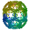 1xi4MC  3iyvMC M: atomic model generated by this map C: citing same article ( |
|---|---|
| Similar structure data |
-Links
| EMDB pages | EMDB (EBI/PDBe) / EMDataResource |
|---|---|
| Related items in Molecule of the Month |

-Map
| File | Download / File: emd_5119.map.gz / Format: CCP4 / Size: 113.1 MB / Type: IMAGE STORED AS FLOATING POINT NUMBER (4 BYTES) | ||||||||||||||||||||||||||||||||||||||||||||||||||||||||||||||||||||
|---|---|---|---|---|---|---|---|---|---|---|---|---|---|---|---|---|---|---|---|---|---|---|---|---|---|---|---|---|---|---|---|---|---|---|---|---|---|---|---|---|---|---|---|---|---|---|---|---|---|---|---|---|---|---|---|---|---|---|---|---|---|---|---|---|---|---|---|---|---|
| Annotation | This is an averaged map of clathrin D6 coat | ||||||||||||||||||||||||||||||||||||||||||||||||||||||||||||||||||||
| Voxel size | X=Y=Z: 2.8 Å | ||||||||||||||||||||||||||||||||||||||||||||||||||||||||||||||||||||
| Density |
| ||||||||||||||||||||||||||||||||||||||||||||||||||||||||||||||||||||
| Symmetry | Space group: 1 | ||||||||||||||||||||||||||||||||||||||||||||||||||||||||||||||||||||
| Details | EMDB XML:
CCP4 map header:
| ||||||||||||||||||||||||||||||||||||||||||||||||||||||||||||||||||||
-Supplemental data
- Sample components
-Entire : Clathrin coats
| Entire | Name: Clathrin coats |
|---|---|
| Components |
|
-Supramolecule #1000: Clathrin coats
| Supramolecule | Name: Clathrin coats / type: sample / ID: 1000 / Details: Clathrin coats assembled from clathrin and AP-2 / Number unique components: 9 |
|---|
-Macromolecule #1: clathrin coat
| Macromolecule | Name: clathrin coat / type: protein_or_peptide / ID: 1 / Name.synonym: clathrin coat / Number of copies: 108 / Oligomeric state: D6 assemble / Recombinant expression: No / Database: NCBI |
|---|---|
| Source (natural) | Organism: Bos taurus (cattle) / synonym: cattle / Tissue: Brain |
-Experimental details
-Structure determination
| Method | cryo EM |
|---|---|
 Processing Processing | single particle reconstruction |
| Aggregation state | particle |
-Sample preparation
| Concentration | 1 mg/mL |
|---|---|
| Buffer | pH: 6.5 / Details: 25mM MES pH 6.5, 2mM DTT |
| Grid | Details: holey carbon grid |
| Vitrification | Cryogen name: ETHANE / Chamber temperature: 93 K / Instrument: OTHER / Details: Vitrification instrument: FEI Vitrobot |
- Electron microscopy
| Microscope | FEI TECNAI F20 |
|---|---|
| Electron beam | Acceleration voltage: 200 kV / Electron source: FIELD EMISSION GUN |
| Electron optics | Calibrated magnification: 51159 / Illumination mode: FLOOD BEAM / Imaging mode: BRIGHT FIELDBright-field microscopy / Cs: 2 mm / Nominal defocus max: 5.0 µm / Nominal defocus min: 2.0 µm / Nominal magnification: 50000 |
| Sample stage | Specimen holder: side entry liquid nitrogen-cooled cryo specimen holder Specimen holder model: GATAN LIQUID NITROGEN |
| Temperature | Average: 93 K |
| Image recording | Category: FILM / Film or detector model: KODAK SO-163 FILM / Digitization - Scanner: ZEISS SCAI / Digitization - Sampling interval: 7 µm / Number real images: 225 / Od range: 1.4 / Bits/pixel: 8 |
| Experimental equipment |  Model: Tecnai F20 / Image courtesy: FEI Company |
-Image processing
| CTF correction | Details: CTF correction of each particle. |
|---|---|
| Final reconstruction | Algorithm: OTHER / Resolution.type: BY AUTHOR / Resolution: 7.9 Å / Resolution method: FSC 0.143 CUT-OFF / Software - Name: Frealign Details: This is an n.c.s averaged map from raw reconstruction Number images used: 1450 |
-Atomic model buiding 1
| Initial model | (PDB ID: , , ) |
|---|---|
| Software | Name: O |
| Details | Protocol: Rigid Body. various segments of clathrin were separately fitted by manual docking using program O, and fitting was improved by MAVE |
| Refinement | Space: REAL / Protocol: RIGID BODY FIT / Target criteria: DENSITY CORRELATION |
| Output model | PDB-1xi4: PDB-3iyv: |