 Movie
Movie Controller

[English] 日本語
 Yorodumi
- EMDB-5115: West Nile virus in complex with a single-chain antibody derivativ... -
+ Open data

- Basic information
| Entry | Database: EMDB / ID: EMD-5115 | |||||||||
|---|---|---|---|---|---|---|---|---|---|---|
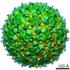
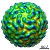
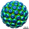
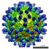
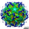
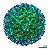
| Title | West Nile virus in complex with a single-chain antibody derivative of the neutralizing monoclonal antibody E16 | |||||||||
 Map data Map data | West Nile virus in complex with a single-chain antibody derivative of the neutralizing monoclonal antibody E16. | |||||||||
 Sample Sample |
| |||||||||
 Keywords Keywords | flavivirus / West Nile Virus / neutralizing antibody / complex / single-chain | |||||||||
| Biological species | unidentified (others) / West Nile Virus NY99 | |||||||||
| Method | single particle reconstruction / cryo EM / Resolution: 22.75 Å | |||||||||
 Authors Authors | Kaufmann B / Chipman PR / Holdaway HA / Johnson S / Kuhn RJ / Diamond MS / Rossmann MG | |||||||||
 Citation Citation | Journal: PLoS Pathog / Year: 2009 Title: Capturing a flavivirus pre-fusion intermediate. Authors: Bärbel Kaufmann / Paul R Chipman / Heather A Holdaway / Syd Johnson / Daved H Fremont / Richard J Kuhn / Michael S Diamond / Michael G Rossmann /  Abstract: During cell entry of flaviviruses, low endosomal pH triggers the rearrangement of the viral surface glycoproteins to a fusion-active state that allows the release of the infectious RNA into the ...During cell entry of flaviviruses, low endosomal pH triggers the rearrangement of the viral surface glycoproteins to a fusion-active state that allows the release of the infectious RNA into the cytoplasm. In this work, West Nile virus was complexed with Fab fragments of the neutralizing mAb E16 and was subsequently exposed to low pH, trapping the virions in a pre-fusion intermediate state. The structure of the complex was studied by cryo-electron microscopy and provides the first structural glimpse of a flavivirus fusion intermediate near physiological conditions. A radial expansion of the outer protein layer of the virion was observed compared to the structure at pH 8. The resulting approximately 60 A-wide shell of low density between lipid bilayer and outer protein layer is likely traversed by the stem region of the E glycoprotein. By using antibody fragments, we have captured a structural intermediate of a virus that likely occurs during cell entry. The trapping of structural transition states by antibody fragments will be applicable for other processes in the flavivirus life cycle and delineating other cellular events that involve conformational rearrangements. | |||||||||
| History |
|
- Structure visualization
| Movie |
 Movie viewer Movie viewer |
|---|---|
| Structure viewer | EM map: SurfViewMolmilJmol/JSmol |
| Supplemental images |

- Downloads & links
-EMDB archive
| Map data | emd_5115.map.gz | 31.7 MB | EMDB map data format | |
|---|---|---|---|---|
| Header (meta data) | emd-5115-v30.xmlemd-5115.xml | 12.2 KB 12.2 KB | Display Display | EMDB header |
| Images | emd_5115_1.tif | 762 KB | ||
| Archive directory |  http://ftp.pdbj.org/pub/emdb/structures/EMD-5115ftp://ftp.pdbj.org/pub/emdb/structures/EMD-5115 http://ftp.pdbj.org/pub/emdb/structures/EMD-5115ftp://ftp.pdbj.org/pub/emdb/structures/EMD-5115 | HTTPS FTP |
-Validation report
| Summary document | emd_5115_validation.pdf.gz | 78.7 KB | Display | EMDB validaton report |
|---|---|---|---|---|
| Full document | emd_5115_full_validation.pdf.gz | 77.8 KB | Display | |
| Data in XML | emd_5115_validation.xml.gz | 492 B | Display | |
| Arichive directory | https://ftp.pdbj.org/pub/emdb/validation_reports/EMD-5115ftp://ftp.pdbj.org/pub/emdb/validation_reports/EMD-5115 | HTTPS FTP |
-Related structure data
| Similar structure data |
|---|
-Links
| EMDB pages | EMDB (EBI/PDBe) / EMDataResource |
|---|
-Map
| File | Download / File: emd_5115.map.gz / Format: CCP4 / Size: 81.8 MB / Type: IMAGE STORED AS FLOATING POINT NUMBER (4 BYTES) | ||||||||||||||||||||||||||||||||||||||||||||||||||||||||||||||||||||
|---|---|---|---|---|---|---|---|---|---|---|---|---|---|---|---|---|---|---|---|---|---|---|---|---|---|---|---|---|---|---|---|---|---|---|---|---|---|---|---|---|---|---|---|---|---|---|---|---|---|---|---|---|---|---|---|---|---|---|---|---|---|---|---|---|---|---|---|---|---|
| Annotation | West Nile virus in complex with a single-chain antibody derivative of the neutralizing monoclonal antibody E16. | ||||||||||||||||||||||||||||||||||||||||||||||||||||||||||||||||||||
| Voxel size | X=Y=Z: 2.967 Å | ||||||||||||||||||||||||||||||||||||||||||||||||||||||||||||||||||||
| Density |
| ||||||||||||||||||||||||||||||||||||||||||||||||||||||||||||||||||||
| Symmetry | Space group: 1 | ||||||||||||||||||||||||||||||||||||||||||||||||||||||||||||||||||||
| Details | EMDB XML:
CCP4 map header:
| ||||||||||||||||||||||||||||||||||||||||||||||||||||||||||||||||||||
-Supplemental data
- Sample components
-Entire : West Nile virus NY99 complexed with single-chain antibody derivat...
| Entire | Name: West Nile virus NY99 complexed with single-chain antibody derivative of neutralizing monoclonal antibody E16 |
|---|---|
| Components |
|
-Supramolecule #1000: West Nile virus NY99 complexed with single-chain antibody derivat...
| Supramolecule | Name: West Nile virus NY99 complexed with single-chain antibody derivative of neutralizing monoclonal antibody E16 type: sample / ID: 1000 Oligomeric state: T1 icosahedron with three E monomers and two scFv molecules per asymmetric unit Number unique components: 2 |
|---|---|
| Molecular weight | Theoretical: 20 MDa |
-Supramolecule #1: West Nile Virus NY99
| Supramolecule | Name: West Nile Virus NY99 / type: virus / ID: 1 / Name.synonym: West Nile Virus Details: the virus is complexed with a single-chain antibody derivative of the neutralizing antibody E16 (120 scFv molecules per virion) Sci species name: West Nile Virus NY99 / Database: NCBI / Virus type: VIRION / Virus isolate: STRAIN / Virus enveloped: Yes / Virus empty: No / Syn species name: West Nile Virus |
|---|---|
| Host (natural) | Organism:  Homo sapiens (human) / synonym: VERTEBRATES Homo sapiens (human) / synonym: VERTEBRATES |
| Molecular weight | Theoretical: 17. MDa |
-Macromolecule #1: E16 scFv derivative
| Macromolecule | Name: E16 scFv derivative / type: protein_or_peptide / ID: 1 Name.synonym: single-chain antibody derivative of neutralizing antibody E16 Details: single-chain antibody derivative of the neutralizing antibody E16 complexed with West Nile virus (120 scFv molecules per virion) Number of copies: 120 / Oligomeric state: Monomer / Recombinant expression: Yes / Database: NCBI |
|---|---|
| Source (natural) | Organism: unidentified (others) |
| Molecular weight | Theoretical: 25 KDa |
-Experimental details
-Structure determination
| Method | cryo EM |
|---|---|
 Processing Processing | single particle reconstruction |
| Aggregation state | particle |
-Sample preparation
| Concentration | 1. mg/mL |
|---|---|
| Buffer | pH: 8 / Details: 12 mM Tris-HCl, 120 mM NaCl, 1 mM EDTA |
| Grid | Details: 400 mesh copper grid |
| Vitrification | Cryogen name: ETHANE / Instrument: HOMEMADE PLUNGER Details: Vitrification instrument: Guillotine-style plunge freezeing device Method: A small vial of ethane is placed inside a larger liquid nitrogen reservoir. The grid holding a few microliters of the sample is held in place at the bottom of a plunger by the means of fine ...Method: A small vial of ethane is placed inside a larger liquid nitrogen reservoir. The grid holding a few microliters of the sample is held in place at the bottom of a plunger by the means of fine tweezers. Once the ethane in the vial is completely frozen, it needs to be slightly melted. When the liquid ethane is ready, a piece of filter paper is then pressed against the sample to blot of excess buffer, sufficient to leave a thin layer on the grid. After a predetermined time, the filter paper is removed, and the plunger is allowed to drop into the liquid ethane. Once the grid enters the liquid ethane, the sample is rapidly frozen, and the grid is transferred under liquid nitrogen to a storage box immersed liquid nitrogen for later use in the microscope. |
- Electron microscopy
| Microscope | FEI/PHILIPS CM300FEG/T |
|---|---|
| Temperature | Average: 98 K |
| Alignment procedure | Legacy - Astigmatism: live FFT at 200K mag |
| Details | low dose imaging |
| Date | Jul 24, 2007 |
| Image recording | Category: FILM / Film or detector model: KODAK SO-163 FILM / Digitization - Scanner: ZEISS SCAI / Digitization - Sampling interval: 7 µm / Number real images: 53 / Average electron dose: 17.22 e/Å2 / Details: scanned images binned 2x2 / Od range: 1 / Bits/pixel: 8 |
| Tilt angle max | 0 |
| Electron beam | Acceleration voltage: 300 kV / Electron source:  FIELD EMISSION GUN FIELD EMISSION GUN |
| Electron optics | Calibrated magnification: 47190 / Illumination mode: FLOOD BEAM / Imaging mode: BRIGHT FIELD / Cs: 2.0 mm / Nominal defocus max: 2.74 µm / Nominal defocus min: 1.35 µm / Nominal magnification: 45000 |
| Sample stage | Specimen holder: Eucentric / Specimen holder model: GATAN LIQUID NITROGEN / Tilt angle min: -0 |
-Image processing
| Details | The particles were selected interactively at the computer terminal. |
|---|---|
| CTF correction | Details: each particle |
| Final reconstruction | Resolution.type: BY AUTHOR / Resolution: 22.75 Å / Resolution method: FSC 0.5 CUT-OFF / Software - Name: PFTSEARCH, PO2R, P3DR / Number images used: 783 |
