 Movie
Movie Controller

[English] 日本語
 Yorodumi
- EMDB-5013: A 3D EM map of the subcomplex Orc1-5 of the yeast origin recognit... -
+ Open data

- Basic information
| Entry | Database: EMDB / ID: EMD-5013 | |||||||||
|---|---|---|---|---|---|---|---|---|---|---|
| Title | A 3D EM map of the subcomplex Orc1-5 of the yeast origin recognition complex (ORC). | |||||||||
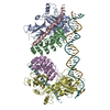
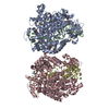
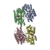
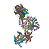
 Map data Map data | This is a 3D negatively stained EM map of a subcomplex (Orc1-5) of the yeast origin recognition complex (ORC - Orc1-6). This subcomplex does not contain the smallest subunit Orc6 of ORC. | |||||||||
 Sample Sample |
| |||||||||
 Keywords Keywords | Negative stain electron microscopy / single particle image reconstruction / yeast replication initiation /  origin recognition complex / DNA binding protein origin recognition complex / DNA binding protein | |||||||||
| Method | single particle reconstruction / cryo EM / negative staining / Resolution: 25.0 Å | |||||||||
 Authors Authors | Chen Z / Speck C / Wendel P / Tang C / Stillman B / Li H | |||||||||
 Citation Citation | Journal: Proc Natl Acad Sci U S A / Year: 2008 Title: The architecture of the DNA replication origin recognition complex in Saccharomyces cerevisiae. Authors: Zhiqiang Chen / Christian Speck / Patricia Wendel / Chunyan Tang / Bruce Stillman / Huilin Li /  Abstract: The origin recognition complex (ORC) is conserved in all eukaryotes. The six proteins of the Saccharomyces cerevisiae ORC that form a stable complex bind to origins of DNA replication and recruit ...The origin recognition complex (ORC) is conserved in all eukaryotes. The six proteins of the Saccharomyces cerevisiae ORC that form a stable complex bind to origins of DNA replication and recruit prereplicative complex (pre-RC) proteins, one of which is Cdc6. To further understand the function of ORC we recently determined by single-particle reconstruction of electron micrographs a low-resolution, 3D structure of S. cerevisiae ORC and the ORC-Cdc6 complex. In this article, the spatial arrangement of the ORC subunits within the ORC structure is described. In one approach, a maltose binding protein (MBP) was systematically fused to the N or the C termini of the five largest ORC subunits, one subunit at a time, generating 10 MBP-fused ORCs, and the MBP density was localized in the averaged, 2D EM images of the MBP-fused ORC particles. Determining the Orc1-5 structure and comparing it with the native ORC structure localized the Orc6 subunit near Orc2 and Orc3. Finally, subunit-subunit interactions were determined by immunoprecipitation of ORC subunits synthesized in vitro. Based on the derived ORC architecture and existing structures of archaeal Orc1-DNA structures, we propose a model for ORC and suggest how ORC interacts with origin DNA and Cdc6. The studies provide a basis for understanding the overall structure of the pre-RC. | |||||||||
| History |
|
- Structure visualization
| Movie |
 Movie viewer Movie viewer |
|---|---|
| Structure viewer | EM map: SurfViewMolmilJmol/JSmol |
| Supplemental images |

- Downloads & links
-EMDB archive
| Map data | emd_5013.map.gz | 3.5 MB | EMDB map data format | |
|---|---|---|---|---|
| Header (meta data) | emd-5013-v30.xmlemd-5013.xml | 15.3 KB 15.3 KB | Display Display | EMDB header |
| Images |  emd_5013_1.gif emd_5013_1.gif | 25.1 KB | ||
| Archive directory |  http://ftp.pdbj.org/pub/emdb/structures/EMD-5013ftp://ftp.pdbj.org/pub/emdb/structures/EMD-5013 http://ftp.pdbj.org/pub/emdb/structures/EMD-5013ftp://ftp.pdbj.org/pub/emdb/structures/EMD-5013 | HTTPS FTP |
-Related structure data
| Similar structure data |
|---|


-Links
| EMDB pages | EMDB (EBI/PDBe) / EMDataResource |
|---|
-Map
| File | Download / File: emd_5013.map.gz / Format: CCP4 / Size: 3.7 MB / Type: IMAGE STORED AS FLOATING POINT NUMBER (4 BYTES) | ||||||||||||||||||||||||||||||||||||||||||||||||||||||||||||||||||||
|---|---|---|---|---|---|---|---|---|---|---|---|---|---|---|---|---|---|---|---|---|---|---|---|---|---|---|---|---|---|---|---|---|---|---|---|---|---|---|---|---|---|---|---|---|---|---|---|---|---|---|---|---|---|---|---|---|---|---|---|---|---|---|---|---|---|---|---|---|---|
| Annotation | This is a 3D negatively stained EM map of a subcomplex (Orc1-5) of the yeast origin recognition complex (ORC - Orc1-6). This subcomplex does not contain the smallest subunit Orc6 of ORC. | ||||||||||||||||||||||||||||||||||||||||||||||||||||||||||||||||||||
| Voxel size | X=Y=Z: 2.54 Å | ||||||||||||||||||||||||||||||||||||||||||||||||||||||||||||||||||||
| Density |
| ||||||||||||||||||||||||||||||||||||||||||||||||||||||||||||||||||||
| Symmetry | Space group: 1 | ||||||||||||||||||||||||||||||||||||||||||||||||||||||||||||||||||||
| Details | EMDB XML:
CCP4 map header:
| ||||||||||||||||||||||||||||||||||||||||||||||||||||||||||||||||||||
-Supplemental data
- Sample components
-Entire : Yeast ORC subcomplex Orc1-5.
| Entire | Name: Yeast ORC subcomplex Orc1-5. |
|---|---|
| Components |
|
-Supramolecule #1000: Yeast ORC subcomplex Orc1-5.
| Supramolecule | Name: Yeast ORC subcomplex Orc1-5. / type: sample / ID: 1000 Details: The sample was purified by gel filtration and was indeed monodisperse. Oligomeric state: One heteropentamer (Orc1-Orc2-Orc3-Orc4-Orc5) Number unique components: 5 |
|---|---|
| Molecular weight | Theoretical: 362 KDa |
-Macromolecule #1: Chromosomal replication origin recognition protein Orc1p
| Macromolecule | Name: Chromosomal replication origin recognition protein Orc1p type: protein_or_peptide / ID: 1 / Name.synonym: Orc1p / Number of copies: 1 / Oligomeric state: monomer / Recombinant expression: Yes |
|---|---|
| Source (natural) | Cell: Yeast / Organelle: Nucleus / Location in cell: Nucleus |
| Molecular weight | Experimental: 120 KDa / Theoretical: 120 KDa |
| Recombinant expression | Organism: Sf9 insect cells / Recombinant plasmid: bvORC1 |
-Macromolecule #2: Orc2
| Macromolecule | Name: Orc2 / type: protein_or_peptide / ID: 2 / Name.synonym: Orc2 / Number of copies: 1 / Oligomeric state: monomer / Recombinant expression: Yes |
|---|---|
| Source (natural) | Strain: S. cerevisiae / Cell: Yeast / Organelle: Nucleus / Location in cell: Nucleus |
| Molecular weight | Experimental: 71 KDa / Theoretical: 71 KDa |
| Recombinant expression | Organism: Sf9 insect cells / Recombinant plasmid: bvORC2 |
-Macromolecule #3: Orc3
| Macromolecule | Name: Orc3 / type: protein_or_peptide / ID: 3 / Name.synonym: Orc3 / Number of copies: 1 / Oligomeric state: Monomer / Recombinant expression: Yes |
|---|---|
| Source (natural) | Strain: S. cerevisiae / Cell: yeast / Organelle: Nucleus / Location in cell: Nucleus |
| Molecular weight | Experimental: 62 KDa / Theoretical: 62 KDa |
| Recombinant expression | Organism: Sf9 insect cells / Recombinant plasmid: bvORC3 |
-Macromolecule #4: Orc4
| Macromolecule | Name: Orc4 / type: protein_or_peptide / ID: 4 / Name.synonym: Orc4 / Number of copies: 1 / Oligomeric state: Monomer / Recombinant expression: Yes |
|---|---|
| Source (natural) | Strain: S. cerevisiae / Cell: yeast / Organelle: Nucleus / Location in cell: Nucleus |
| Molecular weight | Experimental: 56 KDa / Theoretical: 56 KDa |
| Recombinant expression | Organism: bvORC4 / Recombinant plasmid: SF9 insect cells |
-Macromolecule #5: Orc5
| Macromolecule | Name: Orc5 / type: protein_or_peptide / ID: 5 / Name.synonym: Orc5 / Number of copies: 1 / Oligomeric state: Monomer / Recombinant expression: Yes |
|---|---|
| Source (natural) | Strain: S. cerevisiae / Cell: yeast / Organelle: Nucleus / Location in cell: Nucleus |
| Molecular weight | Experimental: 53 KDa / Theoretical: 53 KDa |
| Recombinant expression | Organism: bvORC5 / Recombinant plasmid: SF9 insect cells |
-Experimental details
-Structure determination
| Method | negative staining, cryo EM |
|---|---|
 Processing Processing | single particle reconstruction |
| Aggregation state | particle |
-Sample preparation
| Concentration | 0.1 mg/mL |
|---|---|
| Buffer | pH: 7.6 Details: 50 mM Hepes-KOH pH 7.6, 100 mM potassium glutamate, 5 mM MgCl2, 1 mM EGTA, and 1mM ATP-gamma-S |
| Staining | Type: NEGATIVE Details: A 6.0 micro liter drop of sample solution was applied to a glow-discharged 300-mesh copper grid covered with a thin layer of carbon film, and after a brief incubation of 30 - 60 s, the ...Details: A 6.0 micro liter drop of sample solution was applied to a glow-discharged 300-mesh copper grid covered with a thin layer of carbon film, and after a brief incubation of 30 - 60 s, the excess sample solution was blotted with a small piece of filter paper. The grid was then stained in a deep stain procedure by three consecutive 5 micro liter drops of 2.0% uranyl acetate aqueous solution. Each drop was left on grid for 15 - 20 s at room temperature before blotting. After blotting the last stain drop, the grid was quickly dried by a stream of argon to prevent crystallization of the stain salt. |
| Grid | Details: 300 mesh copper grid covered with continuous carbon film |
| Vitrification | Cryogen name: ETHANE / Instrument: OTHER |
- Electron microscopy
| Microscope | JEOL 1200EX |
|---|---|
| Electron beam | Acceleration voltage: 120 kV / Electron source: LAB6 |
| Electron optics | Illumination mode: FLOOD BEAM / Imaging mode: BRIGHT FIELDBright-field microscopy / Cs: 5.6 mm / Nominal defocus max: 1.5 µm / Nominal defocus min: 1.2 µm / Nominal magnification: 50000 |
| Sample stage | Specimen holder: Eucentric / Specimen holder model: OTHER |
| Alignment procedure | Legacy - Astigmatism: objective lens astigmatism was corrected at 250,000 times magnification Legacy - Electron beam tilt params: 2 |
| Date | Oct 1, 2005 |
| Image recording | Category: FILM / Film or detector model: KODAK SO-163 FILM / Digitization - Scanner: NIKON SUPER COOLSCAN 9000 / Digitization - Sampling interval: 12.7 µm / Number real images: 50 / Average electron dose: 10 e/Å2 / Od range: 1.3 / Bits/pixel: 14 |
-Image processing
| CTF correction | Details: Each micrograph |
|---|---|
| Final two d classification | Number classes: 100 |
| Final reconstruction | Algorithm: OTHER / Resolution.type: BY AUTHOR / Resolution: 25.0 Å / Resolution method: FSC 0.5 CUT-OFF / Software - Name: EMAN, SPIDER / Number images used: 8935 |
