[English] 日本語
Yorodumi
- PDB-4v92: Kluyveromyces lactis 80S ribosome in complex with CrPV-IRES -

+
Open data


ID or keywords:

Loading...

-
Basic information

Entry
Database: PDB / ID: 4v92
TitleKluyveromyces lactis 80S ribosome in complex with CrPV-IRES
Components
  • 18S RRNA18S ribosomal RNA
  • ES1
  • ES10
  • ES12
  • ES17
  • ES19
  • ES21
  • ES24
  • ES25
  • ES26
  • ES27
  • ES28
  • ES30
  • ES31
  • ES4
  • ES6
  • ES7
  • ES8
  • RACK1Receptor for activated C kinase 1
  • RNA OF CRICKET-PARALYSIS-VIRUS-IRES
  • US10
  • US11
  • US12
  • US13
  • US14
  • US15
  • US17
  • US19
  • US2
  • US3
  • US4
  • US5
  • US7
  • US8
  • US9
KeywordsRIBOSOME / EUKARYOTIC / TRANSLATION / INITIATION / IRES
Function / homologyRNA / RNA (> 10) / RNA (> 100) / RNA (> 1000)
Function and homology information
Biological speciesKLUYVEROMYCES LACTIS (yeast)
CRICKET PARALYSIS VIRUS
MethodELECTRON MICROSCOPY / single particle reconstruction / cryo EM / Resolution: 3.7 Å
AuthorsFernandez, I.S. / Bai, X. / Scheres, S.H.W. / Ramakrishnan, V.
CitationJournal: Cell / Year: 2014
Title: Initiation of translation by cricket paralysis virus IRES requires its translocation in the ribosome.
Authors: Israel S Fernández / Xiao-Chen Bai / Garib Murshudov / Sjors H W Scheres / V Ramakrishnan /
Abstract: The cricket paralysis virus internal ribosome entry site (CrPV-IRES) is a folded structure in a viral mRNA that allows initiation of translation in the absence of any host initiation factors. By ...The cricket paralysis virus internal ribosome entry site (CrPV-IRES) is a folded structure in a viral mRNA that allows initiation of translation in the absence of any host initiation factors. By using recent advances in single-particle electron cryomicroscopy, we have solved the structure of CrPV-IRES bound to the ribosome of the yeast Kluyveromyces lactis in both the canonical and rotated states at overall resolutions of 3.7 and 3.8 Å, respectively. In both states, the pseudoknot PKI of the CrPV-IRES mimics a tRNA/mRNA interaction in the decoding center of the A site of the 40S ribosomal subunit. The structure and accompanying factor-binding data show that CrPV-IRES binding mimics a pretranslocation rather than initiation state of the ribosome. Translocation of the IRES by elongation factor 2 (eEF2) is required to bring the first codon of the mRNA into the A site and to allow the start of translation.
History
DepositionMar 21, 2014Deposition site: PDBE / Processing site: PDBE
Revision 1.0Jul 9, 2014Provider: repository / Type: Initial release
SupersessionDec 10, 2014ID: 4CUX, 4CUY
Revision 1.1Dec 10, 2014Group: Other
Revision 1.2Apr 22, 2015Group: Other
Revision 2.0Aug 2, 2017Group: Advisory / Atomic model ...Advisory / Atomic model / Data collection / Derived calculations
Category: atom_site / em_software ...atom_site / em_software / pdbx_validate_close_contact / struct_conn
Item: _atom_site.B_iso_or_equiv / _atom_site.Cartn_x ..._atom_site.B_iso_or_equiv / _atom_site.Cartn_x / _atom_site.Cartn_y / _atom_site.Cartn_z / _em_software.fitting_id / _em_software.image_processing_id / _em_software.name / _pdbx_validate_close_contact.auth_atom_id_1
Revision 2.1Dec 11, 2019Group: Other / Category: atom_sites / pdbx_database_status
Item: _atom_sites.fract_transf_matrix[1][1] / _atom_sites.fract_transf_matrix[2][2] ..._atom_sites.fract_transf_matrix[1][1] / _atom_sites.fract_transf_matrix[2][2] / _atom_sites.fract_transf_matrix[3][3] / _pdbx_database_status.process_site

-
Structure visualization

Movie
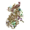
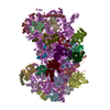
  • Deposited structure unit
  • Imaged by Jmol
  • Download
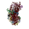
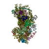
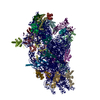
  • Simplified surface model + fitted atomic model
  • EMDB-2603
  • Imaged by Jmol
  • Download
  • Simplified surface model + fitted atomic model
  • EMDB-2604
  • Imaged by Jmol
  • Download
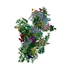
  • Superimposition on EM map
  • EMDB-2603
  • Imaged by UCSF Chimera
  • Download
  • Superimposition on EM map
  • EMDB-2604
  • Imaged by UCSF Chimera
  • Download
Movie viewer
Structure viewerMolecule:
MolmilJmol/JSmol

Downloads & links

-
Assembly

Deposited unit
A2: 18S RRNA
AZ: RNA OF CRICKET-PARALYSIS-VIRUS-IRES
BA: US2
BB: ES1
BC: US5
BD: US3
BE: ES4
BF: US7
BG: ES6
BH: ES7
BI: ES8
BJ: US4
BK: ES10
BL: US17
BM: ES12
BN: US15
BO: US11
BP: US19
BQ: US9
BR: ES17
BS: US13
BT: ES19
BU: US10
BV: ES21
BW: US8
BX: US12
BY: ES24
BZ: ES25
Ba: ES26
Bb: ES27
Bc: ES28
Bd: US14
Be: ES30
Bf: ES31
Bg: RACK1


Theoretical massNumber of molelcules
Total (without water)1,164,74335
Polymers1,164,74335
Non-polymers00
Water0
1


  • Idetical with deposited unit
  • defined by author
TypeNameSymmetry operationNumber
identity operation1_555x,y,z1

-
Components

-
RNA chain , 2 types, 2 molecules A2AZ

#1: RNA chain 18S RRNA / 18S ribosomal RNA


Mass: 569191.250 Da / Num. of mol.: 1 / Source method: isolated from a natural source / Details: 18S RRNA / Source: (natural) KLUYVEROMYCES LACTIS (yeast)
#2: RNA chain RNA OF CRICKET-PARALYSIS-VIRUS-IRES


Mass: 60767.703 Da / Num. of mol.: 1 / Source method: isolated from a natural source / Details: CRICKET-PARALYSIS-VIRUS-IRES / Source: (natural) CRICKET PARALYSIS VIRUS

+
Protein , 33 types, 33 molecules BABBBCBDBEBFBGBHBIBJBKBLBMBNBOBPBQBRBSBTBUBVBWBXBYBZBaBbBcBd...

#3: Protein US2


Mass: 22811.074 Da / Num. of mol.: 1 / Source method: isolated from a natural source / Source: (natural) KLUYVEROMYCES LACTIS (yeast)
#4: Protein ES1


Mass: 23982.734 Da / Num. of mol.: 1 / Source method: isolated from a natural source / Source: (natural) KLUYVEROMYCES LACTIS (yeast)
#5: Protein US5


Mass: 23084.850 Da / Num. of mol.: 1 / Source method: isolated from a natural source / Source: (natural) KLUYVEROMYCES LACTIS (yeast)
#6: Protein US3


Mass: 24631.713 Da / Num. of mol.: 1 / Source method: isolated from a natural source / Source: (natural) KLUYVEROMYCES LACTIS (yeast)
#7: Protein ES4


Mass: 29338.133 Da / Num. of mol.: 1 / Source method: isolated from a natural source / Source: (natural) KLUYVEROMYCES LACTIS (yeast)
#8: Protein US7


Mass: 22822.223 Da / Num. of mol.: 1 / Source method: isolated from a natural source / Source: (natural) KLUYVEROMYCES LACTIS (yeast)
#9: Protein ES6


Mass: 25778.867 Da / Num. of mol.: 1 / Source method: isolated from a natural source / Source: (natural) KLUYVEROMYCES LACTIS (yeast)
#10: Protein ES7


Mass: 21000.492 Da / Num. of mol.: 1 / Source method: isolated from a natural source / Source: (natural) KLUYVEROMYCES LACTIS (yeast)
#11: Protein ES8


Mass: 21018.000 Da / Num. of mol.: 1 / Source method: isolated from a natural source / Source: (natural) KLUYVEROMYCES LACTIS (yeast)
#12: Protein US4


Mass: 20608.047 Da / Num. of mol.: 1 / Source method: isolated from a natural source / Source: (natural) KLUYVEROMYCES LACTIS (yeast)
#13: Protein ES10


Mass: 11054.485 Da / Num. of mol.: 1 / Source method: isolated from a natural source / Source: (natural) KLUYVEROMYCES LACTIS (yeast)
#14: Protein US17


Mass: 16272.149 Da / Num. of mol.: 1 / Source method: isolated from a natural source / Source: (natural) KLUYVEROMYCES LACTIS (yeast)
#15: Protein ES12


Mass: 12926.904 Da / Num. of mol.: 1 / Source method: isolated from a natural source / Source: (natural) KLUYVEROMYCES LACTIS (yeast)
#16: Protein US15


Mass: 16928.748 Da / Num. of mol.: 1 / Source method: isolated from a natural source / Source: (natural) KLUYVEROMYCES LACTIS (yeast)
#17: Protein US11


Mass: 13446.426 Da / Num. of mol.: 1 / Source method: isolated from a natural source / Source: (natural) KLUYVEROMYCES LACTIS (yeast)
#18: Protein US19


Mass: 13060.371 Da / Num. of mol.: 1 / Source method: isolated from a natural source / Source: (natural) KLUYVEROMYCES LACTIS (yeast)
#19: Protein US9


Mass: 15329.792 Da / Num. of mol.: 1 / Source method: isolated from a natural source / Source: (natural) KLUYVEROMYCES LACTIS (yeast)
#20: Protein ES17


Mass: 13882.165 Da / Num. of mol.: 1 / Source method: isolated from a natural source / Source: (natural) KLUYVEROMYCES LACTIS (yeast)
#21: Protein US13


Mass: 16340.733 Da / Num. of mol.: 1 / Source method: isolated from a natural source / Source: (natural) KLUYVEROMYCES LACTIS (yeast)
#22: Protein ES19


Mass: 15713.815 Da / Num. of mol.: 1 / Source method: isolated from a natural source / Source: (natural) KLUYVEROMYCES LACTIS (yeast)
#23: Protein US10


Mass: 11819.814 Da / Num. of mol.: 1 / Source method: isolated from a natural source / Source: (natural) KLUYVEROMYCES LACTIS (yeast)
#24: Protein ES21


Mass: 9758.829 Da / Num. of mol.: 1 / Source method: isolated from a natural source / Source: (natural) KLUYVEROMYCES LACTIS (yeast)
#25: Protein US8


Mass: 14518.867 Da / Num. of mol.: 1 / Source method: isolated from a natural source / Source: (natural) KLUYVEROMYCES LACTIS (yeast)
#26: Protein US12


Mass: 15654.417 Da / Num. of mol.: 1 / Source method: isolated from a natural source / Source: (natural) KLUYVEROMYCES LACTIS (yeast)
#27: Protein ES24


Mass: 15231.650 Da / Num. of mol.: 1 / Source method: isolated from a natural source / Source: (natural) KLUYVEROMYCES LACTIS (yeast)
#28: Protein ES25


Mass: 7366.631 Da / Num. of mol.: 1 / Source method: isolated from a natural source / Source: (natural) KLUYVEROMYCES LACTIS (yeast)
#29: Protein ES26


Mass: 11022.989 Da / Num. of mol.: 1 / Source method: isolated from a natural source / Source: (natural) KLUYVEROMYCES LACTIS (yeast)
#30: Protein ES27


Mass: 8762.195 Da / Num. of mol.: 1 / Source method: isolated from a natural source / Source: (natural) KLUYVEROMYCES LACTIS (yeast)
#31: Protein ES28


Mass: 7116.281 Da / Num. of mol.: 1 / Source method: isolated from a natural source / Source: (natural) KLUYVEROMYCES LACTIS (yeast)
#32: Protein US14


Mass: 6206.189 Da / Num. of mol.: 1 / Source method: isolated from a natural source / Source: (natural) KLUYVEROMYCES LACTIS (yeast)
#33: Protein ES30


Mass: 6284.500 Da / Num. of mol.: 1 / Source method: isolated from a natural source / Source: (natural) KLUYVEROMYCES LACTIS (yeast)
#34: Protein ES31


Mass: 6572.660 Da / Num. of mol.: 1 / Source method: isolated from a natural source / Source: (natural) KLUYVEROMYCES LACTIS (yeast)
#35: Protein RACK1 / Receptor for activated C kinase 1


Mass: 34437.766 Da / Num. of mol.: 1 / Source method: isolated from a natural source / Source: (natural) KLUYVEROMYCES LACTIS (yeast)

-
Experimental details

-
Experiment

ExperimentMethod: ELECTRON MICROSCOPY
EM experimentAggregation state: PARTICLE / 3D reconstruction method: single particle reconstruction

-
Sample preparation

ComponentName: Kluyveromyces lactis 80S ribosome in complex with CrPV-IRES
Type: RIBOSOME
Buffer solutionpH: 6.5
SpecimenEmbedding applied: NO / Shadowing applied: NO / Staining applied: NO / Vitrification applied: YES
Specimen supportDetails: CARBON
VitrificationInstrument: FEI VITROBOT MARK I / Cryogen name: PROPANE / Details: LIQUID ETHANE

-
Electron microscopy imaging

Experimental equipment
Model: Titan Krios / Image courtesy: FEI Company
MicroscopyModel: FEI TITAN KRIOS / Date: Jul 7, 2013
Electron gunElectron source: FIELD EMISSION GUN / Accelerating voltage: 300 kV / Illumination mode: OTHER
Electron lensMode: BRIGHT FIELDBright-field microscopy / Nominal magnification: 47000 X / Nominal defocus max: 3 nm / Nominal defocus min: 1.8 nm / Cs: 2.7 mm
Image recordingElectron dose: 40 e/Å2 / Film or detector model: FEI FALCON II (4k x 4k)
Image scansNum. digital images: 1900
Radiation wavelengthRelative weight: 1

-
Processing

EM software
IDNameCategory
1REFMACmodel fitting
2UCSF Chimeramodel fitting
3RELION3D reconstruction
CTF correctionDetails: EACH PARTICLE
SymmetryPoint symmetry: C1 (asymmetric)
3D reconstructionMethod: RELIONList of Walmart brands / Resolution: 3.7 Å / Num. of particles: 18132 / Nominal pixel size: 1.34 Å / Symmetry type: POINT
Atomic model buildingB value: 60 / Protocol: FLEXIBLE FIT / Space: RECIPROCAL / Target criteria: R-FACTOR, FSC / Details: METHOD--FLEXIBLE
Atomic model buildingPDB-ID: 3B31
RefinementHighest resolution: 3.7 Å
Refinement stepCycle: LAST / Highest resolution: 3.7 Å
ProteinNucleic acidLigandSolventTotal
Num. atoms0 41597 0 0 41597

+
About Yorodumi

-
News

-
Feb 9, 2022. New format data for meta-information of EMDB entries

New format data for meta-information of EMDB entries

  • Version 3 of the EMDB header file is now the official format.
  • The previous official version 1.9 will be removed from the archive.

Related info.:EMDB header

External links:wwPDB to switch to version 3 of the EMDB data model

-
Aug 12, 2020. Covid-19 info

Covid-19 info

URL: https://pdbj.org/emnavi/covid19.php

New page: Covid-19 featured information page in EM Navigator.

Related info.:Covid-19 info / Mar 5, 2020. Novel coronavirus structure data

+
Mar 5, 2020. Novel coronavirus structure data

Novel coronavirus structure data

Related info.:Yorodumi Speices / Aug 12, 2020. Covid-19 info

External links:COVID-19 featured content - PDBj / Molecule of the Month (242):Coronavirus Proteases

+
Jan 31, 2019. EMDB accession codes are about to change! (news from PDBe EMDB page)

EMDB accession codes are about to change! (news from PDBe EMDB page)

  • The allocation of 4 digits for EMDB accession codes will soon come to an end. Whilst these codes will remain in use, new EMDB accession codes will include an additional digit and will expand incrementally as the available range of codes is exhausted. The current 4-digit format prefixed with “EMD-” (i.e. EMD-XXXX) will advance to a 5-digit format (i.e. EMD-XXXXX), and so on. It is currently estimated that the 4-digit codes will be depleted around Spring 2019, at which point the 5-digit format will come into force.
  • The EM Navigator/Yorodumi systems omit the EMD- prefix.

Related info.:Q: What is EMD? / ID/Accession-code notation in Yorodumi/EM Navigator

External links:EMDB Accession Codes are Changing Soon! / Contact to PDBj

+
Jul 12, 2017. Major update of PDB

Major update of PDB

  • wwPDB released updated PDB data conforming to the new PDBx/mmCIF dictionary.
  • This is a major update changing the version number from 4 to 5, and with Remediation, in which all the entries are updated.
  • In this update, many items about electron microscopy experimental information are reorganized (e.g. em_software).
  • Now, EM Navigator and Yorodumi are based on the updated data.

External links:wwPDB Remediation / Enriched Model Files Conforming to OneDep Data Standards Now Available in the PDB FTP Archive

-
Yorodumi

Thousand views of thousand structures

  • Yorodumi is a browser for structure data from EMDB, PDB, SASBDB, etc.
  • This page is also the successor to EM Navigator detail page, and also detail information page/front-end page for Omokage search.
  • The word "yorodu" (or yorozu) is an old Japanese word meaning "ten thousand". "mi" (miru) is to see.

Related info.:EMDB / PDB / SASBDB / Comparison of 3 databanks / Yorodumi Search / Aug 31, 2016. New EM Navigator & Yorodumi / Yorodumi Papers / Jmol/JSmol / Function and homology information / Changes in new EM Navigator and Yorodumi

Read more