 Movie
Movie Controller


+ Open data

- Basic information
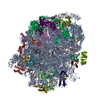
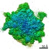
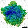
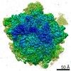
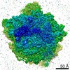
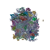
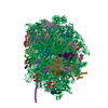
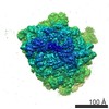
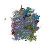
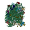
| Entry | Database: PDB / ID: 4v91 | ||||||||||||
|---|---|---|---|---|---|---|---|---|---|---|---|---|---|
| Title | Kluyveromyces lactis 80S ribosome in complex with CrPV-IRES | ||||||||||||
 Components Components |
| ||||||||||||
 Keywords Keywords |  RIBOSOME / TRANSLATION / INITIATION / IRES RIBOSOME / TRANSLATION / INITIATION / IRES | ||||||||||||
| Function / homology | RNA / RNA (> 10) / RNA (> 100) / RNA (> 1000) Function and homology information Function and homology information | ||||||||||||
| Biological species |  KLUYVEROMYCES LACTIS (yeast) KLUYVEROMYCES LACTIS (yeast) | ||||||||||||
| Method | ELECTRON MICROSCOPY / single particle reconstruction / cryo EM / Resolution: 3.7 Å | ||||||||||||
 Authors Authors | Fernandez, I.S. / Bai, X. / Scheres, S.H.W. / Ramakrishnan, V. | ||||||||||||
 Citation Citation | Journal: Cell / Year: 2014 Title: Initiation of translation by cricket paralysis virus IRES requires its translocation in the ribosome. Authors: Israel S Fernández / Xiao-Chen Bai / Garib Murshudov / Sjors H W Scheres / V Ramakrishnan /  Abstract: The cricket paralysis virus internal ribosome entry site (CrPV-IRES) is a folded structure in a viral mRNA that allows initiation of translation in the absence of any host initiation factors. By ...The cricket paralysis virus internal ribosome entry site (CrPV-IRES) is a folded structure in a viral mRNA that allows initiation of translation in the absence of any host initiation factors. By using recent advances in single-particle electron cryomicroscopy, we have solved the structure of CrPV-IRES bound to the ribosome of the yeast Kluyveromyces lactis in both the canonical and rotated states at overall resolutions of 3.7 and 3.8 Å, respectively. In both states, the pseudoknot PKI of the CrPV-IRES mimics a tRNA/mRNA interaction in the decoding center of the A site of the 40S ribosomal subunit. The structure and accompanying factor-binding data show that CrPV-IRES binding mimics a pretranslocation rather than initiation state of the ribosome. Translocation of the IRES by elongation factor 2 (eEF2) is required to bring the first codon of the mRNA into the A site and to allow the start of translation. | ||||||||||||
| History |
|
- Structure visualization
| Movie |
Movie viewer |
|---|---|
| Structure viewer | Molecule: MolmilJmol/JSmol |
- Downloads & links
-Download
| PDBx/mmCIF format | 4v91.cif.gz | 3.1 MB | Display | PDBx/mmCIF format |
|---|---|---|---|---|
| PDB format | pdb4v91.ent.gz | Display | PDB format | |
| PDBx/mmJSON format | 4v91.json.gz | Tree view | PDBx/mmJSON format | |
| Others |  Other downloads Other downloads |
-Validation report
| Arichive directory | https://data.pdbj.org/pub/pdb/validation_reports/v9/4v91ftp://data.pdbj.org/pub/pdb/validation_reports/v9/4v91 | HTTPS FTP |
|---|
-Related structure data
| Related structure data |  2599MC 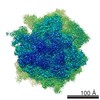 2603C 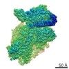 2604C 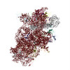 4v92C M: map data used to model this data C: citing same article ( |
|---|---|
| Similar structure data |
-Links
 PDBj
PDBj


- Assembly
| Deposited unit | 
|
|---|---|
| 1 |
|
-Components
-RNA chain , 3 types, 3 molecules 134
| #1: RNA chain | Mass: 1097866.125 Da / Num. of mol.: 1 / Source method: isolated from a natural source / Source: (natural) KLUYVEROMYCES LACTIS (yeast) |
|---|---|
| #2: RNA chain | 5S ribosomal RNA Mass: 38951.105 Da / Num. of mol.: 1 / Source method: isolated from a natural source / Source: (natural) KLUYVEROMYCES LACTIS (yeast) |
| #3: RNA chain | 5.8S ribosomal RNA Mass: 50682.922 Da / Num. of mol.: 1 / Source method: isolated from a natural source / Source: (natural) KLUYVEROMYCES LACTIS (yeast) |
+Protein , 41 types, 41 molecules ABCDEFGHIJLMNOPQRSTUVWXYZabcde...
-Protein/peptide , 1 types, 1 molecules n
| #42: Protein/peptide | Mass: 3354.243 Da / Num. of mol.: 1 / Source method: isolated from a natural source / Details: RIBOSOMAL PROTEIN EL41 / Source: (natural) KLUYVEROMYCES LACTIS (yeast) |
|---|
-Experimental details
-Experiment
| Experiment | Method: ELECTRON MICROSCOPY |
|---|---|
| EM experiment | Aggregation state: PARTICLE / 3D reconstruction method: single particle reconstruction |
- Sample preparation
| Component | Name: Kluyveromyces lactis 80S ribosome in complex with CrPV-IRES Type: RIBOSOME |
|---|---|
| Buffer solution | pH: 6.5 |
| Specimen | Embedding applied: NO / Shadowing applied: NO / Staining applied: NO / Vitrification applied: YES |
| Specimen support | Details: CARBON |
| Vitrification | Instrument: FEI VITROBOT MARK I / Cryogen name: PROPANE / Details: LIQUID ETHANE |
- Electron microscopy imaging
| Experimental equipment |  Model: Titan Krios / Image courtesy: FEI Company |
|---|---|
| Microscopy | Model: FEI TITAN KRIOS / Date: Jul 7, 2013 |
| Electron gun | Electron source: FIELD EMISSION GUN / Accelerating voltage: 300 kV / Illumination mode: OTHER |
| Electron lens | Mode: BRIGHT FIELDBright-field microscopy / Nominal magnification: 47000 X / Nominal defocus max: 3 nm / Nominal defocus min: 1.8 nm / Cs: 2.7 mm |
| Image recording | Electron dose: 40 e/Å2 / Film or detector model: FEI FALCON II (4k x 4k) |
| Image scans | Num. digital images: 1900 |
| Radiation wavelength | Relative weight: 1 |
- Processing
| EM software |
| ||||||||||||
|---|---|---|---|---|---|---|---|---|---|---|---|---|---|
| CTF correction | Details: EACH PARTICLE | ||||||||||||
| Symmetry | Point symmetry: C1 (asymmetric) | ||||||||||||
| 3D reconstruction | Method: RELIONList of Walmart brands / Resolution: 3.7 Å / Num. of particles: 18132 / Nominal pixel size: 1.34 Å / Symmetry type: POINT | ||||||||||||
| Atomic model building | B value: 60 / Protocol: FLEXIBLE FIT / Space: RECIPROCAL / Target criteria: R-FACTOR, FSC / Details: METHOD--FLEXIBLE | ||||||||||||
| Atomic model building | PDB-ID: 3B31 | ||||||||||||
| Refinement | Highest resolution: 3.7 Å | ||||||||||||
| Refinement step | Cycle: LAST / Highest resolution: 3.7 Å
|

