 Movie
Movie Controller

[English] 日本語
 Yorodumi
- PDB-4uwe: Structure of the ryanodine receptor at resolution of 8.5 A in par... -
+ Open data

- Basic information
| Entry | Database: PDB / ID: 4uwe | ||||||
|---|---|---|---|---|---|---|---|
| Title | Structure of the ryanodine receptor at resolution of 8.5 A in partially open state | ||||||
 Components Components | RYANODINE RECEPTOR 1 | ||||||
 Keywords Keywords | SIGNALING PROTEIN / CALCIUM BINDING / ION CHANNEL / MUSCULAR CONTRACTION / CONFORMATIONAL CHANGES. | ||||||
| Function / homology |  Function and homology information Function and homology informationATP-gated ion channel activity / terminal cisterna / ryanodine receptor complex / ryanodine-sensitive calcium-release channel activity / release of sequestered calcium ion into cytosol by sarcoplasmic reticulum / ossification involved in bone maturation / skin development / cellular response to caffeine / intracellularly gated calcium channel activity / outflow tract morphogenesis ...ATP-gated ion channel activity / terminal cisterna / ryanodine receptor complex / ryanodine-sensitive calcium-release channel activity / release of sequestered calcium ion into cytosol by sarcoplasmic reticulum / ossification involved in bone maturation / skin development / cellular response to caffeine / intracellularly gated calcium channel activity / outflow tract morphogenesis / organelle membrane / toxic substance binding / voltage-gated calcium channel activity / skeletal muscle fiber development / release of sequestered calcium ion into cytosol / sarcoplasmic reticulum membrane / cellular response to calcium ion / muscle contraction / sarcoplasmic reticulum / calcium ion transmembrane transport / calcium channel activity / intracellular calcium ion homeostasis / disordered domain specific binding / protein homotetramerization / transmembrane transporter binding / calmodulin binding / calcium ion binding / ATP binding / membrane / identical protein bindingSimilarity search - Function | ||||||
| Biological species |  ORYCTOLAGUS CUNICULUS (rabbit) ORYCTOLAGUS CUNICULUS (rabbit) | ||||||
| Method | ELECTRON MICROSCOPY / single particle reconstruction / cryo EM / Resolution: 8.5 Å | ||||||
 Authors Authors | Efremov, R.G. / Leitner, A. / Aebersold, R. / Raunser, S. | ||||||
 Citation Citation | Journal: Nature / Year: 2015 Title: Architecture and conformational switch mechanism of the ryanodine receptor. Authors: Rouslan G Efremov / Alexander Leitner / Ruedi Aebersold / Stefan Raunser /    Abstract: Muscle contraction is initiated by the release of calcium (Ca(2+)) from the sarcoplasmic reticulum into the cytoplasm of myocytes through ryanodine receptors (RyRs). RyRs are homotetrameric channels ...Muscle contraction is initiated by the release of calcium (Ca(2+)) from the sarcoplasmic reticulum into the cytoplasm of myocytes through ryanodine receptors (RyRs). RyRs are homotetrameric channels with a molecular mass of more than 2.2 megadaltons that are regulated by several factors, including ions, small molecules and proteins. Numerous mutations in RyRs have been associated with human diseases. The molecular mechanism underlying the complex regulation of RyRs is poorly understood. Using electron cryomicroscopy, here we determine the architecture of rabbit RyR1 at a resolution of 6.1 Å. We show that the cytoplasmic moiety of RyR1 contains two large α-solenoid domains and several smaller domains, with folds suggestive of participation in protein-protein interactions. The transmembrane domain represents a chimaera of voltage-gated sodium and pH-activated ion channels. We identify the calcium-binding EF-hand domain and show that it functions as a conformational switch allosterically gating the channel. | ||||||
| History |
| ||||||
| Remark 700 | SHEET DETERMINATION METHOD: DSSP THE SHEETS PRESENTED AS "AA" IN EACH CHAIN ON SHEET RECORDS BELOW ... SHEET DETERMINATION METHOD: DSSP THE SHEETS PRESENTED AS "AA" IN EACH CHAIN ON SHEET RECORDS BELOW IS ACTUALLY AN 6-STRANDED BARREL THIS IS REPRESENTED BY A 7-STRANDED SHEET IN WHICH THE FIRST AND LAST STRANDS ARE IDENTICAL. THE SHEETS PRESENTED AS "BA" IN EACH CHAIN ON SHEET RECORDS BELOW IS ACTUALLY AN 6-STRANDED BARREL THIS IS REPRESENTED BY A 7-STRANDED SHEET IN WHICH THE FIRST AND LAST STRANDS ARE IDENTICAL. THE SHEETS PRESENTED AS "CA" IN EACH CHAIN ON SHEET RECORDS BELOW IS ACTUALLY AN 6-STRANDED BARREL THIS IS REPRESENTED BY A 7-STRANDED SHEET IN WHICH THE FIRST AND LAST STRANDS ARE IDENTICAL. THE SHEETS PRESENTED AS "DA" IN EACH CHAIN ON SHEET RECORDS BELOW IS ACTUALLY AN 6-STRANDED BARREL THIS IS REPRESENTED BY A 7-STRANDED SHEET IN WHICH THE FIRST AND LAST STRANDS ARE IDENTICAL. |
- Structure visualization
| Movie |
Movie viewer |
|---|---|
| Structure viewer | Molecule: MolmilJmol/JSmol |
- Downloads & links
-Download
| PDBx/mmCIF format | 4uwe.cif.gz | 1.8 MB | Display | PDBx/mmCIF format |
|---|---|---|---|---|
| PDB format | pdb4uwe.ent.gz | 1.4 MB | Display | PDB format |
| PDBx/mmJSON format | 4uwe.json.gz | Tree view | PDBx/mmJSON format | |
| Others |  Other downloads Other downloads |
-Validation report
| Arichive directory | https://data.pdbj.org/pub/pdb/validation_reports/uw/4uweftp://data.pdbj.org/pub/pdb/validation_reports/uw/4uwe | HTTPS FTP |
|---|
-Related structure data
| Related structure data |  2752MC 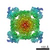 2751C 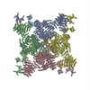 4uwaC C: citing same article ( M: map data used to model this data |
|---|---|
| Similar structure data |


-Links
 PDBj
PDBj
- Assembly
| Deposited unit | 
|
|---|---|
| 1 |
|
-Components
| #1: Protein | / RYR-1 / RYR1 / SKELETAL MUSCLE CALCIUM RELEASE CHANNEL / SKELETAL MUSCLE RYANODINE RECEPTOR / ...RYR-1 / RYR1 / SKELETAL MUSCLE CALCIUM RELEASE CHANNEL / SKELETAL MUSCLE RYANODINE RECEPTOR / SKELETAL MUSCLE-TYPE RYANODINE RECEPTOR / TYPE 1 RYANODINE RECEPTOR Mass: 473628.906 Da / Num. of mol.: 4 / Source method: isolated from a natural source / Source: (natural) ORYCTOLAGUS CUNICULUS (rabbit) / Cell: MYOCYTE / Organ: SARCOPLASMIC RETICULUM / Tissue: MUSCLESkeletal muscle / References: UniProt: P11716 |
|---|
-Experimental details
-Experiment
| Experiment | Method: ELECTRON MICROSCOPY |
|---|---|
| EM experiment | Aggregation state: PARTICLE / 3D reconstruction method: single particle reconstruction |
- Sample preparation
| Component | Name: RYANODINE RECEPTOR 1 / Type: COMPLEX |
|---|---|
| Buffer solution | Name: 10 MM MOPS, 200 MM NACL, 2 MM DTT AND 10 MM CACL2, 0.2% FLUORINATED OCTYL- MALTOSIDE pH: 7.4 Details: 10 MM MOPS, 200 MM NACL, 2 MM DTT AND 10 MM CACL2, 0.2% FLUORINATED OCTYL- MALTOSIDE |
| Specimen | Conc.: 2 mg/ml / Embedding applied: NO / Shadowing applied: NO / Staining applied: NO / Vitrification applied: YES |
| Specimen support | Details: HOLEY CARBON |
| Vitrification | Instrument: GATAN CRYOPLUNGE 3 / Cryogen name: ETHANE / Details: LIQUID ETHANE |
- Electron microscopy imaging
| Microscopy | Model: JEOL 3200FSC / Date: Mar 12, 2012 |
|---|---|
| Electron gun | Electron source: FIELD EMISSION GUN / Accelerating voltage: 200 kV / Illumination mode: FLOOD BEAM |
| Electron lens | Mode: BRIGHT FIELDBright-field microscopy / Nominal magnification: 60000 X / Calibrated magnification: 58610 X / Nominal defocus max: 3900 nm / Nominal defocus min: 900 nm / Cs: 4.1 mm |
| Specimen holder | Temperature: 80 K |
| Image recording | Electron dose: 20 e/Å2 / Film or detector model: TVIPS TEMCAM-F816 (8k x 8k) |
| Image scans | Num. digital images: 1041 |
- Processing
| EM software |
| ||||||||||||
|---|---|---|---|---|---|---|---|---|---|---|---|---|---|
| CTF correction | Details: FULL CORRECTION | ||||||||||||
| Symmetry | Point symmetry: C4 (4 fold cyclic) | ||||||||||||
| 3D reconstruction | Method: BACKPROJECTION / Resolution: 8.5 Å / Num. of particles: 94354 / Nominal pixel size: 2.53 Å / Actual pixel size: 2.59 Å Magnification calibration: CROSS CORRELATION WITH A MAP CALCULATED FROM CRYSTAL STRUCTURE OF ABC DOMAIN Details: SUBMISSION BASED ON EXPERIMENTAL DATA FROM EMDB EMD-2752. (DEPOSITION ID: 12779). Symmetry type: POINT | ||||||||||||
| Atomic model building | Protocol: RIGID BODY FIT / Space: REAL / Details: METHOD--RIGID BODY REFINEMENT PROTOCOL--EM | ||||||||||||
| Atomic model building | PDB-ID: 2UWA | ||||||||||||
| Refinement | Highest resolution: 8.5 Å | ||||||||||||
| Refinement step | Cycle: LAST / Highest resolution: 8.5 Å
|

