 Movie
Movie Controller

[English] 日本語
 Yorodumi
- PDB-4bpq: Structure and substrate induced conformational changes of the sec... -
+ Open data

- Basic information
| Entry | Database: PDB / ID: 4bpq | ||||||
|---|---|---|---|---|---|---|---|
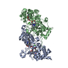
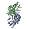
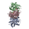
| Title | Structure and substrate induced conformational changes of the secondary citrate-sodium symporter CitS revealed by electron crystallography | ||||||
 Components Components | CITRATE\:SODIUM SYMPORTER | ||||||
 Keywords Keywords |  TRANSPORT PROTEIN / SECONDARY TRANSPORTER / MEMBRANE PROTEIN TRANSPORT PROTEIN / SECONDARY TRANSPORTER / MEMBRANE PROTEIN | ||||||
| Biological species |  KLEBSIELLA PNEUMONIAE (bacteria) KLEBSIELLA PNEUMONIAE (bacteria) | ||||||
| Method | ELECTRON CRYSTALLOGRAPHY / electron crystallography / cryo EM / Resolution: 6 Å | ||||||
 Authors Authors | Kebbel, F. / Kurz, M. / Arheit, M. / Gruetter, M.G. / Stahlberg, H. | ||||||
 Citation Citation | Journal: Structure / Year: 2013 Title: Structure and substrate-induced conformational changes of the secondary citrate/sodium symporter CitS revealed by electron crystallography. Authors: Fabian Kebbel / Mareike Kurz / Marcel Arheit / Markus G Grütter / Henning Stahlberg /  Abstract: The secondary Na+/citrate symporter CitS of Klebsiella pneumoniae is the best-characterized member of the 2-hydroxycarboxylate transporter family. The recent projection structure gave insight into ...The secondary Na+/citrate symporter CitS of Klebsiella pneumoniae is the best-characterized member of the 2-hydroxycarboxylate transporter family. The recent projection structure gave insight into its overall structural organization. Here, we present the three-dimensional map of dimeric CitS obtained with electron crystallography. Each monomer has 13 a-helical transmembrane segments; six are organized in a distal helix cluster and seven in the central dimer interface domain. Based on structural analyses and comparison to VcINDY, we propose a molecular model for CitS, assign the helices, and demonstrate the internal structural symmetry. We also present projections of CitS in several conformational states induced by the presence and absence of sodium and citrate as substrates. Citrate binding induces a defined movement of a helices within the distal helical cluster. Based on this, we propose a substrate translocation site and conformational changes that are in agreement with the transport model of ‘‘alternating access’’. | ||||||
| History |
|
- Structure visualization
| Movie |
 Movie viewer Movie viewer |
|---|---|
| Structure viewer | Molecule: MolmilJmol/JSmol |
- Downloads & links
-Download
| PDBx/mmCIF format | 4bpq.cif.gz | 71.4 KB | Display | PDBx/mmCIF format |
|---|---|---|---|---|
| PDB format | pdb4bpq.ent.gz | 55 KB | Display | PDB format |
| PDBx/mmJSON format | 4bpq.json.gz | Tree view | PDBx/mmJSON format | |
| Others |  Other downloads Other downloads |
-Validation report
| Arichive directory | https://data.pdbj.org/pub/pdb/validation_reports/bp/4bpqftp://data.pdbj.org/pub/pdb/validation_reports/bp/4bpq | HTTPS FTP |
|---|
-Related structure data
| Related structure data | 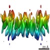 2387MC M: map data used to model this data C: citing same article ( |
|---|---|
| Similar structure data |


-Links
 PDBj
PDBj- Assembly
| Deposited unit | 
| ||||||||
|---|---|---|---|---|---|---|---|---|---|
| 1 |
| ||||||||
| Unit cell |
|
-Components
| #1: Protein | Mass: 27081.221 Da / Num. of mol.: 2 Source method: isolated from a genetically manipulated source Source: (gene. exp.) KLEBSIELLA PNEUMONIAE (bacteria) / Production host: ESCHERICHIA COLI (E. coli)Sequence details | THE SAMPLE SEQUENCE HAS UNIPROT ACCESSION B5Y216. BUT THE REGISTER OF THE RESIDUES ON THE EM VOLUME ...THE SAMPLE SEQUENCE HAS UNIPROT ACCESSION B5Y216. BUT THE REGISTER OF THE RESIDUES ON THE EM VOLUME MAP IS UNKNOWN, HENCE THE MODEL IS BUILT IN AS A POLY-ALA MODEL. THE COORDINATE | |
|---|
-Experimental details
-Experiment
| Experiment | Method: ELECTRON CRYSTALLOGRAPHY / Number of used crystals: 79 |
|---|---|
| EM experiment | Aggregation state: 2D ARRAY / 3D reconstruction method: electron crystallography |
- Sample preparation
| Component | Name: secondary citrate-sodium symporter CitS / Type: COMPLEX |
|---|---|
| Buffer solution | pH: 4.5 |
| Specimen | Embedding applied: NO / Shadowing applied: NO / Staining applied: NO / Vitrification applied: YES |
| Vitrification | Instrument: FEI VITROBOT MARK IV / Cryogen name: ETHANE |
| Crystal grow | pH: 4.5 / Details: pH 4.5 |
-Data collection
| Microscopy | Model: FEI/PHILIPS CM200FEG / Date: Dec 31, 2012 |
|---|---|
| Electron gun | Electron source: FIELD EMISSION GUN / Accelerating voltage: 200 kV / Illumination mode: FLOOD BEAM |
| Electron lens | Mode: BRIGHT FIELDBright-field microscopy / Nominal magnification: 50000 X / Nominal defocus max: 2500 nm / Nominal defocus min: 300 nm |
| Image recording | Electron dose: 6 e/Å2 / Film or detector model: KODAK SO-163 FILM |
| Diffraction | Mean temperature: 87 K |
| Detector | Date: Dec 12, 2012 |
| Radiation wavelength | Relative weight: 1 |
| Reflection | Highest resolution: 6 Å / Num. obs: 11480 / % possible obs: 79 % |
- Processing
| Software |
| ||||||||||||
|---|---|---|---|---|---|---|---|---|---|---|---|---|---|
| 3D reconstruction | Resolution: 6 Å / Resolution method: OTHER / Symmetry type: 2D CRYSTAL | ||||||||||||
| Refinement | Highest resolution: 6 Å | ||||||||||||
| Refinement step | Cycle: LAST / Highest resolution: 6 Å
|
