 Movie
Movie Controller

[English] 日本語
 Yorodumi
- PDB-4bed: Keyhole limpet hemocyanin (KLH): 9A cryoEM structure and molecula... -
+ Open data

- Basic information
| Entry | Database: PDB / ID: 4bed | |||||||||
|---|---|---|---|---|---|---|---|---|---|---|
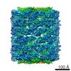
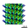
| Title | Keyhole limpet hemocyanin (KLH): 9A cryoEM structure and molecular model of the KLH1 didecamer reveal the interfaces and intricate topology of the 160 functional units | |||||||||
 Components Components | (HEMOCYANIN KLH1) x 2 | |||||||||
 Keywords Keywords |  OXYGEN TRANSPORT / KEYHOLE LIMPET HEMOCYANIN / KLH / GASTROPODA / OXYGEN CARRIER OXYGEN TRANSPORT / KEYHOLE LIMPET HEMOCYANIN / KLH / GASTROPODA / OXYGEN CARRIER | |||||||||
| Function / homology |  Function and homology informationoxygen carrier activity / oxidoreductase activity / extracellular region / metal ion binding Function and homology informationoxygen carrier activity / oxidoreductase activity / extracellular region / metal ion bindingSimilarity search - Function | |||||||||
| Biological species |  MEGATHURA CRENULATA (invertebrata) MEGATHURA CRENULATA (invertebrata) | |||||||||
| Method | ELECTRON MICROSCOPY / single particle reconstruction / cryo EM / Resolution: 9 Å | |||||||||
 Authors Authors | Gatsogiannis, C. / Markl, J. | |||||||||
 Citation Citation | Journal: J Mol Biol / Year: 2009 Title: Keyhole limpet hemocyanin: 9-A CryoEM structure and molecular model of the KLH1 didecamer reveal the interfaces and intricate topology of the 160 functional units. Authors: Christos Gatsogiannis / Jürgen Markl /  Abstract: Hemocyanins are blue copper-containing respiratory proteins in the hemolymph of many arthropods and molluscs. Molluscan hemocyanins are decamers, didecamers, or multidecamers of a 340- to 400-kDa ...Hemocyanins are blue copper-containing respiratory proteins in the hemolymph of many arthropods and molluscs. Molluscan hemocyanins are decamers, didecamers, or multidecamers of a 340- to 400-kDa polypeptide subunit containing seven or eight globular functional units (FUs; FU-a to FU-h), each with an oxygen-binding site. The decamers are short 35-nm hollow cylinders, with their lumen narrowed by a collar complex. Our recently published 9-A cryo-electron microscopy/crystal structure hybrid model of a 3.4-MDa cephalopod hemocyanin decamer [Nautilus pompilius hemocyanin (NpH)] revealed the pathway of the seven-FU subunit (340 kDa), 15 types of inter-FU interface, and an asymmetric collar consisting of five "arcs" (FU-g pairs). We now present a comparable hybrid model of an 8-MDa gastropod hemocyanin didecamer assembled from two asymmetric decamers [isoform keyhole limpet hemocyanin (KLH) 1 of the established immunogen KLH]. Compared to NpH, the KLH1 subunit (400 kDa) is C-terminally elongated by FU-h, which is further extended by a unique tail domain. We have found that the wall-and-arc structure of the KLH1 decamer is very similar to that of NpH. We have traced the subunit pathway and how it continues from KLH1-g to KLH1-h to form an annulus of five "slabs" (FU-h pairs) at one cylinder edge. The 15 types of inter-FU interface detected in NpH are also present in KLH1. Moreover, we have identified one arc/slab interface, two slab/slab interfaces, five slab/wall interfaces, and four decamer/decamer interfaces. The 27 interfaces are described on the basis of two subunit conformers, yielding an asymmetric homodimer. Six protrusions from the cryo-electron microscopy structure per subunit are associated with putative attachment sites for N-linked glycans, indicating a total of 120 sugar trees in KLH1. Also, putative binding sites for divalent cations have been detected. In conclusion, the present 9-A data on KLH1 confirm and substantially broaden our recent analysis of the smaller cephalopod hemocyanin and essentially solve the gastropod hemocyanin structure. #1: Journal: Biochim Biophys Acta / Year: 2013 Title: Evolution of molluscan hemocyanin structures. Authors: Jürgen Markl / Abstract: Hemocyanin transports oxygen in the hemolymph of many molluscs and arthropods and is therefore a central physiological factor in these animals. Molluscan hemocyanin molecules are oligomers composed ...Hemocyanin transports oxygen in the hemolymph of many molluscs and arthropods and is therefore a central physiological factor in these animals. Molluscan hemocyanin molecules are oligomers composed of many protein subunits that in turn encompass subsets of distinct functional units. The structure and evolution of molluscan hemocyanin have been studied for decades, but it required the recent progress in DNA sequencing, X-ray crystallography and 3D electron microscopy to produce a detailed view of their structure and evolution. The basic quaternary structure is a cylindrical decamer 35nm in diameter, consisting of wall and collar (typically at one end of the cylinder). Depending on the animal species, decamers, didecamers and multidecamers occur in the hemolymph. Whereas the wall architecture of the decamer seems to be invariant, four different types of collar have been identified in different molluscan taxa. Correspondingly, there exist four subunit types that differ in their collar functional units and range from 350 to 550kDa. Thus, molluscan hemocyanin subunits are among the largest polypeptides in nature. In this report, recent 3D reconstructions are used to explain and visualize the different functional units, subunits and quaternary structures of molluscan hemocyanins. Moreover, on the basis of DNA analyses and structural considerations, their possible evolution is traced. This article is part of a Special Issue entitled: Oxygen Binding and Sensing Proteins. | |||||||||
| History |
|
- Structure visualization
| Movie |
Movie viewer |
|---|---|
| Structure viewer | Molecule: MolmilJmol/JSmol |
- Downloads & links
-Download
| PDBx/mmCIF format | 4bed.cif.gz | 1.2 MB | Display | PDBx/mmCIF format |
|---|---|---|---|---|
| PDB format | pdb4bed.ent.gz | 975.9 KB | Display | PDB format |
| PDBx/mmJSON format | 4bed.json.gz | Tree view | PDBx/mmJSON format | |
| Others |  Other downloads Other downloads |
-Validation report
| Arichive directory | https://data.pdbj.org/pub/pdb/validation_reports/be/4bedftp://data.pdbj.org/pub/pdb/validation_reports/be/4bed | HTTPS FTP |
|---|
-Related structure data
| Related structure data | 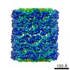 1569MC M: map data used to model this data C: citing same article ( |
|---|---|
| Similar structure data |
-Links
 PDBj
PDBj- Assembly
| Deposited unit | 
|
|---|---|
| 1 | x 10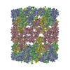
|
| 2 |
|
| 3 | 
|
| Symmetry | Point symmetry: (Schoenflies symbol: D (2xn fold dihedral)) |
-Components
| #1: Protein | Mass: 191623.828 Da / Num. of mol.: 2 / Source method: isolated from a natural source / Source: (natural) MEGATHURA CRENULATA (invertebrata) / References: UniProt: Q53IP9, UniProt: Q10583*PLUS#2: Protein | Mass: 199504.094 Da / Num. of mol.: 2 / Source method: isolated from a natural source / Source: (natural) MEGATHURA CRENULATA (invertebrata) / References: UniProt: Q53IP9, UniProt: Q10583*PLUS#3: Chemical | ChemComp-CUO /   Mass: 159.091 Da / Num. of mol.: 16 / Source method: obtained synthetically / Formula: Cu2O2 Mass: 159.091 Da / Num. of mol.: 16 / Source method: obtained synthetically / Formula: Cu2O2Sequence details | THIS PDB ENTRY IS BASED ON AN UPDATED SEQUENCE | |
|---|
-Experimental details
-Experiment
| Experiment | Method: ELECTRON MICROSCOPY |
|---|---|
| EM experiment | Aggregation state: PARTICLE / 3D reconstruction method: single particle reconstruction |
- Sample preparation
| Component | Name: KEYHOLE LIMPET HEMOCYANIN ISOFORM 1 (KLH1) / Type: COMPLEX |
|---|---|
| Buffer solution | Name: 50 MM TRIS-HCL, 150 MM NACL, 5MM CACL, 5MM MGCL2 / pH: 7.4 / Details: 50 MM TRIS-HCL, 150 MM NACL, 5MM CACL, 5MM MGCL2 |
| Specimen | Conc.: 0.7 mg/ml / Embedding applied: NO / Shadowing applied: NO / Staining applied: NO / Vitrification applied: YES |
| Specimen support | Details: HOLEY CARBON |
| Vitrification | Instrument: HOMEMADE PLUNGER / Cryogen name: ETHANE Details: VITRIFICATION INSTRUMENT - HOME MADE. VITRIFICATION CARRIED OUT IN 25 PERCENT OXYGEN ATMOSPHERE. SINGLE- SIDED BLOTTING |
- Electron microscopy imaging
| Experimental equipment |  Model: Tecnai F20 / Image courtesy: FEI Company |
|---|---|
| Microscopy | Model: FEI TECNAI F20 / Date: Jul 25, 2007 |
| Electron gun | Electron source: FIELD EMISSION GUN / Accelerating voltage: 200 kV / Illumination mode: FLOOD BEAM |
| Electron lens | Mode: BRIGHT FIELDBright-field microscopy / Nominal magnification: 50000 X / Calibrated magnification: 50000 X / Nominal defocus max: 4000 nm / Nominal defocus min: 800 nm / Cs: 2 mm |
| Specimen holder | Temperature: 68 K |
| Image recording | Film or detector model: KODAK SO-163 FILM |
| Image scans | Num. digital images: 98 |
- Processing
| EM software | Name: IMAGIC / Version: 5 / Category: 3D reconstruction | ||||||||||||
|---|---|---|---|---|---|---|---|---|---|---|---|---|---|
| CTF correction | Details: PER MICROGRAPH | ||||||||||||
| Symmetry | Point symmetry: D5 (2x5 fold dihedral) | ||||||||||||
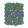
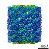
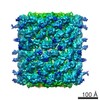
| 3D reconstruction | Resolution: 9 Å / Num. of particles: 4762 / Nominal pixel size: 1 Å / Actual pixel size: 1 Å Details: KLH1 IS A CYLINDRICAL 8 MDA PROTEIN WITH D5 SYMMETRY AND COMPOSED OF 20 IDENTICAL 400 KDA POLYPEPTIDE SUBUNITS. THE ASYMMETRIC UNIT IS A SUBUNIT DIMER (800 KDA). THE TWO CONSTITUENT ...Details: KLH1 IS A CYLINDRICAL 8 MDA PROTEIN WITH D5 SYMMETRY AND COMPOSED OF 20 IDENTICAL 400 KDA POLYPEPTIDE SUBUNITS. THE ASYMMETRIC UNIT IS A SUBUNIT DIMER (800 KDA). THE TWO CONSTITUENT POLYPEPTIDES ARE REPRESENTED HERE BY FOUR CHAINS (A, B, C, D). THIS ALLOWS A QUICK VISUALIZATION OF TWO ALTERNATIVE SUBUNIT PATHWAYS THAT HAVE BEEN PROPOSED: A-B + C-D (THIS STUDY) AND A-D + C-B (PDB-ID 3J32). THE 400 KDA POLYPEPTIDE IS FOLDED INTO EIGHT FUNCTIONAL UNITS, TERMED KLH1-A TO KLH1-H (1-421, 422-835, 836-1255, 1256-1664, 1665-2081, 2082-2488, 2489-2905, 2906-3398). THEY ARE CONCATENATED VIA LONG LINKERS. FOR THIS MODEL, THE STRUCTURE OF KLH1-H WAS AVAILABLE (PDB-ID 3QJO). HOMOLOGY MODELING OF THE OTHER FUNCTIONAL UNITS WAS DONE USING PDB-ID 1JS8 AS TEMPLATE. EACH FUNCTIONAL UNIT IS ABLE TO BIND A DIOXYGEN MOLECULE BETWEEN TWO COPPER IONS COORDINATED BY SIX HISTIDINES: 42, 60, 69, 179, 183, 210; 462, 482, 491, 602, 606, 633; 876, 896, 905, 1015, 1019, 1046; 1293, 1311, 1320, 1424, 1428, 1455; 1705, 1725, 1734, 1847, 1851, 1878; 2122, 2141, 2150, 2260, 2264, 2291; 2542, 2561, 2570, 2670, 2674, 2701; 2946, 2965, 2974, 3075, 3079, 3106. SUBMISSION BASED ON EXPERIMENTAL DATA FROM EMDB EMD-1569. (DEPOSITION ID: 6428). Symmetry type: POINT | ||||||||||||
| Atomic model building | Protocol: RIGID BODY FIT / Details: METHOD--RIGID-BODY FITTING AND LOOP REFINEMENT | ||||||||||||
| Refinement | Highest resolution: 9 Å | ||||||||||||
| Refinement step | Cycle: LAST / Highest resolution: 9 Å
|
