Movie
Movie Controller

[English] 日本語
 Yorodumi
- PDB-4ard: Structure of the immature retroviral capsid at 8A resolution by c... -
+ Open data

- Basic information
| Entry | Database: PDB / ID: 4ard | ||||||
|---|---|---|---|---|---|---|---|
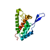
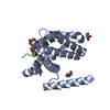
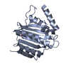
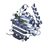
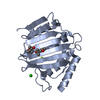
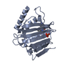
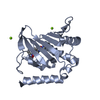
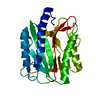
| Title | Structure of the immature retroviral capsid at 8A resolution by cryo- electron microscopy | ||||||
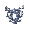
 Components Components | CAPSID PROTEIN P27 | ||||||
 Keywords Keywords | VIRAL PROTEIN / RETROVIRUS / GAG | ||||||
| Function / homology |  Function and homology information Function and homology informationviral budding via host ESCRT complex / viral nucleocapsid / host cell cytoplasm / structural constituent of virion / nucleic acid binding / zinc ion binding / metal ion bindingSimilarity search - Function | ||||||
| Biological species |  MASON-PFIZER MONKEY VIRUS MASON-PFIZER MONKEY VIRUS | ||||||
| Method | ELECTRON MICROSCOPY / helical reconstruction / cryo EM / Resolution: 7 Å | ||||||
| Model type details | CA ATOMS ONLY, CHAIN A, B | ||||||
 Authors Authors | Bharat, T.A.M. / Davey, N.E. / Ulbrich, P. / Riches, J.D. / Marco, A.D. / Rumlova, M. / Sachse, C. / Ruml, T. / Briggs, J.A.G. | ||||||
 Citation Citation | Journal: Nature / Year: 2012 Title: Structure of the immature retroviral capsid at 8 Å resolution by cryo-electron microscopy. Authors: Tanmay A M Bharat / Norman E Davey / Pavel Ulbrich / James D Riches / Alex de Marco / Michaela Rumlova / Carsten Sachse / Tomas Ruml / John A G Briggs /  Abstract: The assembly of retroviruses such as HIV-1 is driven by oligomerization of their major structural protein, Gag. Gag is a multidomain polyprotein including three conserved folded domains: MA (matrix), ...The assembly of retroviruses such as HIV-1 is driven by oligomerization of their major structural protein, Gag. Gag is a multidomain polyprotein including three conserved folded domains: MA (matrix), CA (capsid) and NC (nucleocapsid). Assembly of an infectious virion proceeds in two stages. In the first stage, Gag oligomerization into a hexameric protein lattice leads to the formation of an incomplete, roughly spherical protein shell that buds through the plasma membrane of the infected cell to release an enveloped immature virus particle. In the second stage, cleavage of Gag by the viral protease leads to rearrangement of the particle interior, converting the non-infectious immature virus particle into a mature infectious virion. The immature Gag shell acts as the pivotal intermediate in assembly and is a potential target for anti-retroviral drugs both in inhibiting virus assembly and in disrupting virus maturation. However, detailed structural information on the immature Gag shell has not previously been available. For this reason it is unclear what protein conformations and interfaces mediate the interactions between domains and therefore the assembly of retrovirus particles, and what structural transitions are associated with retrovirus maturation. Here we solve the structure of the immature retroviral Gag shell from Mason-Pfizer monkey virus by combining cryo-electron microscopy and tomography. The 8-Å resolution structure permits the derivation of a pseudo-atomic model of CA in the immature retrovirus, which defines the protein interfaces mediating retrovirus assembly. We show that transition of an immature retrovirus into its mature infectious form involves marked rotations and translations of CA domains, that the roles of the amino-terminal and carboxy-terminal domains of CA in assembling the immature and mature hexameric lattices are exchanged, and that the CA interactions that stabilize the immature and mature viruses are almost completely distinct. | ||||||
| History |
|
- Structure visualization
| Movie |
Movie viewer |
|---|---|
| Structure viewer | Molecule: MolmilJmol/JSmol |
- Downloads & links
-Download
| PDBx/mmCIF format | 4ard.cif.gz | 13.9 KB | Display | PDBx/mmCIF format |
|---|---|---|---|---|
| PDB format | pdb4ard.ent.gz | 7.3 KB | Display | PDB format |
| PDBx/mmJSON format | 4ard.json.gz | Tree view | PDBx/mmJSON format | |
| Others |  Other downloads Other downloads |
-Validation report
| Arichive directory | https://data.pdbj.org/pub/pdb/validation_reports/ar/4ardftp://data.pdbj.org/pub/pdb/validation_reports/ar/4ard | HTTPS FTP |
|---|
-Related structure data
| Related structure data | 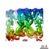 2090MC 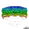 2089C  4argC C: citing same article ( M: map data used to model this data |
|---|---|
| Similar structure data |


-Links
 PDBj
PDBj

- Assembly
| Deposited unit | 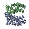
|
|---|---|
| 1 |
|
-Components
| #1: Protein | / M-PMV DPRO CANC PROTEIN / Coordinate model: Cα atoms only Mass: 12885.230 Da / Num. of mol.: 2 / Fragment: M-PMV CA-NTD, RESIDUES 318-433 Source method: isolated from a genetically manipulated source Source: (gene. exp.) MASON-PFIZER MONKEY VIRUS / Production host:  ESCHERICHIA COLI (E. coli) / References: UniProt: P07567 ESCHERICHIA COLI (E. coli) / References: UniProt: P07567 |
|---|
-Experimental details
-Experiment
| Experiment | Method: ELECTRON MICROSCOPY |
|---|---|
| EM experiment | Aggregation state: HELICAL ARRAY / 3D reconstruction method: helical reconstruction |
- Sample preparation
| Component | Name: M-PMV CANC GAG TUBES / Type: COMPLEX |
|---|---|
| Buffer solution | Name: 100MM NACL, 50MM TRIS-HCL, 1UM ZN / pH: 7.7 / Details: 100MM NACL, 50MM TRIS-HCL, 1UM ZN |
| Specimen | Embedding applied: NO / Shadowing applied: NO / Staining applied: NO / Vitrification applied: YES |
| Specimen support | Details: HOLEY CARBON |
| Vitrification | Cryogen name: ETHANE / Details: LIQUID ETHANE |
- Electron microscopy imaging
| Experimental equipment |  Model: Titan Krios / Image courtesy: FEI Company |
|---|---|
| Microscopy | Model: FEI TITAN KRIOS / Date: Jul 5, 2011 |
| Electron gun | Electron source: FIELD EMISSION GUN / Accelerating voltage: 300 kV / Illumination mode: FLOOD BEAM |
| Electron lens | Mode: BRIGHT FIELDBright-field microscopy / Nominal magnification: 47000 X / Nominal defocus max: 4000 nm / Nominal defocus min: 1000 nm / Cs: 2.7 mm |
| Image recording | Electron dose: 0.2 e/Å2 / Film or detector model: KODAK SO-163 FILM |
| Image scans | Num. digital images: 46 |
| Radiation wavelength | Relative weight: 1 |
- Processing
| EM software |
| ||||||||||||
|---|---|---|---|---|---|---|---|---|---|---|---|---|---|
| CTF correction | Details: DIVISION BY 3D CTF SQ | ||||||||||||
| 3D reconstruction | Method: HELICAL RECONSTRUCTION WITH 3D ASYMMETRIC UNIT AVERAGING Resolution: 7 Å / Nominal pixel size: 1.53 Å / Actual pixel size: 1.53 Å Details: HELICAL RECONSTRUCTION WITH 3D ASYMMETRIC UNIT AVERAGING SUBMISSION BASED ON EXPERIMENTAL DATA FROM EMDB EMD-2090. (DEPOSITION ID: 10768). Symmetry type: HELICAL | ||||||||||||
| Atomic model building | Protocol: RIGID BODY FIT / Space: REAL / Details: METHOD--RIGID BODY REFINEMENT PROTOCOL--NMR | ||||||||||||
| Atomic model building | PDB-ID: 2KGF | ||||||||||||
| Refinement | Highest resolution: 7 Å | ||||||||||||
| Refinement step | Cycle: LAST / Highest resolution: 7 Å
|