Movie
Movie Controller

[English] 日本語
 Yorodumi
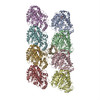
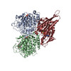
- PDB-4abo: Mal3 CH domain homology model and mammalian tubulin (2XRP) docked... -
+ Open data

- Basic information
| Entry | Database: PDB / ID: 4abo | ||||||
|---|---|---|---|---|---|---|---|
| Title | Mal3 CH domain homology model and mammalian tubulin (2XRP) docked into the 8.6-Angstrom cryo-EM map of Mal3-GTPgammaS-microtubules | ||||||
 Components Components |
| ||||||
 Keywords Keywords |  STRUCTURAL PROTEIN / CYTOSKELETON / GTPASE / END BINDING STRUCTURAL PROTEIN / CYTOSKELETON / GTPASE / END BINDING | ||||||
| Function / homology |  Function and homology information Function and homology informationdynein-driven meiotic oscillatory nuclear movement / post-anaphase array microtubule end / nuclear migration involved in conjugation with cellular fusion / cell cortex of cell tip / cortical microtubule / mitotic spindle astral microtubule / karyogamy involved in conjugation with cellular fusion / mitotic spindle pole body / nuclear microtubule / mitotic spindle midzone ...dynein-driven meiotic oscillatory nuclear movement / post-anaphase array microtubule end / nuclear migration involved in conjugation with cellular fusion / cell cortex of cell tip / cortical microtubule / mitotic spindle astral microtubule / karyogamy involved in conjugation with cellular fusion / mitotic spindle pole body / nuclear microtubule / mitotic spindle midzone / astral microtubule / protein localization to microtubule / microtubule plus-end / cytoskeletal anchor activity / attachment of mitotic spindle microtubules to kinetochore / microtubule plus-end binding / microtubule organizing center / microtubule lateral binding / ATPase activator activity / spindle assembly / regulation of microtubule polymerization or depolymerization / spindle midzone / cytoplasmic microtubule / molecular condensate scaffold activity / Hydrolases; Acting on acid anhydrides; Acting on GTP to facilitate cellular and subcellular movement / structural constituent of cytoskeleton / microtubule cytoskeleton organization / microtubule cytoskeleton / mitotic cell cycle / microtubule binding / microtubule / hydrolase activity / cell division / GTPase activity / GTP binding / metal ion binding / nucleus / cytoplasmSimilarity search - Function | ||||||
| Biological species |  SCHIZOSACCHAROMYCES POMBE (fission yeast) SCHIZOSACCHAROMYCES POMBE (fission yeast) SUS SCROFA (pig) SUS SCROFA (pig) | ||||||
| Method | ELECTRON MICROSCOPY / single particle reconstruction / cryo EM / Resolution: 8.6 Å | ||||||
 Authors Authors | Maurer, S.P. / Fourniol, F.J. / Bohner, G. / Moores, C.A. / Surrey, T. | ||||||
 Citation Citation | Journal: Cell / Year: 2012 Title: EBs recognize a nucleotide-dependent structural cap at growing microtubule ends. Authors: Sebastian P Maurer / Franck J Fourniol / Gergő Bohner / Carolyn A Moores / Thomas Surrey /  Abstract: Growing microtubule ends serve as transient binding platforms for essential proteins that regulate microtubule dynamics and their interactions with cellular substructures. End-binding proteins (EBs) ...Growing microtubule ends serve as transient binding platforms for essential proteins that regulate microtubule dynamics and their interactions with cellular substructures. End-binding proteins (EBs) autonomously recognize an extended region at growing microtubule ends with unknown structural characteristics and then recruit other factors to the dynamic end structure. Using cryo-electron microscopy, subnanometer single-particle reconstruction, and fluorescence imaging, we present a pseudoatomic model of how the calponin homology (CH) domain of the fission yeast EB Mal3 binds to the end regions of growing microtubules. The Mal3 CH domain bridges protofilaments except at the microtubule seam. By binding close to the exchangeable GTP-binding site, the CH domain is ideally positioned to sense the microtubule's nucleotide state. The same microtubule-end region is also a stabilizing structural cap protecting the microtubule from depolymerization. This insight supports a common structural link between two important biological phenomena, microtubule dynamic instability and end tracking. | ||||||
| History |
|
- Structure visualization
| Movie |
Movie viewer |
|---|---|
| Structure viewer | Molecule: MolmilJmol/JSmol |
- Downloads & links
-Download
| PDBx/mmCIF format | 4abo.cif.gz | 673 KB | Display | PDBx/mmCIF format |
|---|---|---|---|---|
| PDB format | pdb4abo.ent.gz | 552.9 KB | Display | PDB format |
| PDBx/mmJSON format | 4abo.json.gz | Tree view | PDBx/mmJSON format | |
| Others |  Other downloads Other downloads |
-Validation report
| Arichive directory | https://data.pdbj.org/pub/pdb/validation_reports/ab/4aboftp://data.pdbj.org/pub/pdb/validation_reports/ab/4abo | HTTPS FTP |
|---|
-Related structure data
| Related structure data |  2005MC  2004C  2006C M: map data used to model this data C: citing same article ( |
|---|---|
| Similar structure data |


-Links
 PDBj
PDBj


- Assembly
| Deposited unit | 
|
|---|---|
| 1 |
|
-Components
| #1: Protein | Mass: 49907.770 Da / Num. of mol.: 4 / Source method: isolated from a natural source / Source: (natural) SUS SCROFA (pig) / Organ: BRAIN / References: UniProt: P02554*PLUS, EC: 3.6.5.6#2: Protein | / ALPHA-TUBULIN 1 / TUBULIN ALPHA-1 CHAINMass: 50107.238 Da / Num. of mol.: 4 / Source method: isolated from a natural source / Source: (natural) SUS SCROFA (pig) / Organ: BRAIN / References: UniProt: P02550*PLUS, EC: 3.6.5.6#3: Protein | | Mass: 16754.949 Da / Num. of mol.: 1 / Fragment: CALPONIN HOMOLOGY DOMAIN, RESIDUES 2-142 Source method: isolated from a genetically manipulated source Source: (gene. exp.) SCHIZOSACCHAROMYCES POMBE (fission yeast)Production host:  ESCHERICHIA COLI (E. coli) / Strain (production host): BL21 / References: UniProt: Q10113 ESCHERICHIA COLI (E. coli) / Strain (production host): BL21 / References: UniProt: Q10113#4: Chemical | ChemComp-GSP /   Mass: 539.246 Da / Num. of mol.: 4 / Source method: obtained synthetically / Formula: C10H16N5O13P3S Mass: 539.246 Da / Num. of mol.: 4 / Source method: obtained synthetically / Formula: C10H16N5O13P3S#5: Chemical | ChemComp-GTP / Guanosine triphosphate  Mass: 523.180 Da / Num. of mol.: 4 / Source method: obtained synthetically / Formula: C10H16N5O14P3 / Comment: GTP, energy-carrying molecule*YM Mass: 523.180 Da / Num. of mol.: 4 / Source method: obtained synthetically / Formula: C10H16N5O14P3 / Comment: GTP, energy-carrying molecule*YMNonpolymer details | 5'-GUANOSINE-DIPHOSPHATE-MONOTHIOPHOSPHATE (GSP): GTPGAMMAS WAS DOCKED IN THE STRUCTURE BY ...5'-GUANOSINE-DIPHOSPHAT | Sequence details | THIS IS A CHIMERIC STRUCTURE BASED ON PDB ENTRIES 1JFF AND 3HKE (ALSO USED IN 2XRP) CLONING ...THIS IS A CHIMERIC STRUCTURE BASED ON PDB ENTRIES 1JFF AND 3HKE (ALSO USED IN 2XRP) CLONING INTRODUCED | |
|---|
-Experimental details
-Experiment
| Experiment | Method: ELECTRON MICROSCOPY |
|---|---|
| EM experiment | Aggregation state: FILAMENT / 3D reconstruction method: single particle reconstruction |
- Sample preparation
| Component | Name: GTPGAMMAS MICROTUBULES DECORATED WITH MONOMERIC MAL3 / Type: COMPLEX |
|---|---|
| Buffer solution | Name: 40MM PIPES, 1MM MGCL2, 1MM EGTA / pH: 6.8 / Details: 40MM PIPES, 1MM MGCL2, 1MM EGTA |
| Specimen | Embedding applied: NO / Shadowing applied: NO / Staining applied: NO / Vitrification applied: YES |
| Specimen support | Details: HOLEY CARBON |
| Vitrification | Instrument: FEI VITROBOT MARK I / Cryogen name: ETHANE Details: VITRIFICATION 1 -- CRYOGEN- ETHANE, HUMIDITY- 90, INSTRUMENT- VITROBOT (FEI), METHOD- CHAMBER AT 37 DEGREES C, BLOT 2S, |
- Electron microscopy imaging
| Experimental equipment |  Model: Tecnai F20 / Image courtesy: FEI Company |
|---|---|
| Microscopy | Model: FEI TECNAI F20 |
| Electron gun | Electron source: FIELD EMISSION GUN / Accelerating voltage: 200 kV / Illumination mode: FLOOD BEAM |
| Electron lens | Mode: BRIGHT FIELDBright-field microscopy / Nominal magnification: 68000 X / Nominal defocus max: 3600 nm / Nominal defocus min: 700 nm / Cs: 2 mm |
| Specimen holder | Temperature: 93 K |
| Image recording | Electron dose: 17 e/Å2 / Film or detector model: GATAN ULTRASCAN 4000 (4k x 4k) |
| Image scans | Num. digital images: 162 |
| Radiation wavelength | Relative weight: 1 |
- Processing
| EM software |
| ||||||||||||
|---|---|---|---|---|---|---|---|---|---|---|---|---|---|
| CTF correction | Details: FREALIGN | ||||||||||||
| 3D reconstruction | Method: SINGLE PARTICLESingle particle analysis / Resolution: 8.6 Å / Num. of particles: 129000 / Nominal pixel size: 2.2 Å / Actual pixel size: 2.2 Å Details: SUBMISSION BASED ON EXPERIMENTAL DATA FROM EMDB EMD-2005.(DEPOSITION ID: 10422). Symmetry type: HELICAL | ||||||||||||
| Atomic model building | Protocol: FLEXIBLE FIT / Space: REAL / Target criteria: Cross-correlation coefficient Details: METHOD--FLEXIBLE FITTING REFINEMENT PROTOCOL--FLEXIBLE FITTING IN CRYOEM MAP | ||||||||||||
| Atomic model building | PDB-ID: 2XRP | ||||||||||||
| Refinement | Highest resolution: 8.6 Å | ||||||||||||
| Refinement step | Cycle: LAST / Highest resolution: 8.6 Å
|