[English] 日本語
Yorodumi
- EMDB-4230: Single particle cryo em structure of Mycobacterium tuberculosis R... -

+
Open data


ID or keywords:

Loading...

-
Basic information

Entry
Database: EMDB / ID: EMD-4230
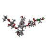
TitleSingle particle cryo em structure of Mycobacterium tuberculosis RNA polymerase in complex with Fidaxomicin
Map data
Sample
  • Complex: structure of Mycobacterium tuberculosis RNA polymerase in complex with Fidaxomicin
    • Protein or peptide: DNA-directed RNA polymerase subunit alphaPolymerase
    • Protein or peptide: DNA-directed RNA polymerase subunit betaPolymerase
    • Protein or peptide: DNA-directed RNA polymerase subunit beta'Polymerase
    • Protein or peptide: DNA-directed RNA polymerase subunit omegaPolymerase
    • Protein or peptide: RNA polymerase sigma factor SigA
  • Ligand: ZINC ION
  • Ligand: MAGNESIUM ION
  • Ligand: Fidaxomicin
  • Ligand: water
KeywordsLipiarmycin / RNA pol / RNAP / inhibitor / drug / Clostridium difficile / ANTIBIOTIC / Tiacumicin B / CCDC 114782 / transcription
Function / homology
Function and homology information


response to water / Antimicrobial action and antimicrobial resistance in Mtb / sigma factor activity / peptidoglycan-based cell wall / DNA-directed RNA polymerase complex / DNA-templated transcription initiation / ribonucleoside binding / DNA-directed 5'-3' RNA polymerase activity / DNA-directed RNA polymerase / protein dimerization activity ...response to water / Antimicrobial action and antimicrobial resistance in Mtb / sigma factor activity / peptidoglycan-based cell wall / DNA-directed RNA polymerase complex / DNA-templated transcription initiation / ribonucleoside binding / DNA-directed 5'-3' RNA polymerase activity / DNA-directed RNA polymerase / protein dimerization activity / response to antibiotic / DNA-templated transcription / magnesium ion binding / DNA binding / zinc ion binding / plasma membrane / cytosol / cytoplasm
Similarity search - Function
Sigma-70 factors family signature 1. / RNA polymerase sigma factor RpoD, C-terminal / RNA polymerase sigma factor RpoD / RNA polymerase sigma-70 region 1.2 / Sigma-70 factor, region 1.2 / RNA polymerase sigma-70 region 3 / Sigma-70 region 3 / Sigma-70 factors family signature 2. / RNA polymerase sigma-70 / RNA polymerase sigma-70 region 4 ...Sigma-70 factors family signature 1. / RNA polymerase sigma factor RpoD, C-terminal / RNA polymerase sigma factor RpoD / RNA polymerase sigma-70 region 1.2 / Sigma-70 factor, region 1.2 / RNA polymerase sigma-70 region 3 / Sigma-70 region 3 / Sigma-70 factors family signature 2. / RNA polymerase sigma-70 / RNA polymerase sigma-70 region 4 / Sigma-70, region 4 / RNA polymerase sigma-70 region 2 / RNA polymerase sigma-70 like domain / Sigma-70 region 2 / RNA polymerase sigma factor, region 2 / RNA polymerase sigma factor, region 3/4-like / DNA-directed RNA polymerase, omega subunit / DNA-directed RNA polymerase, subunit beta-prime, bacterial type / DNA-directed RNA polymerase, beta subunit, external 1 domain superfamily / DNA-directed RNA polymerase, beta subunit, external 1 domain / RNA polymerase beta subunit external 1 domain / RNA polymerase, alpha subunit, C-terminal / Bacterial RNA polymerase, alpha chain C terminal domain / DNA-directed RNA polymerase, alpha subunit / DNA-directed RNA polymerase beta subunit, bacterial-type / RNA polymerase Rpb6 / RNA polymerase, subunit omega/Rpo6/RPB6 / RNA polymerase Rpb6 / RNA polymerase Rpb1, domain 3 superfamily / RNA polymerase Rpb1, clamp domain superfamily / RPB6/omega subunit-like superfamily / DNA-directed RNA polymerase, subunit beta-prime / RNA polymerase Rpb1, domain 3 / RNA polymerase Rpb1, domain 3 / RNA polymerase Rpb2, domain 2 superfamily / RNA polymerase Rpb1, domain 1 / RNA polymerase Rpb1, domain 1 / RNA polymerase, alpha subunit / RNA polymerase Rpb1, domain 4 / RNA polymerase Rpb1, domain 2 / RNA polymerase Rpb1, domain 4 / RNA polymerase, N-terminal / RNA polymerase Rpb1, funnel domain superfamily / RNA polymerase I subunit A N-terminus / RNA polymerase Rpb1, domain 5 / RNA polymerase Rpb1, domain 5 / RNA polymerase, beta subunit, protrusion / RNA polymerase beta subunit / DNA-directed RNA polymerase, insert domain / DNA-directed RNA polymerase, RpoA/D/Rpb3-type / RNA polymerase Rpb3/RpoA insert domain / RNA polymerase Rpb3/Rpb11 dimerisation domain / RNA polymerases D / DNA-directed RNA polymerase, insert domain superfamily / RNA polymerase, RBP11-like subunit / RNA polymerase Rpb2, domain 2 / RNA polymerase Rpb2, domain 2 / RNA polymerase, beta subunit, conserved site / RNA polymerase Rpb2, domain 7 / RNA polymerase Rpb2, domain 3 / RNA polymerase Rpb2, OB-fold / RNA polymerase Rpb2, domain 7 / RNA polymerase Rpb2, domain 3 / RNA polymerases beta chain signature. / DNA-directed RNA polymerase, subunit 2, hybrid-binding domain / DNA-directed RNA polymerase, subunit 2 / DNA-directed RNA polymerase, subunit 2, hybrid-binding domain superfamily / RNA polymerase Rpb2, domain 6 / Winged helix-like DNA-binding domain superfamily
Similarity search - Domain/homology
RNA polymerase sigma factor SigA / DNA-directed RNA polymerase subunit omega / DNA-directed RNA polymerase subunit beta' / DNA-directed RNA polymerase subunit beta / DNA-directed RNA polymerase subunit alpha
Similarity search - Component
Biological speciesMycobacterium tuberculosis (strain ATCC 25618 / H37Rv) (bacteria)
Methodsingle particle reconstruction / cryo EM / Resolution: 3.52 Å
AuthorsDas K
Funding support Belgium, 1 items
OrganizationGrant numberCountry
Rega Foundation Belgium
CitationJournal: Mol Cell / Year: 2018
Title: Structural Basis of Transcription Inhibition by Fidaxomicin (Lipiarmycin A3).
Authors: Wei Lin / Kalyan Das / David Degen / Abhishek Mazumder / Diego Duchi / Dongye Wang / Yon W Ebright / Richard Y Ebright / Elena Sineva / Matthew Gigliotti / Aashish Srivastava / Sukhendu ...Authors: Wei Lin / Kalyan Das / David Degen / Abhishek Mazumder / Diego Duchi / Dongye Wang / Yon W Ebright / Richard Y Ebright / Elena Sineva / Matthew Gigliotti / Aashish Srivastava / Sukhendu Mandal / Yi Jiang / Yu Liu / Ruiheng Yin / Zhening Zhang / Edward T Eng / Dennis Thomas / Stefano Donadio / Haibo Zhang / Changsheng Zhang / Achillefs N Kapanidis / Richard H Ebright /
Abstract: Fidaxomicin is an antibacterial drug in clinical use for treatment of Clostridium difficile diarrhea. The active ingredient of fidaxomicin, lipiarmycin A3 (Lpm), functions by inhibiting bacterial ...Fidaxomicin is an antibacterial drug in clinical use for treatment of Clostridium difficile diarrhea. The active ingredient of fidaxomicin, lipiarmycin A3 (Lpm), functions by inhibiting bacterial RNA polymerase (RNAP). Here we report a cryo-EM structure of Mycobacterium tuberculosis RNAP holoenzyme in complex with Lpm at 3.5-Å resolution. The structure shows that Lpm binds at the base of the RNAP "clamp." The structure exhibits an open conformation of the RNAP clamp, suggesting that Lpm traps an open-clamp state. Single-molecule fluorescence resonance energy transfer experiments confirm that Lpm traps an open-clamp state and define effects of Lpm on clamp dynamics. We suggest that Lpm inhibits transcription by trapping an open-clamp state, preventing simultaneous interaction with promoter -10 and -35 elements. The results account for the absence of cross-resistance between Lpm and other RNAP inhibitors, account for structure-activity relationships of Lpm derivatives, and enable structure-based design of improved Lpm derivatives.
History
DepositionDec 19, 2017-
Header (metadata) releaseFeb 28, 2018-
Map releaseFeb 28, 2018-
UpdateMay 15, 2024-
Current statusMay 15, 2024Processing site: PDBe / Status: Released

-
Structure visualization

Movie
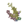
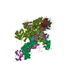
  • Surface view with section colored by density value
  • Surface level: 0.03
  • Imaged by UCSF Chimera
  • Download
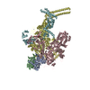
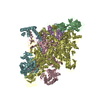
  • Surface view colored by radius
  • Surface level: 0.03
  • Imaged by UCSF Chimera
  • Download
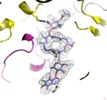
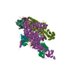
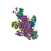
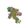
  • Surface view with fitted model
  • Atomic models: PDB-6fbv
  • Surface level: 0.03
  • Imaged by UCSF Chimera
  • Download
Movie viewer
Structure viewerEM map:
SurfViewMolmilJmol/JSmol
Supplemental images

Downloads & links

-
Map

FileDownload / File: emd_4230.map.gz / Format: CCP4 / Size: 27 MB / Type: IMAGE STORED AS FLOATING POINT NUMBER (4 BYTES)
Voxel sizeX=Y=Z: 1.061 Å
Density
Contour LevelBy AUTHOR: 0.03 / Movie #1: 0.03
Minimum - Maximum-0.15635955 - 0.2548963
Average (Standard dev.)0.0016079389 (±0.012585061)
SymmetrySpace group: 1
Details

EMDB XML:

Map geometry
Axis orderXYZ
Origin000
Dimensions192192192
Spacing192192192
CellA=B=C: 203.712 Å
α=β=γ: 90.0 °

CCP4 map header:

modeImage stored as Reals
Å/pix. X/Y/Z1.0611.0611.061
M x/y/z192192192
origin x/y/z0.0000.0000.000
length x/y/z203.712203.712203.712
α/β/γ90.00090.00090.000
MAP C/R/S123
start NC/NR/NS000
NC/NR/NS192192192
D min/max/mean-0.1560.2550.002

-
Supplemental data

-
Half map: #1

Fileemd_4230_half_map_1.map
Projections & Slices
AxesZYX

Projections

Slices (1/2)
Density Histograms

-
Half map: #2

Fileemd_4230_half_map_2.map
Projections & Slices
AxesZYX

Projections

Slices (1/2)
Density Histograms

-
Sample components

+
Entire : structure of Mycobacterium tuberculosis RNA polymerase in complex...

EntireName: structure of Mycobacterium tuberculosis RNA polymerase in complex with Fidaxomicin
Components
  • Complex: structure of Mycobacterium tuberculosis RNA polymerase in complex with Fidaxomicin
    • Protein or peptide: DNA-directed RNA polymerase subunit alphaPolymerase
    • Protein or peptide: DNA-directed RNA polymerase subunit betaPolymerase
    • Protein or peptide: DNA-directed RNA polymerase subunit beta'Polymerase
    • Protein or peptide: DNA-directed RNA polymerase subunit omegaPolymerase
    • Protein or peptide: RNA polymerase sigma factor SigA
  • Ligand: ZINC ION
  • Ligand: MAGNESIUM ION
  • Ligand: Fidaxomicin
  • Ligand: water

+
Supramolecule #1: structure of Mycobacterium tuberculosis RNA polymerase in complex...

SupramoleculeName: structure of Mycobacterium tuberculosis RNA polymerase in complex with Fidaxomicin
type: complex / ID: 1 / Parent: 0 / Macromolecule list: #1-#5 / Details: RNA polymerase inhibitor complex
Source (natural)Organism: Mycobacterium tuberculosis (strain ATCC 25618 / H37Rv) (bacteria)

+
Macromolecule #1: DNA-directed RNA polymerase subunit alpha

MacromoleculeName: DNA-directed RNA polymerase subunit alpha / type: protein_or_peptide / ID: 1 / Number of copies: 2 / Enantiomer: LEVO / EC number: DNA-directed RNA polymerase
Source (natural)Organism: Mycobacterium tuberculosis (strain ATCC 25618 / H37Rv) (bacteria)
Molecular weightTheoretical: 37.745328 KDa
Recombinant expressionOrganism: Escherichia coli (E. coli)
SequenceString: MLLSQRPTLS EDVLTDNRSQ FVIEPLEPGF GYTLGNSLRR TLLSSIPGAA VTSIRIDGVL HEFTTVPGVK EDVTEIILNL KSLVVSSEE DEPVTMYLRK QGPGEVTAGD IVPPAGVTVH NPGMHIATLN DKGKLEVELV VERGRGYVPA VQNRASGAEI G RIPVDSIY ...String:
MLLSQRPTLS EDVLTDNRSQ FVIEPLEPGF GYTLGNSLRR TLLSSIPGAA VTSIRIDGVL HEFTTVPGVK EDVTEIILNL KSLVVSSEE DEPVTMYLRK QGPGEVTAGD IVPPAGVTVH NPGMHIATLN DKGKLEVELV VERGRGYVPA VQNRASGAEI G RIPVDSIY SPVLKVTYKV DATRVEQRTD FDKLILDVET KNSISPRDAL ASAGKTLVEL FGLARELNVE AEGIEIGPSP AE ADHIASF ALPIDDLDLT VRSYNCLKRE GVHTVGELVA RTESDLLDIR NFGQKSIDEV KIKLHQLGLS LKDSPPSFDP SEV AGYDVA TGTWSTEGAY DEQDYAETEQ L

UniProtKB: DNA-directed RNA polymerase subunit alpha

+
Macromolecule #2: DNA-directed RNA polymerase subunit beta

MacromoleculeName: DNA-directed RNA polymerase subunit beta / type: protein_or_peptide / ID: 2 / Number of copies: 1 / Enantiomer: LEVO / EC number: DNA-directed RNA polymerase
Source (natural)Organism: Mycobacterium tuberculosis (strain ATCC 25618 / H37Rv) (bacteria)
Molecular weightTheoretical: 130.018828 KDa
Recombinant expressionOrganism: Escherichia coli (E. coli)
SequenceString: MLEGCILADS RQSKTAASPS PSRPQSSSNN SVPGAPNRVS FAKLREPLEV PGLLDVQTDS FEWLIGSPRW RESAAERGDV NPVGGLEEV LYELSPIEDF SGSMSLSFSD PRFDDVKAPV DECKDKDMTY AAPLFVTAEF INNNTGEIKS QTVFMGDFPM M TEKGTFII ...String:
MLEGCILADS RQSKTAASPS PSRPQSSSNN SVPGAPNRVS FAKLREPLEV PGLLDVQTDS FEWLIGSPRW RESAAERGDV NPVGGLEEV LYELSPIEDF SGSMSLSFSD PRFDDVKAPV DECKDKDMTY AAPLFVTAEF INNNTGEIKS QTVFMGDFPM M TEKGTFII NGTERVVVSQ LVRSPGVYFD ETIDKSTDKT LHSVKVIPSR GAWLEFDVDK RDTVGVRIDR KRRQPVTVLL KA LGWTSEQ IVERFGFSEI MRSTLEKDNT VGTDEALLDI YRKLRPGEPP TKESAQTLLE NLFFKEKRYD LARVGRYKVN KKL GLHVGE PITSSTLTEE DVVATIEYLV RLHEGQTTMT VPGGVEVPVE TDDIDHFGNR RLRTVGELIQ NQIRVGMSRM ERVV RERMT TQDVEAITPQ TLINIRPVVA AIKEFFGTSQ LSQFMDQNNP LSGLTHKRRL SALGPGGLSR ERAGLEVRDV HPSHY GRMC PIETPEGPNI GLIGSLSVYA RVNPFGFIET PYRKVVDGVV SDEIVYLTAD EEDRHVVAQA NSPIDADGRF VEPRVL VRR KAGEVEYVPS SEVDYMDVSP RQMVSVATAM IPFLEHDDAN RALMGANMQR QAVPLVRSEA PLVGTGMELR AAIDAGD VV VAEESGVIEE VSADYITVMH DNGTRRTYRM RKFARSNHGT CANQCPIVDA GDRVEAGQVI ADGPCTDDGE MALGKNLL V AIMPWEGHNY EDAIILSNRL VEEDVLTSIH IEEHEIDARD TKLGAEEITR DIPNISDEVL ADLDERGIVR IGAEVRDGD ILVGKVTPKG ETELTPEERL LRAIFGEKAR EVRDTSLKVP HGESGKVIGI RVFSREDEDE LPAGVNELVR VYVAQKRKIS DGDKLAGRH GNKGVIGKIL PVEDMPFLAD GTPVDIILNT HGVPRRMNIG QILETHLGWC AHSGWKVDAA KGVPDWAARL P DELLEAQP NAIVSTPVFD GAQEAELQGL LSCTLPNRDG DVLVDADGKA MLFDGRSGEP FPYPVTVGYM YIMKLHHLVD DK IHARSTG PYSMITQQPL GGKAQFGGQR FGEMECWAMQ AYGAAYTLQE LLTIKSDDTV GRVKVYEAIV KGENIPEPGI PES FKVLLK ELQSLCLNVE VLSSDGAAIE LREGEDEDLE RAAANLGINL SRNESASVED LA

UniProtKB: DNA-directed RNA polymerase subunit beta

+
Macromolecule #3: DNA-directed RNA polymerase subunit beta'

MacromoleculeName: DNA-directed RNA polymerase subunit beta' / type: protein_or_peptide / ID: 3 / Number of copies: 1 / Enantiomer: LEVO / EC number: DNA-directed RNA polymerase
Source (natural)Organism: Mycobacterium tuberculosis (strain ATCC 25618 / H37Rv) (bacteria)
Molecular weightTheoretical: 146.968969 KDa
Recombinant expressionOrganism: Escherichia coli (E. coli)
SequenceString: MLDVNFFDEL RIGLATAEDI RQWSYGEVKK PETINYRTLK PEKDGLFCEK IFGPTRDWEC YCGKYKRVRF KGIICERCGV EVTRAKVRR ERMGHIELAA PVTHIWYFKG VPSRLGYLLD LAPKDLEKII YFAAYVITSV DEEMRHNELS TLEAEMAVER K AVEDQRDG ...String:
MLDVNFFDEL RIGLATAEDI RQWSYGEVKK PETINYRTLK PEKDGLFCEK IFGPTRDWEC YCGKYKRVRF KGIICERCGV EVTRAKVRR ERMGHIELAA PVTHIWYFKG VPSRLGYLLD LAPKDLEKII YFAAYVITSV DEEMRHNELS TLEAEMAVER K AVEDQRDG ELEARAQKLE ADLAELEAEG AKADARRKVR DGGEREMRQI RDRAQRELDR LEDIWSTFTK LAPKQLIVDE NL YRELVDR YGEYFTGAMG AESIQKLIEN FDIDAEAESL RDVIRNGKGQ KKLRALKRLK VVAAFQQSGN SPMGMVLDAV PVI PPELRP MVQLDGGRFA TSDLNDLYRR VINRNNRLKR LIDLGAPEII VNNEKRMLQE SVDALFDNGR RGRPVTGPGN RPLK SLSDL LKGKQGRFRQ NLLGKRVDYS GRSVIVVGPQ LKLHQCGLPK LMALELFKPF VMKRLVDLNH AQNIKSAKRM VERQR PQVW DVLEEVIAEH PVLLNRAPTL HRLGIQAFEP MLVEGKAIQL HPLVCEAFNA DFDGDQMAVH LPLSAEAQAE ARILML SSN NILSPASGRP LAMPRLDMVT GLYYLTTEVP GDTGEYQPAS GDHPETGVYS SPAEAIMAAD RGVLSVRAKI KVRLTQL RP PVEIEAELFG HSGWQPGDAW MAETTLGRVM FNELLPLGYP FVNKQMHKKV QAAIINDLAE RYPMIVVAQT VDKLKDAG F YWATRSGVTV SMADVLVPPR KKEILDHYEE RADKVEKQFQ RGALNHDERN EALVEIWKEA TDEVGQALRE HYPDDNPII TIVDSGATGN FTQTRTLAGM KGLVTNPKGE FIPRPVKSSF REGLTVLEYF INTHGARKGL ADTALRTADS GYLTRRLVDV SQDVIVREH DCQTERGIVV ELAERAPDGT LIRDPYIETS AYARTLGTDA VDEAGNVIVE RGQDLGDPEI DALLAAGITQ V KVRSVLTC ATSTGVCATC YGRSMATGKL VDIGEAVGIV AAQSIGEPGT QLTMRTFHQG GVGEDITGGL PRVQELFEAR VP RGKAPIA DVTGRVRLED GERFYKITIV PDDGGEEVVY DKISKRQRLR VFKHEDGSER VLSDGDHVEV GQQLMEGSAD PHE VLRVQG PREVQIHLVR EVQEVYRAQG VSIHDKHIEV IVRQMLRRVT IIDSGSTEFL PGSLIDRAEF EAENRRVVAE GGEP AAGRP VLMGITKASL ATDSWLSAAS FQETTRVLTD AAINCRSDKL NGLKENVIIG KLIPAGTGIN RYRNIAVQPT EEARA AAYT IPSYEDQYYS PDFGAATGAA VPLDDYGYSD YR

UniProtKB: DNA-directed RNA polymerase subunit beta'

+
Macromolecule #4: DNA-directed RNA polymerase subunit omega

MacromoleculeName: DNA-directed RNA polymerase subunit omega / type: protein_or_peptide / ID: 4 / Number of copies: 1 / Enantiomer: LEVO / EC number: DNA-directed RNA polymerase
Source (natural)Organism: Mycobacterium tuberculosis (strain ATCC 25618 / H37Rv) (bacteria)
Molecular weightTheoretical: 11.85114 KDa
Recombinant expressionOrganism: Escherichia coli (E. coli)
SequenceString:
MSISQSDASL AAVPAVDQFD PSSGASGGYD TPLGITNPPI DELLDRVSSK YALVIYAAKR ARQINDYYNQ LGEGILEYVG PLVEPGLQE KPLSIALREI HADLLEHTEG E

UniProtKB: DNA-directed RNA polymerase subunit omega

+
Macromolecule #5: RNA polymerase sigma factor SigA

MacromoleculeName: RNA polymerase sigma factor SigA / type: protein_or_peptide / ID: 5 / Number of copies: 1 / Enantiomer: LEVO
Source (natural)Organism: Mycobacterium tuberculosis (strain ATCC 25618 / H37Rv) (bacteria)
Molecular weightTheoretical: 57.87716 KDa
Recombinant expressionOrganism: Escherichia coli (E. coli)
SequenceString: MAATKASTAT DEPVKRTATK SPAASASGAK TGAKRTAAKS ASGSPPAKRA TKPAARSVKP ASAPQDTTTS TIPKRKTRAA AKSAAAKAP SARGHATKPR APKDAQHEAA TDPEDALDSV EELDAEPDLD VEPGEDLDLD AADLNLDDLE DDVAPDADDD L DSGDDEDH ...String:
MAATKASTAT DEPVKRTATK SPAASASGAK TGAKRTAAKS ASGSPPAKRA TKPAARSVKP ASAPQDTTTS TIPKRKTRAA AKSAAAKAP SARGHATKPR APKDAQHEAA TDPEDALDSV EELDAEPDLD VEPGEDLDLD AADLNLDDLE DDVAPDADDD L DSGDDEDH EDLEAEAAVA PGQTADDDEE IAEPTEKDKA SGDFVWDEDE SEALRQARKD AELTASADSV RAYLKQIGKV AL LNAEEEV ELAKRIEAGL YATQLMTELS ERGEKLPAAQ RRDMMWICRD GDRAKNHLLE ANLRLVVSLA KRYTGRGMAF LDL IQEGNL GLIRAVEKFD YTKGYKFSTY ATWWIRQAIT RAMADQARTI RIPVHMVEVI NKLGRIQREL LQDLGREPTP EELA KEMDI TPEKVLEIQQ YAREPISLDQ TIGDEGDSQL GDFIEDSEAV VAVDAVSFTL LQDQLQSVLD TLSEREAGVV RLRFG LTDG QPRTLDEIGQ VYGVTRERIR QIESKTMSKL RHPSRSQVLR DYLD

UniProtKB: RNA polymerase sigma factor SigA

+
Macromolecule #6: ZINC ION

MacromoleculeName: ZINC ION / type: ligand / ID: 6 / Number of copies: 2 / Formula: ZN
Molecular weightTheoretical: 65.409 Da

+
Macromolecule #7: MAGNESIUM ION

MacromoleculeName: MAGNESIUM ION / type: ligand / ID: 7 / Number of copies: 1 / Formula: MG
Molecular weightTheoretical: 24.305 Da

+
Macromolecule #8: Fidaxomicin

MacromoleculeName: Fidaxomicin / type: ligand / ID: 8 / Number of copies: 1 / Formula: FI8
Molecular weightTheoretical: 1.058039 KDa
Chemical component information

ChemComp-FI8:
Fidaxomicin / antibiotic*YM / Fidaxomicin

+
Macromolecule #9: water

MacromoleculeName: water / type: ligand / ID: 9 / Number of copies: 3 / Formula: HOH
Molecular weightTheoretical: 18.015 Da
Chemical component information

ChemComp-HOH:
WATER / Water

-
Experimental details

-
Structure determination

Methodcryo EM
Processingsingle particle reconstruction
Aggregation stateparticle

-
Sample preparation

BufferpH: 8
Details: 3.5 microliter 1 microM Mtb RNAP-Lpm and 50 microMolar Lpm in 20 mM Tris-HCl, pH 8.0, 75 mM NaCl, 5 mM MgCl2, 5 mM dithiothreitol, and 0.1% n-octyl-beta-D-glucopyranoside
GridMaterial: COPPER / Mesh: 300 / Support film - Material: CARBON / Support film - topology: LACEY / Support film - Film thickness: 100 / Pretreatment - Type: GLOW DISCHARGE / Pretreatment - Time: 25 sec. / Pretreatment - Atmosphere: AIR / Pretreatment - Pressure: 0.039 kPa
VitrificationCryogen name: ETHANE / Chamber humidity: 95 % / Chamber temperature: 291 K

-
Electron microscopy

MicroscopeFEI TITAN KRIOS
Electron beamAcceleration voltage: 300 kV / Electron source: FIELD EMISSION GUN
Electron opticsIllumination mode: FLOOD BEAM / Imaging mode: BRIGHT FIELDBright-field microscopy / Nominal defocus max: -0.002 µm / Nominal defocus min: -0.001 µm / Nominal magnification: 130000
Sample stageSpecimen holder model: FEI TITAN KRIOS AUTOGRID HOLDER
Image recordingFilm or detector model: GATAN K2 SUMMIT (4k x 4k) / Detector mode: COUNTING / Digitization - Dimensions - Width: 3838 pixel / Digitization - Dimensions - Height: 3710 pixel / Digitization - Frames/image: 1-35 / Number grids imaged: 2 / Number real images: 2458 / Average exposure time: 0.2 sec. / Average electron dose: 1.4 e/Å2
Details: Movies were recorded at 200 ms/frame for 10s (50 frames total), resulting in a total radiation dose of 72.05 electrons/A**2 per movie Defocus range was varied between 1.0 - 2.0 micrometer.
Experimental equipment
Model: Titan Krios / Image courtesy: FEI Company

-
Image processing

Particle selectionNumber selected: 819506 / Details: Autopick
Startup modelType of model: PDB ENTRY
PDB model - PDB ID:

Details: Protein atoms only from the structure 5UH5; 60 A lowpass filter applied.
Initial angle assignmentType: ANGULAR RECONSTITUTION / Software - Name: RELION (ver. 2.0.5)
Final 3D classificationSoftware - Name: RELION (ver. 2.0.5)
Final angle assignmentType: ANGULAR RECONSTITUTION / Software - Name: RELION (ver. 2.0.5)
Final reconstructionApplied symmetry - Point group: C1 (asymmetric) / Resolution.type: BY AUTHOR / Resolution: 3.52 Å / Resolution method: FSC 0.143 CUT-OFF / Software - Name: RELION (ver. 2.0.5) / Number images used: 68895
FSC plot (resolution estimation)

-
Atomic model buiding 1

RefinementProtocol: OTHER / Overall B value: 95.3 / Target criteria: Cross-correlation coefficient
Output model

PDB-6fbv:
Single particle cryo em structure of Mycobacterium tuberculosis RNA polymerase in complex with Fidaxomicin

+
About Yorodumi

-
News

-
Feb 9, 2022. New format data for meta-information of EMDB entries

New format data for meta-information of EMDB entries

  • Version 3 of the EMDB header file is now the official format.
  • The previous official version 1.9 will be removed from the archive.

Related info.:EMDB header

External links:wwPDB to switch to version 3 of the EMDB data model

-
Aug 12, 2020. Covid-19 info

Covid-19 info

URL: https://pdbj.org/emnavi/covid19.php

New page: Covid-19 featured information page in EM Navigator.

Related info.:Covid-19 info / Mar 5, 2020. Novel coronavirus structure data

+
Mar 5, 2020. Novel coronavirus structure data

Novel coronavirus structure data

Related info.:Yorodumi Speices / Aug 12, 2020. Covid-19 info

External links:COVID-19 featured content - PDBj / Molecule of the Month (242):Coronavirus Proteases

+
Jan 31, 2019. EMDB accession codes are about to change! (news from PDBe EMDB page)

EMDB accession codes are about to change! (news from PDBe EMDB page)

  • The allocation of 4 digits for EMDB accession codes will soon come to an end. Whilst these codes will remain in use, new EMDB accession codes will include an additional digit and will expand incrementally as the available range of codes is exhausted. The current 4-digit format prefixed with “EMD-” (i.e. EMD-XXXX) will advance to a 5-digit format (i.e. EMD-XXXXX), and so on. It is currently estimated that the 4-digit codes will be depleted around Spring 2019, at which point the 5-digit format will come into force.
  • The EM Navigator/Yorodumi systems omit the EMD- prefix.

Related info.:Q: What is EMD? / ID/Accession-code notation in Yorodumi/EM Navigator

External links:EMDB Accession Codes are Changing Soon! / Contact to PDBj

+
Jul 12, 2017. Major update of PDB

Major update of PDB

  • wwPDB released updated PDB data conforming to the new PDBx/mmCIF dictionary.
  • This is a major update changing the version number from 4 to 5, and with Remediation, in which all the entries are updated.
  • In this update, many items about electron microscopy experimental information are reorganized (e.g. em_software).
  • Now, EM Navigator and Yorodumi are based on the updated data.

External links:wwPDB Remediation / Enriched Model Files Conforming to OneDep Data Standards Now Available in the PDB FTP Archive

-
Yorodumi

Thousand views of thousand structures

  • Yorodumi is a browser for structure data from EMDB, PDB, SASBDB, etc.
  • This page is also the successor to EM Navigator detail page, and also detail information page/front-end page for Omokage search.
  • The word "yorodu" (or yorozu) is an old Japanese word meaning "ten thousand". "mi" (miru) is to see.

Related info.:EMDB / PDB / SASBDB / Comparison of 3 databanks / Yorodumi Search / Aug 31, 2016. New EM Navigator & Yorodumi / Yorodumi Papers / Jmol/JSmol / Function and homology information / Changes in new EM Navigator and Yorodumi

Read more