 Movie
Movie Controller


+ Open data

- Basic information
| Entry |  | ||||||||||||
|---|---|---|---|---|---|---|---|---|---|---|---|---|---|
| Title | Cryo-EM structure of the PP2A:B55-ARPP19 complex | ||||||||||||
 Map data Map data | Relion 3D auto refine full map. This map was used for Phenix real-space refinement of model coordinates and ADPs. | ||||||||||||
 Sample Sample |
| ||||||||||||
 Keywords Keywords | Protein Phosphatase 2A:B55 holoenzyme / ARPP19 inhibitor /  cell cycle regulation / SIGNALING PROTEIN / HYDROLASE cell cycle regulation / SIGNALING PROTEIN / HYDROLASE | ||||||||||||
| Function / homology |  Function and homology informationphosphatase inhibitor activity / negative regulation of protein dephosphorylation / meiotic spindle elongation / Integration of energy metabolism / PP2A-mediated dephosphorylation of key metabolic factors / regulation of microtubule binding / MASTL Facilitates Mitotic Progression / mitotic sister chromatid separation / regulation of meiotic cell cycle process involved in oocyte maturation / protein phosphatase type 2A complex ...phosphatase inhibitor activity / negative regulation of protein dephosphorylation / meiotic spindle elongation / Integration of energy metabolism / PP2A-mediated dephosphorylation of key metabolic factors / regulation of microtubule binding / MASTL Facilitates Mitotic Progression / mitotic sister chromatid separation / regulation of meiotic cell cycle process involved in oocyte maturation / protein phosphatase type 2A complex / meiotic sister chromatid cohesion, centromeric / peptidyl-serine dephosphorylation / peptidyl-threonine dephosphorylation / : / positive regulation of microtubule binding / negative regulation of tyrosine phosphorylation of STAT protein / Regulation of glycolysis by fructose 2,6-bisphosphate metabolism / Inhibition of replication initiation of damaged DNA by RB1/E2F1 / female meiotic nuclear division / protein antigen binding / protein phosphatase regulator activity / ceramide metabolic process / GABA receptor binding / negative regulation of epithelial to mesenchymal transition / APC truncation mutants have impaired AXIN binding / AXIN missense mutants destabilize the destruction complex / Truncations of AMER1 destabilize the destruction complex / Initiation of Nuclear Envelope (NE) Reformation / ERKs are inactivated / response to morphine / positive regulation of extrinsic apoptotic signaling pathway in absence of ligand / Beta-catenin phosphorylation cascade / Signaling by GSK3beta mutants / CTNNB1 S33 mutants aren't phosphorylated / CTNNB1 S37 mutants aren't phosphorylated / CTNNB1 S45 mutants aren't phosphorylated / CTNNB1 T41 mutants aren't phosphorylated / regulation of Wnt signaling pathway / Disassembly of the destruction complex and recruitment of AXIN to the membrane / regulation of growth / protein phosphatase inhibitor activity / negative regulation of glycolytic process through fructose-6-phosphate / positive regulation of NLRP3 inflammasome complex assembly / myosin phosphatase activity / protein serine/threonine phosphatase activity / CTLA4 inhibitory signaling / Platelet sensitization by LDL / negative regulation of MAPK cascade / protein-serine/threonine phosphatase / regulation of cell differentiation / T cell homeostasis / ERK/MAPK targets / regulation of G1/S transition of mitotic cell cycle / phosphoprotein phosphatase activity / regulation of DNA replication / mesoderm development / chromosome, centromeric region / DARPP-32 events / negative regulation of phosphatidylinositol 3-kinase/protein kinase B signal transduction / lateral plasma membrane / potassium channel regulator activity / Nonsense Mediated Decay (NMD) enhanced by the Exon Junction Complex (EJC) / Amplification of signal from unattached kinetochores via a MAD2 inhibitory signal / regulation of cell adhesion / Cyclin A/B1/B2 associated events during G2/M transition / Mitotic Prometaphase / EML4 and NUDC in mitotic spindle formation / positive regulation of gluconeogenesis / Loss of Nlp from mitotic centrosomes / Loss of proteins required for interphase microtubule organization from the centrosome / Recruitment of mitotic centrosome proteins and complexes / Resolution of Sister Chromatid Cohesion / Recruitment of NuMA to mitotic centrosomes / Anchoring of the basal body to the plasma membrane / AURKA Activation by TPX2 / protein dephosphorylation / RNA splicing / meiotic cell cycle / protein phosphatase 2A binding / response to organic substance / protein tyrosine phosphatase activity / chromosome segregation / RHO GTPases Activate Formins / positive regulation of glucose import / response to lead ion / regulation of protein phosphorylation / Spry regulation of FGF signaling / RAF activation / PKR-mediated signaling / Degradation of beta-catenin by the destruction complex / tau protein binding / positive regulation of protein serine/threonine kinase activity / negative regulation of cell growth / spindle pole / Negative regulation of MAPK pathway / Separation of Sister Chromatids / Cyclin D associated events in G1 / microtubule cytoskeleton / G2/M transition of mitotic cell cycle / Regulation of PLK1 Activity at G2/M Transition Function and homology informationphosphatase inhibitor activity / negative regulation of protein dephosphorylation / meiotic spindle elongation / Integration of energy metabolism / PP2A-mediated dephosphorylation of key metabolic factors / regulation of microtubule binding / MASTL Facilitates Mitotic Progression / mitotic sister chromatid separation / regulation of meiotic cell cycle process involved in oocyte maturation / protein phosphatase type 2A complex ...phosphatase inhibitor activity / negative regulation of protein dephosphorylation / meiotic spindle elongation / Integration of energy metabolism / PP2A-mediated dephosphorylation of key metabolic factors / regulation of microtubule binding / MASTL Facilitates Mitotic Progression / mitotic sister chromatid separation / regulation of meiotic cell cycle process involved in oocyte maturation / protein phosphatase type 2A complex / meiotic sister chromatid cohesion, centromeric / peptidyl-serine dephosphorylation / peptidyl-threonine dephosphorylation / : / positive regulation of microtubule binding / negative regulation of tyrosine phosphorylation of STAT protein / Regulation of glycolysis by fructose 2,6-bisphosphate metabolism / Inhibition of replication initiation of damaged DNA by RB1/E2F1 / female meiotic nuclear division / protein antigen binding / protein phosphatase regulator activity / ceramide metabolic process / GABA receptor binding / negative regulation of epithelial to mesenchymal transition / APC truncation mutants have impaired AXIN binding / AXIN missense mutants destabilize the destruction complex / Truncations of AMER1 destabilize the destruction complex / Initiation of Nuclear Envelope (NE) Reformation / ERKs are inactivated / response to morphine / positive regulation of extrinsic apoptotic signaling pathway in absence of ligand / Beta-catenin phosphorylation cascade / Signaling by GSK3beta mutants / CTNNB1 S33 mutants aren't phosphorylated / CTNNB1 S37 mutants aren't phosphorylated / CTNNB1 S45 mutants aren't phosphorylated / CTNNB1 T41 mutants aren't phosphorylated / regulation of Wnt signaling pathway / Disassembly of the destruction complex and recruitment of AXIN to the membrane / regulation of growth / protein phosphatase inhibitor activity / negative regulation of glycolytic process through fructose-6-phosphate / positive regulation of NLRP3 inflammasome complex assembly / myosin phosphatase activity / protein serine/threonine phosphatase activity / CTLA4 inhibitory signaling / Platelet sensitization by LDL / negative regulation of MAPK cascade / protein-serine/threonine phosphatase / regulation of cell differentiation / T cell homeostasis / ERK/MAPK targets / regulation of G1/S transition of mitotic cell cycle / phosphoprotein phosphatase activity / regulation of DNA replication / mesoderm development / chromosome, centromeric region / DARPP-32 events / negative regulation of phosphatidylinositol 3-kinase/protein kinase B signal transduction / lateral plasma membrane / potassium channel regulator activity / Nonsense Mediated Decay (NMD) enhanced by the Exon Junction Complex (EJC) / Amplification of signal from unattached kinetochores via a MAD2 inhibitory signal / regulation of cell adhesion / Cyclin A/B1/B2 associated events during G2/M transition / Mitotic Prometaphase / EML4 and NUDC in mitotic spindle formation / positive regulation of gluconeogenesis / Loss of Nlp from mitotic centrosomes / Loss of proteins required for interphase microtubule organization from the centrosome / Recruitment of mitotic centrosome proteins and complexes / Resolution of Sister Chromatid Cohesion / Recruitment of NuMA to mitotic centrosomes / Anchoring of the basal body to the plasma membrane / AURKA Activation by TPX2 / protein dephosphorylation / RNA splicing / meiotic cell cycle / protein phosphatase 2A binding / response to organic substance / protein tyrosine phosphatase activity / chromosome segregation / RHO GTPases Activate Formins / positive regulation of glucose import / response to lead ion / regulation of protein phosphorylation / Spry regulation of FGF signaling / RAF activation / PKR-mediated signaling / Degradation of beta-catenin by the destruction complex / tau protein binding / positive regulation of protein serine/threonine kinase activity / negative regulation of cell growth / spindle pole / Negative regulation of MAPK pathway / Separation of Sister Chromatids / Cyclin D associated events in G1 / microtubule cytoskeleton / G2/M transition of mitotic cell cycle / Regulation of PLK1 Activity at G2/M TransitionSimilarity search - Function | ||||||||||||
| Biological species |  Homo sapiens (human) Homo sapiens (human) | ||||||||||||
| Method | single particle reconstruction / cryo EM / Resolution: 2.77 Å | ||||||||||||
 Authors Authors | Fuller JR / Padi SKR / Peti W / Page R | ||||||||||||
| Funding support |  United States, 3 items United States, 3 items
| ||||||||||||
 Citation Citation | Journal: Nature / Year: 2024 Title: Cryo-EM structures of PP2A:B55-FAM122A and PP2A:B55-ARPP19. Authors: Sathish K R Padi / Margaret R Vos / Rachel J Godek / James R Fuller / Thomas Kruse / Jamin B Hein / Jakob Nilsson / Matthew S Kelker / Rebecca Page / Wolfgang Peti /  Abstract: Progression through the cell cycle is controlled by regulated and abrupt changes in phosphorylation. Mitotic entry is initiated by increased phosphorylation of mitotic proteins, a process driven by ...Progression through the cell cycle is controlled by regulated and abrupt changes in phosphorylation. Mitotic entry is initiated by increased phosphorylation of mitotic proteins, a process driven by kinases, whereas mitotic exit is achieved by counteracting dephosphorylation, a process driven by phosphatases, especially PP2A:B55. Although the role of kinases in mitotic entry is well established, recent data have shown that mitosis is only successfully initiated when the counterbalancing phosphatases are also inhibited. Inhibition of PP2A:B55 is achieved by the intrinsically disordered proteins ARPP19 and FAM122A. Despite their critical roles in mitosis, the mechanisms by which they achieve PP2A:B55 inhibition is unknown. Here, we report the single-particle cryo-electron microscopy structures of PP2A:B55 bound to phosphorylated ARPP19 and FAM122A. Consistent with our complementary NMR spectroscopy studies, both intrinsically disordered proteins bind PP2A:B55, but do so in highly distinct manners, leveraging multiple distinct binding sites on B55. Our extensive structural, biophysical and biochemical data explain how substrates and inhibitors are recruited to PP2A:B55 and provide a molecular roadmap for the development of therapeutic interventions for PP2A:B55-related diseases. | ||||||||||||
| History |
|
- Structure visualization
| Supplemental images |
|---|

- Downloads & links
-EMDB archive
| Map data | emd_41604.map.gz | 131.2 MB | EMDB map data format | |
|---|---|---|---|---|
| Header (meta data) | emd-41604-v30.xmlemd-41604.xml | 27 KB 27 KB | Display Display | EMDB header |
| FSC (resolution estimation) | emd_41604_fsc.xml | 12.5 KB | Display | FSC data file |
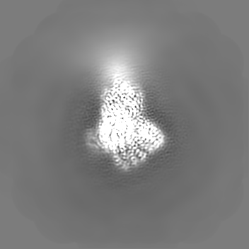
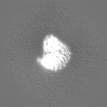
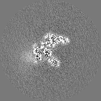
| Images | 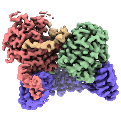 emd_41604.png emd_41604.png | 167.1 KB | ||
| Masks | emd_41604_msk_1.map | 166.4 MB | Mask map | |
| Filedesc metadata | emd-41604.cif.gz | 7.8 KB | ||
| Others | emd_41604_additional_1.map.gzemd_41604_half_map_1.map.gzemd_41604_half_map_2.map.gz | 101 MB 131.5 MB 131.5 MB | ||
| Archive directory |  http://ftp.pdbj.org/pub/emdb/structures/EMD-41604ftp://ftp.pdbj.org/pub/emdb/structures/EMD-41604 http://ftp.pdbj.org/pub/emdb/structures/EMD-41604ftp://ftp.pdbj.org/pub/emdb/structures/EMD-41604 | HTTPS FTP |
-Related structure data
| Related structure data |  8ttbMC 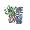 8so0C  8tweC  8twiC M: atomic model generated by this map C: citing same article ( |
|---|---|
| Similar structure data |


-Links
| EMDB pages | EMDB (EBI/PDBe) / EMDataResource |
|---|---|
| Related items in Molecule of the Month |


-Map
| File | Download / File: emd_41604.map.gz / Format: CCP4 / Size: 166.4 MB / Type: IMAGE STORED AS FLOATING POINT NUMBER (4 BYTES) | ||||||||||||||||||||
|---|---|---|---|---|---|---|---|---|---|---|---|---|---|---|---|---|---|---|---|---|---|
| Annotation | Relion 3D auto refine full map. This map was used for Phenix real-space refinement of model coordinates and ADPs. | ||||||||||||||||||||
| Voxel size | X=Y=Z: 0.827 Å | ||||||||||||||||||||
| Density |
| ||||||||||||||||||||
| Symmetry | Space group: 1 | ||||||||||||||||||||
| Details | EMDB XML:
|
-Supplemental data
-Mask #1
| File | emd_41604_msk_1.map | ||||||||||||
|---|---|---|---|---|---|---|---|---|---|---|---|---|---|
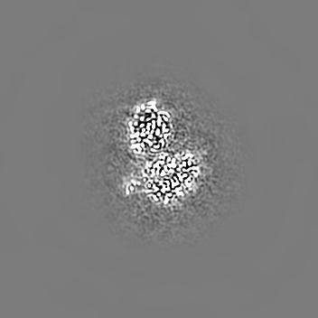
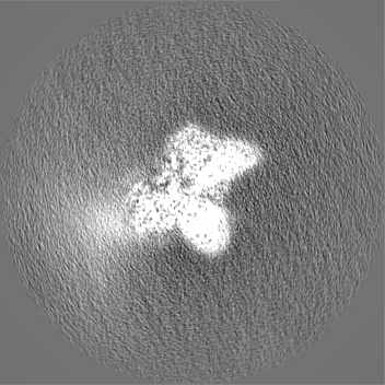
| Projections & Slices |
| ||||||||||||
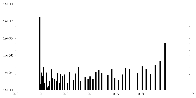
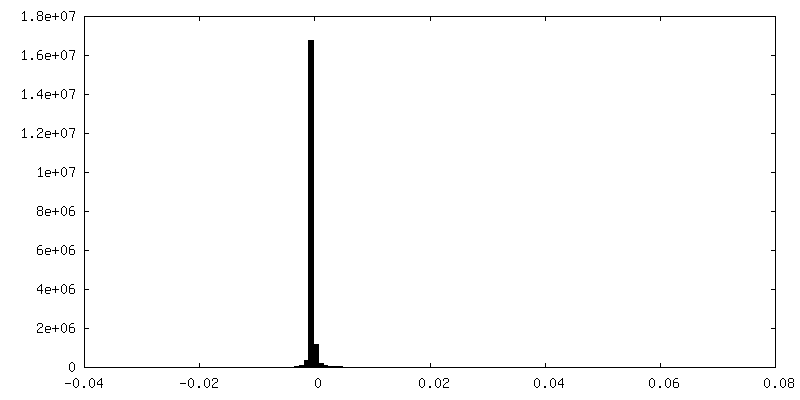
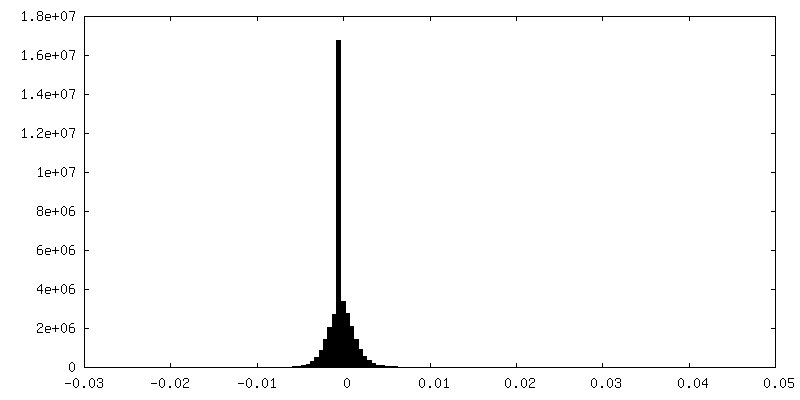
| Density Histograms |
 Z
Z Y
Y X
X


-Additional map: Map version that was globally sharpened by a...
| File | emd_41604_additional_1.map | ||||||||||||
|---|---|---|---|---|---|---|---|---|---|---|---|---|---|
| Annotation | Map version that was globally sharpened by a B-factor of -40.0 and filtered to local resolution by the implementation in Relion. This was used to guide manual model building. | ||||||||||||
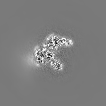
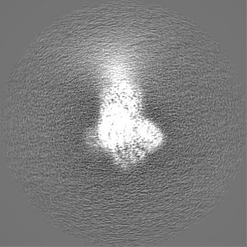
| Projections & Slices |
| ||||||||||||
| Density Histograms |


-Half map: Relion 3D auto refine half map 1
| File | emd_41604_half_map_1.map | ||||||||||||
|---|---|---|---|---|---|---|---|---|---|---|---|---|---|
| Annotation | Relion 3D auto refine half map 1 | ||||||||||||
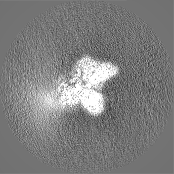
| Projections & Slices |
| ||||||||||||
| Density Histograms |


-Half map: Relion 3D auto refine half map 2
| File | emd_41604_half_map_2.map | ||||||||||||
|---|---|---|---|---|---|---|---|---|---|---|---|---|---|
| Annotation | Relion 3D auto refine half map 2 | ||||||||||||
| Projections & Slices |
| ||||||||||||
| Density Histograms |


- Sample components
-Entire : Quadruple complex of PP2A:B55 (PP2Aa:PP2Ac:B55) bound to thiophos...
| Entire | Name: Quadruple complex of PP2A:B55 (PP2Aa:PP2Ac:B55) bound to thiophosphorylated ARPP19 |
|---|---|
| Components |
|
-Supramolecule #1: Quadruple complex of PP2A:B55 (PP2Aa:PP2Ac:B55) bound to thiophos...
| Supramolecule | Name: Quadruple complex of PP2A:B55 (PP2Aa:PP2Ac:B55) bound to thiophosphorylated ARPP19 type: complex / ID: 1 / Parent: 0 / Macromolecule list: #1-#4 |
|---|---|
| Molecular weight | Theoretical: 165 KDa |
-Macromolecule #1: Serine/threonine-protein phosphatase 2A 65 kDa regulatory subunit...
| Macromolecule | Name: Serine/threonine-protein phosphatase 2A 65 kDa regulatory subunit A alpha isoform type: protein_or_peptide / ID: 1 / Number of copies: 1 / Enantiomer: LEVO |
|---|---|
| Source (natural) | Organism: Homo sapiens (human) |
| Molecular weight | Theoretical: 64.95798 KDa |
| Recombinant expression | Organism:  Escherichia coli (E. coli) Escherichia coli (E. coli) |
| Sequence | String: GHMSLYPIAV LIDELRNEDV QLRLNSIKKL STIALALGVE RTRSELLPFL TDTIYDEDEV LLALAEQLGT FTTLVGGPEY VHCLLPPLE SLATVEETVV RDKAVESLRA ISHEHSPSDL EAHFVPLVKR LAGGDWFTSR TSACGLFSVC YPRVSSAVKA E LRQYFRNL ...String: GHMSLYPIAV LIDELRNEDV QLRLNSIKKL STIALALGVE RTRSELLPFL TDTIYDEDEV LLALAEQLGT FTTLVGGPEY VHCLLPPLE SLATVEETVV RDKAVESLRA ISHEHSPSDL EAHFVPLVKR LAGGDWFTSR TSACGLFSVC YPRVSSAVKA E LRQYFRNL CSDDTPMVRR AAASKLGEFA KVLELDNVKS EIIPMFSNLA SDEQDSVRLL AVEACVNIAQ LLPQEDLEAL VM PTLRQAA EDKSWRVRYM VADKFTELQK AVGPEITKTD LVPAFQNLMK DCEAEVRAAA SHKVKEFCEN LSADCRENVI MSQ ILPCIK ELVSDANQHV KSALASVIMG LSPILGKDNT IEHLLPLFLA QLKDECPEVR LNIISNLDCV NEVIGIRQLS QSLL PAIVE LAEDAKWRVR LAIIEYMPLL AGQLGVEFFD EKLNSLCMAW LVDHVYAIRE AATSNLKKLV EKFGKEWAHA TIIPK VLAM SGDPNYLHRM TTLFCINVLS EVCGQDITTK HMLPTVLRMA GDPVANVRFN VAKSLQKIGP ILDNSTLQSE VKPILE KLT QDQDVDVKYF AQEALTVLSL A UniProtKB: Serine/threonine-protein phosphatase 2A 65 kDa regulatory subunit A alpha isoform |
-Macromolecule #2: Serine/threonine-protein phosphatase 2A 55 kDa regulatory subunit...
| Macromolecule | Name: Serine/threonine-protein phosphatase 2A 55 kDa regulatory subunit B alpha isoform type: protein_or_peptide / ID: 2 / Number of copies: 1 / Enantiomer: LEVO |
|---|---|
| Source (natural) | Organism: Homo sapiens (human) |
| Molecular weight | Theoretical: 52.044289 KDa |
| Recombinant expression | Organism: Homo sapiens (human) |
| Sequence | String: HMGSAGAGGG NDIQWCFSQV KGAVDDDVAE ADIISTVEFN HSGELLATGD KGGRVVIFQQ EQENKIQSHS RGEYNVYSTF QSHEPEFDY LKSLEIEEKI NKIRWLPQKN AAQFLLSTND KTIKLWKISE RDKRPEGYNL KEEDGRYRDP TTVTTLRVPV F RPMDLMVE ...String: HMGSAGAGGG NDIQWCFSQV KGAVDDDVAE ADIISTVEFN HSGELLATGD KGGRVVIFQQ EQENKIQSHS RGEYNVYSTF QSHEPEFDY LKSLEIEEKI NKIRWLPQKN AAQFLLSTND KTIKLWKISE RDKRPEGYNL KEEDGRYRDP TTVTTLRVPV F RPMDLMVE ASPRRIFANA HTYHINSISI NSDYETYLSA DDLRINLWHL EITDRSFNIV DIKPANMEEL TEVITAAEFH PN SCNTFVY SSSKGTIRLC DMRASALCDR HSKLFEEPED PSNRSFFSEI ISSISDVKFS HSGRYMMTRD YLSVKIWDLN MEN RPVETY QVHEYLRSKL CSLYENDCIF DKFECCWNGS DSVVMTGSYN NFFRMFDRNT KRDITLEASR ENNKPRTVLK PRKV CASGK RKKDEISVDS LDFNKKILHT AWHPKENIIA VATTNNLYIF QDKVN UniProtKB: Serine/threonine-protein phosphatase 2A 55 kDa regulatory subunit B alpha isoform |
-Macromolecule #3: Serine/threonine-protein phosphatase 2A catalytic subunit alpha i...
| Macromolecule | Name: Serine/threonine-protein phosphatase 2A catalytic subunit alpha isoform type: protein_or_peptide / ID: 3 / Number of copies: 1 / Enantiomer: LEVO / EC number: protein-serine/threonine phosphatase |
|---|---|
| Source (natural) | Organism: Homo sapiens (human) |
| Molecular weight | Theoretical: 35.845375 KDa |
| Recombinant expression | Organism: Homo sapiens (human) |
| Sequence | String: GHMDEKVFTK ELDQWIEQLN ECKQLSESQV KSLCEKAKEI LTKESNVQEV RCPVTVCGDV HGQFHDLMEL FRIGGKSPDT NYLFMGDYV DRGYYSVETV TLLVALKVRY RERITILRGN HESRQITQVY GFYDECLRKY GNANVWKYFT DLFDYLPLTA L VDGQIFCL ...String: GHMDEKVFTK ELDQWIEQLN ECKQLSESQV KSLCEKAKEI LTKESNVQEV RCPVTVCGDV HGQFHDLMEL FRIGGKSPDT NYLFMGDYV DRGYYSVETV TLLVALKVRY RERITILRGN HESRQITQVY GFYDECLRKY GNANVWKYFT DLFDYLPLTA L VDGQIFCL HGGLSPSIDT LDHIRALDRL QEVPHEGPMC DLLWSDPDDR GGWGISPRGA GYTFGQDISE TFNHANGLTL VS RAHQLVM EGYNWCHDRN VVTIFSAPNY CYRCGNQAAI MELDDTLKYS FLQFDPAPRR GEPHVTRRTP DYF(MLL) UniProtKB: Serine/threonine-protein phosphatase 2A catalytic subunit alpha isoform |
-Macromolecule #4: cAMP-regulated phosphoprotein 19
| Macromolecule | Name: cAMP-regulated phosphoprotein 19 / type: protein_or_peptide / ID: 4 / Number of copies: 1 / Enantiomer: LEVO |
|---|---|
| Source (natural) | Organism: Homo sapiens (human) |
| Molecular weight | Theoretical: 12.620241 KDa |
| Recombinant expression | Organism: Escherichia coli (E. coli) |
| Sequence | String: GHMSAEVPEA ASAEEQKEME DKVTSPEKAE EAKLKARYPH LGQKPGGSDF LRKRLQKGQK YFD(2RX)GDYNMA KAKMKN KQL PTAAPDKTEV TGDHIPTPQD LPQRKPALVA SKLAG UniProtKB: cAMP-regulated phosphoprotein 19 |
-Macromolecule #5: FE (III) ION
| Macromolecule | Name: FE (III) ION / type: ligand / ID: 5 / Number of copies: 1 / Formula: FE |
|---|---|
| Molecular weight | Theoretical: 55.845 Da |
-Macromolecule #6: ZINC ION
| Macromolecule | Name: ZINC ION / type: ligand / ID: 6 / Number of copies: 1 / Formula: ZN |
|---|---|
| Molecular weight | Theoretical: 65.409 Da |
-Experimental details
-Structure determination
| Method | cryo EM |
|---|---|
 Processing Processing | single particle reconstruction |
| Aggregation state | particle |
-Sample preparation
| Concentration | 1.2 mg/mL | ||||||||||||||||||
|---|---|---|---|---|---|---|---|---|---|---|---|---|---|---|---|---|---|---|---|
| Buffer | pH: 8 Component:
Details: CHAPSO was added only immediately prior to vitrification | ||||||||||||||||||
| Vitrification | Cryogen name: ETHANE / Chamber humidity: 100 % / Chamber temperature: 291 K / Instrument: FEI VITROBOT MARK IV |
- Electron microscopy
| Microscope | FEI TITAN KRIOS |
|---|---|
| Electron beam | Acceleration voltage: 300 kV / Electron source: FIELD EMISSION GUN |
| Electron optics | C2 aperture diameter: 50.0 µm / Calibrated defocus max: 2.6 µm / Calibrated defocus min: 0.39 µm / Illumination mode: FLOOD BEAM / Imaging mode: BRIGHT FIELDBright-field microscopy / Cs: 2.7 mm / Nominal defocus max: 1.9000000000000001 µm / Nominal defocus min: 0.7000000000000001 µm / Nominal magnification: 105000 |
| Specialist optics | Energy filter - Name: GIF Bioquantum / Energy filter - Slit width: 20 eV |
| Sample stage | Specimen holder model: FEI TITAN KRIOS AUTOGRID HOLDER / Cooling holder cryogen: NITROGEN |
| Image recording | Film or detector model: GATAN K3 BIOQUANTUM (6k x 4k) / Number grids imaged: 1 / Average electron dose: 70.0 e/Å2 Details: Camera was operated in CDS mode, with hardware binning of super-resolution pixels, writing movies with 62 frames |
| Experimental equipment |  Model: Titan Krios / Image courtesy: FEI Company |
-Image processing
| Particle selection | Number selected: 1170216 |
|---|---|
| Startup model | Type of model: OTHER Details: Initial model was generated directly from particles images using the RELION 4.0.1 3D initial model algorithm |
| Initial angle assignment | Type: MAXIMUM LIKELIHOOD / Software - Name: RELION (ver. 4.0.1) |
| Final angle assignment | Type: MAXIMUM LIKELIHOOD / Software - Name: RELION (ver. 4.0) |
| Final reconstruction | Resolution.type: BY AUTHOR / Resolution: 2.77 Å / Resolution method: FSC 0.143 CUT-OFF / Software - Name: RELION (ver. 4.0.1) / Number images used: 52934 |
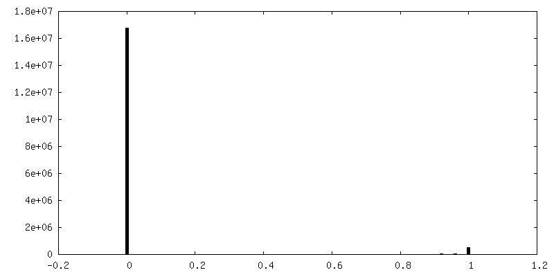
| FSC plot (resolution estimation) | 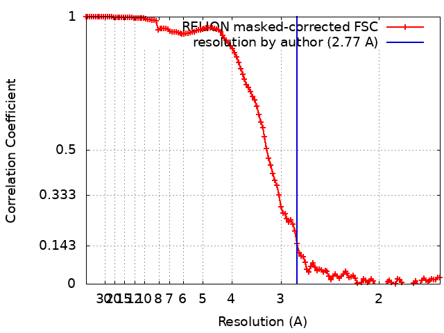 |
-Atomic model buiding 1
| Initial model |
| ||||||
|---|---|---|---|---|---|---|---|
| Details | Iterating between manual refinement in Coot and automated real-space refinement in Phenix | ||||||
| Refinement | Space: REAL / Protocol: FLEXIBLE FIT / Target criteria: Cross-correlation | ||||||
| Output model | PDB-8ttb: |


