 Movie
Movie Controller

[English] 日本語
 Yorodumi
- PDB-3m9i: Electron crystallographic structure of lens Aquaporin-0 (AQP0) (l... -
+ Open data

- Basic information
| Entry | Database: PDB / ID: 3m9i | ||||||
|---|---|---|---|---|---|---|---|
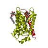
| Title | Electron crystallographic structure of lens Aquaporin-0 (AQP0) (lens MIP) in E. coli polar lipids | ||||||
 Components Components | Lens fiber major intrinsic protein | ||||||
 Keywords Keywords | MEMBRANE PROTEIN / water channel / lens / lipid-protein interactions | ||||||
| Function / homology |  Function and homology information Function and homology informationgap junction-mediated intercellular transport / water channel activity / water transport / structural constituent of eye lens / gap junction / response to stimulus / lens development in camera-type eye / positive regulation of cell adhesion / visual perception / protein homotetramerization ...gap junction-mediated intercellular transport / water channel activity / water transport / structural constituent of eye lens / gap junction / response to stimulus / lens development in camera-type eye / positive regulation of cell adhesion / visual perception / protein homotetramerization / calmodulin binding / endoplasmic reticulum / plasma membraneSimilarity search - Function | ||||||
| Biological species |  Ovis aries (sheep) Ovis aries (sheep) | ||||||
| Method | ELECTRON CRYSTALLOGRAPHY / electron crystallography / MOLECULAR REPLACEMENT / cryo EM / Resolution: 2.5 Å | ||||||
 Authors Authors | Hite, R.K. / Li, Z. / Walz, T. | ||||||
 Citation Citation | Journal: EMBO J / Year: 2010 Title: Principles of membrane protein interactions with annular lipids deduced from aquaporin-0 2D crystals. Authors: Richard K Hite / Zongli Li / Thomas Walz /  Abstract: We have previously described the interactions of aquaporin-0 (AQP0) with dimyristoyl phosphatidylcholine (DMPC) lipids. We have now determined the 2.5 A structure of AQP0 in two-dimensional (2D) ...We have previously described the interactions of aquaporin-0 (AQP0) with dimyristoyl phosphatidylcholine (DMPC) lipids. We have now determined the 2.5 A structure of AQP0 in two-dimensional (2D) crystals formed with Escherichia coli polar lipids (EPLs), which differ from DMPC both in headgroups and acyl chains. Comparison of the two structures shows that AQP0 does not adapt to the different length of the acyl chains in EPLs and that the distance between the phosphodiester groups in the two leaflets of the DMPC and EPL bilayers is almost identical. The EPL headgroups interact differently with AQP0 than do those of DMPC, but the acyl chains in the EPL and DMPC bilayers occupy similar positions. The interactions of annular lipids with membrane proteins seem to be driven by the propensity of the acyl chains to fill gaps in the protein surface. Interactions of the lipid headgroups may be responsible for the specific interactions found in tightly bound lipids but seem to have a negligible effect on interactions of generic annular lipids with membrane proteins. | ||||||
| History |
|
- Structure visualization
| Movie |
Movie viewer |
|---|---|
| Structure viewer | Molecule: MolmilJmol/JSmol |
- Downloads & links
-Download
| PDBx/mmCIF format | 3m9i.cif.gz | 63.3 KB | Display | PDBx/mmCIF format |
|---|---|---|---|---|
| PDB format | pdb3m9i.ent.gz | 45.9 KB | Display | PDB format |
| PDBx/mmJSON format | 3m9i.json.gz | Tree view | PDBx/mmJSON format | |
| Others |  Other downloads Other downloads |
-Validation report
| Arichive directory | https://data.pdbj.org/pub/pdb/validation_reports/m9/3m9iftp://data.pdbj.org/pub/pdb/validation_reports/m9/3m9i | HTTPS FTP |
|---|
-Related structure data
| Related structure data |  2b6oS S: Starting model for refinement |
|---|---|
| Similar structure data |


-Links
 PDBj
PDBj
- Assembly
| Deposited unit | 
| ||||||||
|---|---|---|---|---|---|---|---|---|---|
| 1 | x 8
| ||||||||
| Unit cell |
| ||||||||
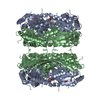
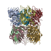
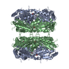
| Details | The octamer can be generated by applying P422 symmetry. |
-Components
| #1: Protein | / Aquaporin-0 Mass: 23514.447 Da / Num. of mol.: 1 / Fragment: UNP residues 7 to 226 / Source method: isolated from a natural source / Source: (natural) Ovis aries (sheep) / References: UniProt: Q6J8I9 | ||
|---|---|---|---|
| #2: Chemical | ChemComp-3PE / Phosphatidylethanolamine  Mass: 748.065 Da / Num. of mol.: 7 / Source method: obtained synthetically / Formula: C41H82NO8P / Comment: phospholipid*YM Mass: 748.065 Da / Num. of mol.: 7 / Source method: obtained synthetically / Formula: C41H82NO8P / Comment: phospholipid*YM#3: Water | ChemComp-HOH / | Water Mass: 18.015 Da / Num. of mol.: 8 / Source method: isolated from a natural source / Formula: H2O Mass: 18.015 Da / Num. of mol.: 8 / Source method: isolated from a natural source / Formula: H2O |
-Experimental details
-Experiment
| Experiment | Method: ELECTRON CRYSTALLOGRAPHY / Number of used crystals: 281 |
|---|---|
| EM experiment | Aggregation state: 2D ARRAY / 3D reconstruction method: electron crystallography |
- Sample preparation
| Component | Name: lens Aquaporin 0 / Type: COMPLEX |
|---|---|
| Buffer solution | pH: 8 |
| Specimen | Embedding applied: NO / Shadowing applied: NO / Staining applied: NO / Vitrification applied: YES Details: Purified membranes were solubilized in 4% (w/v) octyl glucoside in 10 mM Tris (pH 8.0) for 30 min at 22C |
| Crystal grow | Temperature: 300 K / Method: microdialysis / pH: 6 Details: 100 mM NaCl, 50mM MgCl2, 10mM MES pH 6.0, MICRODIALYSIS, temperature 300K |
-Data collection
| Experimental equipment |  Model: Tecnai Polara / Image courtesy: FEI Company |
|---|---|
| Microscopy | Model: FEI POLARA 300 |
| Electron gun | Electron source: FIELD EMISSION GUN / Accelerating voltage: 300 kV / Illumination mode: FLOOD BEAM |
| Electron lens | Mode: DIFFRACTION |
| Image recording | Film or detector model: GENERIC GATAN (4k x 4k) |
| Diffraction | Mean temperature: 279 K |
| Diffraction source | Source: ELECTRON MICROSCOPE / Type: OTHER |
| Detector | Type: Gatan / Detector: CCD / Date: Feb 24, 2009 |
| Reflection | Resolution: 2.5→15.89 Å / Num. all: 14417 / Num. obs: 14417 / % possible obs: 92.3 % / Observed criterion σ(F): 0 / Observed criterion σ(I): 0 / Redundancy: 8.1 % / Biso Wilson estimate: 14.9 Å2 / Rmerge(I) obs: 0.226 / Rsym value: 0.189 |
| Reflection shell | Resolution: 2.5→2.66 Å / Redundancy: 4 % / Num. unique all: 1935 / % possible all: 83.7 |
- Processing
| Software |
| |||||||||||||||||||||||||
|---|---|---|---|---|---|---|---|---|---|---|---|---|---|---|---|---|---|---|---|---|---|---|---|---|---|---|
| Refinement | Method to determine structure: MOLECULAR REPLACEMENT Starting model: PDB entry 2B6O Resolution: 2.5→15.89 Å / Rfactor Rfree error: 0.007 / Data cutoff high absF: 2341911.44 / Data cutoff low absF: 0 / Isotropic thermal model: RESTRAINED / Cross valid method: THROUGHOUT / σ(F): 0 / Stereochemistry target values: Engh & Huber / Details: BULK SOLVENT MODEL USED
| |||||||||||||||||||||||||
| Solvent computation | Solvent model: FLAT MODEL / Bsol: 44.0682 Å2 / ksol: 0.1 e/Å3 | |||||||||||||||||||||||||
| Displacement parameters | Biso mean: 48.6 Å2
| |||||||||||||||||||||||||
| Refine analyze |
| |||||||||||||||||||||||||
| Refinement step | Cycle: LAST / Resolution: 2.5→15.89 Å
| |||||||||||||||||||||||||
| Refine LS restraints |
| |||||||||||||||||||||||||
| Refine LS restraints NCS | NCS model details: NONE | |||||||||||||||||||||||||
| LS refinement shell | Resolution: 2.5→2.66 Å / Rfactor Rfree error: 0.029 / Total num. of bins used: 6
| |||||||||||||||||||||||||
| Xplor file |
|
