Movie
Movie Controller

[English] 日本語
 Yorodumi
- PDB-3jbb: Characterization of red-shifted phycobiliprotein complexes isolat... -
+ Open data

- Basic information
| Entry | Database: PDB / ID: 3jbb | ||||||
|---|---|---|---|---|---|---|---|
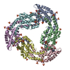
| Title | Characterization of red-shifted phycobiliprotein complexes isolated from the chlorophyll f-containing cyanobacterium Halomicronema hongdechloris | ||||||
 Components Components |
| ||||||
 Keywords Keywords |  PHOTOSYNTHESIS / alpha-helical phycobiliprotein / light harvesting / phycocyano methylation on ASN71 in APCB SUBUNIT / phycobilisome PHOTOSYNTHESIS / alpha-helical phycobiliprotein / light harvesting / phycocyano methylation on ASN71 in APCB SUBUNIT / phycobilisome | ||||||
| Function / homology |  Function and homology information Function and homology information | ||||||
| Biological species |  Halomicronema hongdechloris (bacteria) Halomicronema hongdechloris (bacteria) | ||||||
| Method | ELECTRON MICROSCOPY / single particle reconstruction / negative staining / Resolution: 26 Å | ||||||
 Authors Authors | Li, Y. / Lin, Y. / Garvey, C. / Birch, D. / Corkery, R.W. / Loughlin, P.C. / Scheer, H. / Willows, R.D. / Chen, M. | ||||||
 Citation Citation | Journal: Biochim Biophys Acta / Year: 2016 Title: Characterization of red-shifted phycobilisomes isolated from the chlorophyll f-containing cyanobacterium Halomicronema hongdechloris. Authors: Yaqiong Li / Yuankui Lin / Christopher J Garvey / Debra Birch / Robert W Corkery / Patrick C Loughlin / Hugo Scheer / Robert D Willows / Min Chen /    Abstract: Phycobilisomes are the main light-harvesting protein complexes in cyanobacteria and some algae. It is commonly accepted that these complexes only absorb green and orange light, complementing ...Phycobilisomes are the main light-harvesting protein complexes in cyanobacteria and some algae. It is commonly accepted that these complexes only absorb green and orange light, complementing chlorophyll absorbance. Here, we present a new phycobilisome derived complex that consists only of allophycocyanin core subunits, having red-shifted absorption peaks of 653 and 712 nm. These red-shifted phycobiliprotein complexes were isolated from the chlorophyll f-containing cyanobacterium, Halomicronema hongdechloris, grown under monochromatic 730 nm-wavelength (far-red) light. The 3D model obtained from single particle analysis reveals a double disk assembly of 120-145 Å with two α/β allophycocyanin trimers fitting into the two separated disks. They are significantly smaller than typical phycobilisomes formed from allophycocyanin subunits and core-membrane linker proteins, which fit well with a reduced distance between thylakoid membranes observed from cells grown under far-red light. Spectral analysis of the dissociated and denatured phycobiliprotein complexes grown under both these light conditions shows that the same bilin chromophore, phycocyanobilin, is exclusively used. Our findings show that red-shifted phycobilisomes are required for assisting efficient far-red light harvesting. Their discovery provides new insights into the molecular mechanisms of light harvesting under extreme conditions for photosynthesis, as well as the strategies involved in flexible chromatic acclimation to diverse light conditions. #1: Journal: Acta Crystallogr D Biol Crystallogr / Year: 2014Title: The structure of allophycocyanin B from Synechocystis PCC 6803 reveals the structural basis for the extreme redshift of the terminal emitter in phycobilisomes. Authors: Pan Pan Peng / Liang Liang Dong / Ya Fang Sun / Xiao Li Zeng / Wen Long Ding / Hugo Scheer / Xiaojing Yang / Kai Hong Zhao /   Abstract: Allophycocyanin B (AP-B) is one of the two terminal emitters in phycobilisomes, the unique light-harvesting complexes of cyanobacteria and red algae. Its low excitation-energy level and the ...Allophycocyanin B (AP-B) is one of the two terminal emitters in phycobilisomes, the unique light-harvesting complexes of cyanobacteria and red algae. Its low excitation-energy level and the correspondingly redshifted absorption and fluorescence emission play an important role in funnelling excitation energy from the hundreds of chromophores of the extramembraneous phycobilisome to the reaction centres within the photosynthetic membrane. In the absence of crystal structures of these low-abundance terminal emitters, the molecular basis for the extreme redshift and directional energy transfer is largely unknown. Here, the crystal structure of trimeric AP-B [(ApcD/ApcB)3] from Synechocystis sp. PCC 6803 at 1.75 Å resolution is reported. In the crystal lattice, eight trimers of AP-B form a porous, spherical, 48-subunit assembly of 193 Å in diameter with an internal cavity of 1.1 × 10(6) Å(3). While the overall structure of trimeric AP-B is similar to those reported for many other phycobiliprotein trimers, the chromophore pocket of the α-subunit, ApcD, has more bulky residues that tightly pack the phycocyanobilin (PCB). Ring D of the chromophores is further stabilized by close interactions with ApcB from the adjacent monomer. The combined contributions from both subunits render the conjugated rings B, C and D of the PCB in ApcD almost perfectly coplanar. Together with mutagenesis data, it is proposed that the enhanced planarity effectively extends the conjugation system of PCB and leads to the redshifted absorption (λmax = 669 nm) and fluorescence emission (679 nm) of the ApcD chromophore in AP-B, thereby enabling highly efficient energy transfer from the phycobilisome core to the reaction centres. | ||||||
| History |
|
- Structure visualization
| Movie |
Movie viewer |
|---|---|
| Structure viewer | Molecule: MolmilJmol/JSmol |
- Downloads & links
-Download
| PDBx/mmCIF format | 3jbb.cif.gz | 820.9 KB | Display | PDBx/mmCIF format |
|---|---|---|---|---|
| PDB format | pdb3jbb.ent.gz | 713.4 KB | Display | PDB format |
| PDBx/mmJSON format | 3jbb.json.gz | Tree view | PDBx/mmJSON format | |
| Others |  Other downloads Other downloads |
-Validation report
| Arichive directory | https://data.pdbj.org/pub/pdb/validation_reports/jb/3jbbftp://data.pdbj.org/pub/pdb/validation_reports/jb/3jbb | HTTPS FTP |
|---|
-Related structure data
| Related structure data | 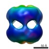 6430MC M: map data used to model this data C: citing same article ( |
|---|---|
| Similar structure data |
-Links
 PDBj
PDBj- Assembly
| Deposited unit | 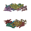
|
|---|---|
| 1 |
|
-Components
| #1: Protein | Mass: 18769.273 Da / Num. of mol.: 6 / Fragment: APCD subunit (SEE REMARK 999) / Source method: isolated from a natural source / Source: (natural) Halomicronema hongdechloris (bacteria) / References: UniProt: A0A0R4I959*PLUS#2: Protein | Mass: 17245.629 Da / Num. of mol.: 6 / Fragment: APCB subunit (SEE REMARK 999) / Source method: isolated from a natural source / Source: (natural) Halomicronema hongdechloris (bacteria) / References: UniProt: A0A0R4I960*PLUS#3: Chemical | ChemComp-CYC / Phycocyanobilin  Mass: 588.694 Da / Num. of mol.: 12 / Source method: obtained synthetically / Formula: C33H40N4O6 Mass: 588.694 Da / Num. of mol.: 12 / Source method: obtained synthetically / Formula: C33H40N4O6#4: Chemical | ChemComp-SO4 / Sulfate  Mass: 96.063 Da / Num. of mol.: 42 / Source method: obtained synthetically / Formula: SO4 Mass: 96.063 Da / Num. of mol.: 42 / Source method: obtained synthetically / Formula: SO4#5: Water | ChemComp-HOH / | Water Mass: 18.015 Da / Num. of mol.: 2396 / Source method: isolated from a natural source / Formula: H2O Mass: 18.015 Da / Num. of mol.: 2396 / Source method: isolated from a natural source / Formula: H2OSequence details | THE IMAGED PROTEINS ARE FROM HALOMICRONEMA HONGDECHLORIS, BUT THE MODELED SEQUENCES ARE FROM ...THE IMAGED PROTEINS ARE FROM HALOMICRON | |
|---|
-Experimental details
-Experiment
| Experiment | Method: ELECTRON MICROSCOPY / Number of used crystals: 1 |
|---|---|
| EM experiment | Aggregation state: PARTICLE / 3D reconstruction method: single particle reconstruction |
- Sample preparation
| Component |
| |||||||||||||||
|---|---|---|---|---|---|---|---|---|---|---|---|---|---|---|---|---|
| Buffer solution | Name: 0.8 M phosphate buffer / pH: 7.5 / Details: 0.8 M phosphate buffer | |||||||||||||||
| Specimen | Embedding applied: NO / Shadowing applied: NO / Staining applied: YES / Vitrification applied: NO / Details: 2% uranyl acetate for 2-3 seconds | |||||||||||||||
| EM staining | Type: NEGATIVE / Material: uranyl acetate | |||||||||||||||
| Specimen support | Details: 200 mesh gold grid with thin carbon support |
- Electron microscopy imaging
| Microscopy | Model: FEI/PHILIPS CM10 / Date: Jun 8, 2015 / Details: PHILIPS CM10 |
|---|---|
| Electron gun | Electron source: TUNGSTEN HAIRPIN / Accelerating voltage: 100 kV / Illumination mode: FLOOD BEAM |
| Electron lens | Mode: BRIGHT FIELDBright-field microscopy / Nominal magnification: 180000 X / Nominal defocus max: 3000 nm |
| Specimen holder | Specimen holder model: PHILIPS ROTATION HOLDER |
| Image recording | Film or detector model: GENERIC IMAGE PLATES |
| Image scans | Num. digital images: 12 |
- Processing
| EM software |
| ||||||||||||
|---|---|---|---|---|---|---|---|---|---|---|---|---|---|
| CTF correction | Details: Each particle | ||||||||||||
| Symmetry | Point symmetry: D3 (2x3 fold dihedral) | ||||||||||||
| 3D reconstruction | Method: Cross-common lines / Resolution: 26 Å / Resolution method: FSC 0.33 CUT-OFF / Num. of particles: 420 / Nominal pixel size: 3.8 Å / Actual pixel size: 3.8 Å Details: (Single particle details: Semiautomatic selection using e2boxer.py swarm function) (Single particle--Applied symmetry: D3) Num. of class averages: 32 / Symmetry type: POINT | ||||||||||||
| Atomic model building | Protocol: RIGID BODY FIT / Space: REAL Details: REFINEMENT PROTOCOL--rigid body DETAILS--Two trimers were fitted to each end of the EM map. 99.5% of atoms fit within the map at contour level 0.87 | ||||||||||||
| Atomic model building |
| ||||||||||||
| Refinement step | Cycle: LAST
|