 Movie
Movie Controller

[English] 日本語
 Yorodumi
- PDB-3j9c: CryoEM single particle reconstruction of anthrax toxin protective... -
+ Open data

- Basic information
| Entry | Database: PDB / ID: 3j9c | ||||||
|---|---|---|---|---|---|---|---|
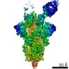
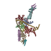
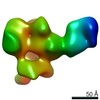
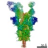
| Title | CryoEM single particle reconstruction of anthrax toxin protective antigen pore at 2.9 Angstrom resolution | ||||||
 Components Components | Protective antigen PA-63 | ||||||
 Keywords Keywords | TOXIN / TRANSPORT PROTEIN / bacterial toxin / anthrax toxin / protective antigen / protein translocation channel | ||||||
| Function / homology |  Function and homology information Function and homology informationpositive regulation of apoptotic process in another organism / host cell cytosol / negative regulation of MAPK cascade / Uptake and function of anthrax toxins / host cell endosome membrane / protein homooligomerization / toxin activity / host cell plasma membrane / extracellular region / identical protein binding ...positive regulation of apoptotic process in another organism / host cell cytosol / negative regulation of MAPK cascade / Uptake and function of anthrax toxins / host cell endosome membrane / protein homooligomerization / toxin activity / host cell plasma membrane / extracellular region / identical protein binding / membrane / metal ion binding Similarity search - Function | ||||||
| Biological species |  | ||||||
| Method | ELECTRON MICROSCOPY / single particle reconstruction / cryo EM / Resolution: 2.9 Å | ||||||
 Authors Authors | Jiang, J. / Pentelute, B.L. / Collier, R.J. / Zhou, Z.H. | ||||||
 Citation Citation | Journal: Nature / Year: 2015 Title: Atomic structure of anthrax protective antigen pore elucidates toxin translocation. Authors: Jiansen Jiang / Bradley L Pentelute / R John Collier / Z Hong Zhou /  Abstract: Anthrax toxin, comprising protective antigen, lethal factor, and oedema factor, is the major virulence factor of Bacillus anthracis, an agent that causes high mortality in humans and animals. ...Anthrax toxin, comprising protective antigen, lethal factor, and oedema factor, is the major virulence factor of Bacillus anthracis, an agent that causes high mortality in humans and animals. Protective antigen forms oligomeric prepores that undergo conversion to membrane-spanning pores by endosomal acidification, and these pores translocate the enzymes lethal factor and oedema factor into the cytosol of target cells. Protective antigen is not only a vaccine component and therapeutic target for anthrax infections but also an excellent model system for understanding the mechanism of protein translocation. On the basis of biochemical and electrophysiological results, researchers have proposed that a phi (Φ)-clamp composed of phenylalanine (Phe)427 residues of protective antigen catalyses protein translocation via a charge-state-dependent Brownian ratchet. Although atomic structures of protective antigen prepores are available, how protective antigen senses low pH, converts to active pore, and translocates lethal factor and oedema factor are not well defined without an atomic model of its pore. Here, by cryo-electron microscopy with direct electron counting, we determine the protective antigen pore structure at 2.9-Å resolution. The structure reveals the long-sought-after catalytic Φ-clamp and the membrane-spanning translocation channel, and supports the Brownian ratchet model for protein translocation. Comparisons of four structures reveal conformational changes in prepore to pore conversion that support a multi-step mechanism by which low pH is sensed and the membrane-spanning channel is formed. | ||||||
| History |
|
- Structure visualization
| Movie |
Movie viewer |
|---|---|
| Structure viewer | Molecule: MolmilJmol/JSmol |
- Downloads & links
-Download
| PDBx/mmCIF format | 3j9c.cif.gz | 99.9 KB | Display | PDBx/mmCIF format |
|---|---|---|---|---|
| PDB format | pdb3j9c.ent.gz | 73.5 KB | Display | PDB format |
| PDBx/mmJSON format | 3j9c.json.gz | Tree view | PDBx/mmJSON format | |
| Others |  Other downloads Other downloads |
-Validation report
| Summary document | 3j9c_validation.pdf.gz | 876.9 KB | Display | wwPDB validaton report |
|---|---|---|---|---|
| Full document | 3j9c_full_validation.pdf.gz | 881.3 KB | Display | |
| Data in XML | 3j9c_validation.xml.gz | 18 KB | Display | |
| Data in CIF | 3j9c_validation.cif.gz | 25 KB | Display | |
| Arichive directory | https://data.pdbj.org/pub/pdb/validation_reports/j9/3j9cftp://data.pdbj.org/pub/pdb/validation_reports/j9/3j9c | HTTPS FTP |
-Related structure data
| Related structure data | 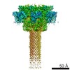 6224MC  6225C M: map data used to model this data C: citing same article ( |
|---|---|
| Similar structure data |

-Links
 PDBj
PDBj


- Assembly
| Deposited unit | 
|
|---|---|
| 1 | x 7
|
| 2 |
|
| 3 | 
|
| Symmetry | Point symmetry: (Schoenflies symbol: C7 (7 fold cyclic)) |
-Components
| #1: Protein | Mass: 63019.012 Da / Num. of mol.: 1 / Fragment: C-terminal 63-kDa fragment (UNP residues 203-764) Source method: isolated from a genetically manipulated source Source: (gene. exp.) |
|---|---|
| #2: Chemical |   Mass: 40.078 Da / Num. of mol.: 2 / Source method: obtained synthetically / Formula: Ca Mass: 40.078 Da / Num. of mol.: 2 / Source method: obtained synthetically / Formula: Ca |
-Experimental details
-Experiment
| Experiment | Method: ELECTRON MICROSCOPY |
|---|---|
| EM experiment | Aggregation state: PARTICLE / 3D reconstruction method: single particle reconstruction |
- Sample preparation
| Component |
| |||||||||||||||
|---|---|---|---|---|---|---|---|---|---|---|---|---|---|---|---|---|
| Molecular weight | Value: 0.44 MDa / Experimental value: NO | |||||||||||||||
| Buffer solution | Name: 50 mM NaOAc, pH 5.0, 0.05% Igepal CA-630 / pH: 5 / Details: 50 mM NaOAc, pH 5.0, 0.05% Igepal CA-630 | |||||||||||||||
| Specimen | Conc.: 0.05 mg/ml / Embedding applied: NO / Shadowing applied: NO / Staining applied: NO / Vitrification applied: YES | |||||||||||||||
| Specimen support | Details: Quantifoil R1.2/1.3 grid with thin carbon support, glow discharged | |||||||||||||||
| Vitrification | Instrument: FEI VITROBOT MARK IV / Cryogen name: ETHANE / Humidity: 100 % Details: Plunged into liguid nitrogen (FEI VITROBOT MARK IV). |
- Electron microscopy imaging
| Experimental equipment |  Model: Titan Krios / Image courtesy: FEI Company |
|---|---|
| Microscopy | Model: FEI TITAN KRIOS / Date: Jan 8, 2014 |
| Electron gun | Electron source:  FIELD EMISSION GUN / Accelerating voltage: 300 kV / Illumination mode: FLOOD BEAM FIELD EMISSION GUN / Accelerating voltage: 300 kV / Illumination mode: FLOOD BEAM |
| Electron lens | Mode: BRIGHT FIELD / Nominal magnification: 22500 X / Calibrated magnification: 39062 X / Nominal defocus max: 5100 nm / Nominal defocus min: 1800 nm / Cs: 2.7 mm / Camera length: 0 mm |
| Specimen holder | Specimen holder model: FEI TITAN KRIOS AUTOGRID HOLDER / Specimen holder type: Liquid nitrogen cooled |
| Image recording | Electron dose: 30 e/Å2 / Film or detector model: GATAN K2 (4k x 4k) / Details: Counting mode of the detector was used. |
| Image scans | Num. digital images: 7062 |
| Radiation | Protocol: SINGLE WAVELENGTH / Monochromatic (M) / Laue (L): M / Scattering type: x-ray |
| Radiation wavelength | Relative weight: 1 |
- Processing
| EM software | Name: RELION / Category: 3D reconstruction | ||||||||||||
|---|---|---|---|---|---|---|---|---|---|---|---|---|---|
| CTF correction | Details: Each particle | ||||||||||||
| Symmetry | Point symmetry: C7 (7 fold cyclic) | ||||||||||||
| 3D reconstruction | Method: Maximum a posteriori optimization / Resolution: 2.9 Å / Resolution method: FSC 0.143 CUT-OFF / Num. of particles: 60455 / Nominal pixel size: 1.28 Å / Actual pixel size: 1.28 Å / Details: (Single particle--Applied symmetry: C7) / Symmetry type: POINT | ||||||||||||
| Refinement step | Cycle: LAST
|
