Movie
Movie Controller


+ Open data

- Basic information
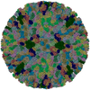
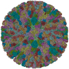
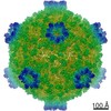
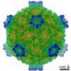
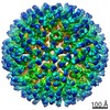
| Entry | Database: PDB / ID: 3j8d | ||||||
|---|---|---|---|---|---|---|---|
| Title | Cryoelectron microscopy of dengue-Fab E104 complex at pH 5.5 | ||||||
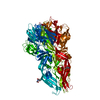
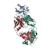
 Components Components |
| ||||||
 Keywords Keywords | VIRUS/IMMUNE SYSTEM /  Dengue virus / DENV2 Fab E104 / Low pH / fusion trimer / VIRUS-IMMUNE SYSTEM complex Dengue virus / DENV2 Fab E104 / Low pH / fusion trimer / VIRUS-IMMUNE SYSTEM complex | ||||||
| Function / homology |  Function and homology information Function and homology informationsymbiont-mediated suppression of host JAK-STAT cascade via inhibition of host TYK2 activity / host cell mitochondrion / flavivirin / symbiont-mediated suppression of host JAK-STAT cascade via inhibition of STAT2 activity / symbiont-mediated suppression of host cytoplasmic pattern recognition receptor signaling pathway via inhibition of MAVS activity / : / viral capsid / nucleoside-triphosphate phosphatase / double-stranded RNA binding / protein complex oligomerization ...symbiont-mediated suppression of host JAK-STAT cascade via inhibition of host TYK2 activity / host cell mitochondrion / flavivirin / symbiont-mediated suppression of host JAK-STAT cascade via inhibition of STAT2 activity / symbiont-mediated suppression of host cytoplasmic pattern recognition receptor signaling pathway via inhibition of MAVS activity / : / viral capsid / nucleoside-triphosphate phosphatase / double-stranded RNA binding / protein complex oligomerization / monoatomic ion channel activity / mRNA (guanine-N7)-methyltransferase / methyltransferase cap1 / clathrin-dependent endocytosis of virus by host cell / mRNA (nucleoside-2'-O-)-methyltransferase activity / mRNA 5'-cap (guanine-N7-)-methyltransferase activity / RNA helicase activity / host cell endoplasmic reticulum membrane / host cell perinuclear region of cytoplasm / protein dimerization activity / RNA helicase / induction by virus of host autophagy / RNA-directed RNA polymerase / viral RNA genome replication / RNA-dependent RNA polymerase activity / serine-type endopeptidase activity / fusion of virus membrane with host endosome membrane / viral envelope / symbiont-mediated suppression of host type I interferon-mediated signaling pathway / host cell nucleus / structural molecule activity / virion attachment to host cell / virion membrane / ATP hydrolysis activity / proteolysis / extracellular region / ATP binding / membrane / metal ion bindingSimilarity search - Function | ||||||
| Biological species |  Dengue virus 2 Thailand/16681/84 Dengue virus 2 Thailand/16681/84 Mus musculus (house mouse) Mus musculus (house mouse) | ||||||
| Method | ELECTRON MICROSCOPY / single particle reconstruction / cryo EM / Resolution: 26 Å | ||||||
 Authors Authors | Zhang, X.Z. / Sheng, J. / Austin, S.K. / Hoornweg, T. / Smit, J.M. / Kuhn, R.J. / Diamond, M.S. / Rossmann, M.G. | ||||||
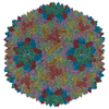
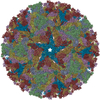
 Citation Citation | Journal: J Virol / Year: 2015 Title: Structure of acidic pH dengue virus showing the fusogenic glycoprotein trimers. Authors: Xinzheng Zhang / Ju Sheng / S Kyle Austin / Tabitha E Hoornweg / Jolanda M Smit / Richard J Kuhn / Michael S Diamond / Michael G Rossmann /   Abstract: Flaviviruses undergo large conformational changes during their life cycle. Under acidic pH conditions, the mature virus forms transient fusogenic trimers of E glycoproteins that engage the lipid ...Flaviviruses undergo large conformational changes during their life cycle. Under acidic pH conditions, the mature virus forms transient fusogenic trimers of E glycoproteins that engage the lipid membrane in host cells to initiate viral fusion and nucleocapsid penetration into the cytoplasm. However, the dynamic nature of the fusogenic trimer has made the determination of its structure a challenge. Here we have used Fab fragments of the neutralizing antibody DV2-E104 to stop the conformational change of dengue virus at an intermediate stage of the fusion process. Using cryo-electron microscopy, we show that in this intermediate stage, the E glycoproteins form 60 trimers that are similar to the predicted "open" fusogenic trimer. IMPORTANCE: The structure of a dengue virus has been captured during the formation of fusogenic trimers. This was accomplished by binding Fab fragments of the neutralizing antibody DV2-E104 to the ...IMPORTANCE: The structure of a dengue virus has been captured during the formation of fusogenic trimers. This was accomplished by binding Fab fragments of the neutralizing antibody DV2-E104 to the virus at neutral pH and then decreasing the pH to 5.5. These trimers had an "open" conformation, which is distinct from the "closed" conformation of postfusion trimers. Only two of the three E proteins within each spike are bound by a Fab molecule at domain III. Steric hindrance around the icosahedral 3-fold axes prevents binding of a Fab to the third domain III of each E protein spike. Binding of the DV2-E104 Fab fragments prevents domain III from rotating by about 130° to the postfusion orientation and thus precludes the stem region from "zipping" together the three E proteins along the domain II boundaries into the "closed" postfusion conformation, thus inhibiting fusion. | ||||||
| History |
|
- Structure visualization
| Movie |
Movie viewer |
|---|---|
| Structure viewer | Molecule: MolmilJmol/JSmol |
- Downloads & links
-Download
| PDBx/mmCIF format | 3j8d.cif.gz | 82.2 KB | Display | PDBx/mmCIF format |
|---|---|---|---|---|
| PDB format | pdb3j8d.ent.gz | 48.3 KB | Display | PDB format |
| PDBx/mmJSON format | 3j8d.json.gz | Tree view | PDBx/mmJSON format | |
| Others |  Other downloads Other downloads |
-Validation report
| Arichive directory | https://data.pdbj.org/pub/pdb/validation_reports/j8/3j8dftp://data.pdbj.org/pub/pdb/validation_reports/j8/3j8d | HTTPS FTP |
|---|
-Related structure data
| Related structure data |  6146MC M: map data used to model this data C: citing same article ( |
|---|---|
| Similar structure data |

-Links
 PDBj
PDBj


- Assembly
| Deposited unit | 
|
|---|---|
| 1 | x 60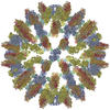
|
| 2 |
|
| 3 | x 5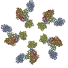
|
| 4 | x 6
|
| 5 | 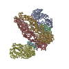
|
| Symmetry | Point symmetry: (Schoenflies symbol: I (icosahedral)) |
-Components
| #1: Protein | Mass: 10616.091 Da / Num. of mol.: 2 / Fragment: SEE REMARK 999 Source method: isolated from a genetically manipulated source Source: (gene. exp.) Dengue virus 2 Thailand/16681/84 / Production host: unidentified (others) / References: UniProt: P17763*PLUS#2: Antibody | Mass: 46637.730 Da / Num. of mol.: 2 / Source method: isolated from a natural source / Source: (natural) Mus musculus (house mouse)#3: Protein | / Coordinate model: Cα atoms onlyMass: 43819.391 Da / Num. of mol.: 3 / Fragment: UNP residues 281-674 Source method: isolated from a genetically manipulated source Source: (gene. exp.) Dengue virus 2 Thailand/16681/84 / Production host: unidentified (others) / References: UniProt: P12823, UniProt: P29990*PLUSSequence details | THE MODELED SEQUENCE FOR GLYCOPROTE | |
|---|
-Experimental details
-Experiment
| Experiment | Method: ELECTRON MICROSCOPY |
|---|---|
| EM experiment | Aggregation state: PARTICLE / 3D reconstruction method: single particle reconstruction |
- Sample preparation
| Component |
| ||||||||||||||||||||
|---|---|---|---|---|---|---|---|---|---|---|---|---|---|---|---|---|---|---|---|---|---|
| Details of virus | Empty: NO / Enveloped: YES / Host category: VERTEBRATES / Isolate: STRAIN / Type: VIRION | ||||||||||||||||||||
| Natural host | Organism: Homo sapiens | ||||||||||||||||||||
| Buffer solution | Name: 120 mM NaCl, 20 mM Tris HCl, 1 mM EDTA / pH: 5.5 / Details: 120 mM NaCl, 20 mM Tris HCl, 1 mM EDTA | ||||||||||||||||||||
| Specimen | Conc.: 1 mg/ml / Embedding applied: NO / Shadowing applied: NO / Staining applied: NO / Vitrification applied: YES | ||||||||||||||||||||
| Vitrification | Instrument: HOMEMADE PLUNGER / Cryogen name: ETHANE / Details: Plunged into liquid ethane |
- Electron microscopy imaging
| Experimental equipment |  Model: Titan Krios / Image courtesy: FEI Company |
|---|---|
| Microscopy | Model: FEI TITAN KRIOS / Date: Jan 1, 2013 |
| Electron gun | Electron source: FIELD EMISSION GUN / Accelerating voltage: 300 kV / Illumination mode: FLOOD BEAM |
| Electron lens | Mode: BRIGHT FIELDBright-field microscopy / Cs: 2.7 mm / Camera length: 0 mm |
| Specimen holder | Specimen holder model: FEI TITAN KRIOS AUTOGRID HOLDER |
| Image recording | Electron dose: 24 e/Å2 / Film or detector model: GATAN ULTRASCAN 4000 (4k x 4k) |
| Image scans | Num. digital images: 1000 |
| Radiation | Protocol: SINGLE WAVELENGTH / Monochromatic (M) / Laue (L): M / Scattering type: x-ray |
| Radiation wavelength | Relative weight: 1 |
- Processing
| EM software |
| |||||||||||||||||||||
|---|---|---|---|---|---|---|---|---|---|---|---|---|---|---|---|---|---|---|---|---|---|---|
| Symmetry | Point symmetry: I (icosahedral) | |||||||||||||||||||||
| 3D reconstruction | Resolution: 26 Å / Resolution method: FSC 0.143 CUT-OFF / Num. of particles: 528 / Nominal pixel size: 5.2 Å / Actual pixel size: 5.2 Å / Details: (Single particle--Applied symmetry: I) / Symmetry type: POINT | |||||||||||||||||||||
| Atomic model building | Protocol: RIGID BODY FIT / Space: RECIPROCAL / Details: REFINEMENT PROTOCOL--rigid body | |||||||||||||||||||||
| Atomic model building |
| |||||||||||||||||||||
| Refinement step | Cycle: LAST
|